Myoepithelioma of
minor salivary gland - An
immunohistochemical analysis
of four cases
Summary
Éricka Janine Dantas da Silveira1, Antonio Luiz
Amaral Pereira2, Maria Carmen Fontora3, Lélia
Batista de Souza4, Roseana de Almeida Freitas5
1 MS in Oral Pathology -UFRN, PhD student in Oral Pathology - UFRN.
2 PhD student at the Postgraduate Program in Oral Pathology - Federal University of Rio Grande do Norte. 3 PhD student at the Postgraduate Program in Oral Pathology - Federal University of Rio Grande do Norte. 4 PhD, Professor - Postgraduate Program in Oral Pathology - Federal University of Rio Grande do Norte. 5 PhD, Professor - Postgraduate Program in Oral Pathology - Federal University of Rio Grande do Norte.
Federal University of Rio Grande do Norte.
Mailing Address: Roseana de Almeida Freitas - Universidade Federal do Rio Grande do Norte Departamento de Odontologia Programa de Pós-Graduação em Patologia Oral - Av. Senador Salgado Filho 1787 Lagoa Nova 59056-000 Natal RN.
Tel/Fax: (0xx84) 3215-4138 - E-mail: [email protected]
Paper submitted to the ABORL-CCF SGP (Management Publications System) on December 7th, 2005 and accepted for publication on May 10th, 2006.
I
ntroduction and Methods: We performed an immunohistochemical study in four cases of myopitheliomas with objective to realize a profile in respect of differentiation grade by the monoclonal antibodies CK14, vimentin and alph-SMA, besides to investigate the cell proliferation by anti-PCNA, besides, we compare the immunoreactive with glandular normal tissue. Results: In the glandular normal tissue the myoepithelials cells had shown expression for alpha-SMA and CK 14, while that in the ductals cells, only the presence of CK 14 was verified. All the cases was verified positivy for CK 14 and vimentin, however, CK 14 had been present only in epithelioid and fusiform cells, while that the vimentin revealed positive also in the cytoplasm of the plasmocytoid cells. alpha-SMA was not detected in the neoplasic cells. Immunopositivity for the PCNA was observed in more than 75% of the cellular component of the analyzed tumors, independent of the cellular type. Conclusions: We concluded that it did not have difference in the proliferative activity among the cellular types presents in the myoepitheliomas and, still, the results of this study suggest that the constituent cells of this neoplasia one really represent cells of the mioepitelial ancestry, but in different stages of differentiation.Keywords: minor salivary gland, immunohistochemical, myo-epithelioma.
ORIGINAL ARTICLE
INTRODUCTION
Myoepitheliomas are rare benign neoplasias of the
salivary gland, more commonly found in the parotid1,2,
be-ing responsible for less than 7% of salivary gland tumors.
It was first described in 19433,4. Such lesion shows a varied
pattern of morphological growth, it may be solid, myxoid or reticular. It differs from the pleomorphic adenoma
be-cause it does not bear any ductal component5.
This tumor has varied cell morphology, being
fu-siform, plasmocytoid, epidermoid or clear cells6. Some
studies have shown that fusiform cells have some muscular
differentiation because they react to a-SMA and vimentin4,
while other investigations did not show any muscular
differentiation in the plasmocytoid cells4,7. According to
Jaeger et al. (1997), these cells could have origins other than myoepithelial, and they lost or had changed their capacity to express muscular evidence markers.
Besides the many phenotypes myoepithelioma cells may have, some authors report that fusiform and clear cells have a higher prolipherative capacity, when compared to the plasmocytoid cells, and they also stated that the pro-duction of myxoid material would be related to the low
prolipherative activity of these tumors9.
Medical literature mentions the use of different markers for histogenetic-related prolipherative activity both in the normal salivary gland and also in the glandu-lar benign and malignant tumors. Thus, we should use immunohistochemistry to analyze differentiation patterns and the prolipherative activity of the different cell types present in salivary gland myoepitheliomas.
MATERIALS AND METHODS
We selected 4 cases of small salivary gland myoepi-theliomas from the files of the Pathology lab of the Oral Pathology Department of the Federal University of Rio Grande do Norte. Paraffin-bounded specimens were cut in 5mm thickness slices and hematoxylin-eosin was used for the cellular morphology analysis.
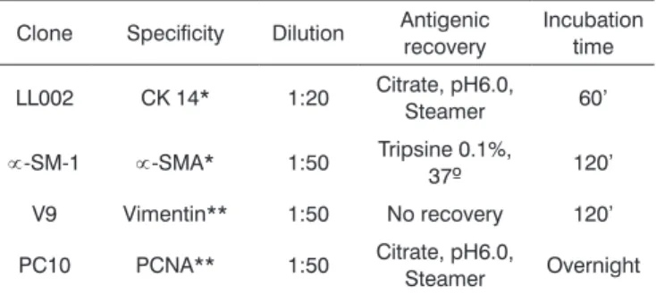
An immunohistochemical study by the strepta-vidine-biotin technique was carried out using antibodies against vimentin, a-SMA, CK 14 and PCNA (Cell Prolifera-tion Nuclear Antigen). Chart 1 lists the clones, antigenic recovery, dilution, incubation time and manufacturers of the antibodies used.
All the material selected was fixed in formaldehyde and embedded in paraffin, histological cross-sections of
3μm were made and placed on slides adhered by
3-amino-propyltriethoxy-silane (Sigma Chemical CO., St. Louis, MO, USA). The histology cross-sections were deparaffined in xylol, rehydrated in an alcohol sequence up to water and washed in two distilled water vials for 5 minutes each. En-dogenous peroxidase was blocked by hydrogen peroxide 20vol, flushed with water and incubated in TRIS-HCL
(Tris-Chart 1. Antibodies used.
Clone Specificity Dilution Antigenicrecovery Incubation time
LL002 CK 14* 1:20 Citrate, pH6.0, Steamer 60’
∝-SM-1 ∝-SMA* 1:50 Tripsine 0.1%, 37º 120’ V9 Vimentin** 1:50 No recovery 120’
PC10 PCNA** 1:50 Citrate, pH6.0,
Steamer Overnight
hydroximethil-aminomethane), pH 7.4 for 10 minutes. The cross-sections were incubated with anti-mouse monoclonal antibody, diluted in a TRIS-HCL buffer solution (Chart 1), for incubation with the streptoavidine-Biotin complex, in a 1:100 dilution for 30 minutes. For development purposes, we used a 0.03% diaminebenzidine cromogen solution, diluted in TRIS-HCL added to 0.6ml of 20vol hydrogen peroxide in a dark chamber for 3 minutes. For counter-coloring we used Mayer Hematoxylin for 10 min, flushing in water after each step. To finish the process we used alcohol for dehydration and diaphanization in xylol for slide preparation with Permount.
Fragments of normal salivary gland were used as internal positive control and for comparative purposes.
The immunopositiviness analysis was carried out by two examiners at different times, in a double-blinded study through light microscopy, and all the Brown col-ored cells in their cytoplasm or nucleus were considered positive (PCNA). Thus we investigated the presence or absence of markers, assigning the following scores: - (no marker); + (focal marker, less than 10% of cells marked) and ++ (diffuse marking).
RESULTS
Table 1 lists the patients’ clinical data.
Morphological Results
Tumors were well circumscribed, and we frequently found a fibrous connective tissue capsule surrounding the specimens. The four tumors presented a predominantly solid growth and organizational patterns. The specimens were made up of nests of cohesive and non-cohesive cells in a matrix that varied between hyaline and myxoid. Tumor cells showed different morphologies, frequently fusiform, polygonal of eosinophilic cytoplasm (epithelioid) and, sometimes, with hyper chromatic nucleus. No tumor had necrotic areas; although in one case we did find squamous metaplasia and calcifications.
Immunohistochemical results
had immunopositiviness for CK 14 and vimentin. How-ever, CK 14 was present only in epithelioid and fusiform cells, while vimentin was also present in the cytoplasm of plasmocytoid cells. a-SMA was not detected in neoplastic cells, although it was present in the blood vessels, and they were used as internal positive control.
Immunopositiviness for PCNA was seen in more than 75% of cell components belonging to the analyzed tumors, regardless of cell type and the amount of stroma present.
Next to 2 of the 4 myoepitheliomas analyzed there was normal glandular tissue, predominantly made of ducts and mucous acini. In this tissue, the myoepithelial cells showed markers for a-SMA and CK 14. Immune mark-ing for CK 14 was detected in ductal cells. Table 2 lists the immunoreactions for the analyzed myoepithelioma antibodies.
DISCUSSION
The benign myoepithelioma of salivary gland is a rare neoplasia of myoepithelial nature, made up of
fusiform, epithelioid, plasmocytoid and clear cells10. For
some authors such as Simpson et al. (1995) this neoplasia
Table 1. Clinical data of the myoepithelioma cases studied.
Case Gender Age Anatomic location diagnosisClinical Size
1 Male years39 Hard palate pleomorphicAdenoma 2,5 cm
2 Male years80 Upper lip Lipoma 0,5 cm
3 Female 30
years Hard palate
Adenoma
pleomorphic 0,5 cm
4 Male 31
years Hard palate
Adenoma
pleomorphic 2,0 cm
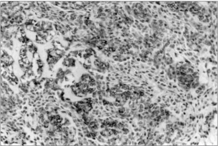
Figure 1. CK 14 expression in epithelioid and fusiform cells in small salivary gland myoepitheliomas (SABC, 200x).
Figure 2. Vimentin immonomarking in small salivary gland myoepi-thelioma (SABC, 200x).
is but a rare type of pleomorphic adenoma. However, according to Dardick et al. (1995), it is different from the pleomorphic adenoma because it bears little or no ductal component.
An interesting aspect about myoepitheliomas is the scarce information we have about its biological behavior, because of its low incidence rates. Some authors consider
it to be more aggressive than pleomorphic adenomas11,
while another investigation noticed, through PCNA expres-sion, that there were no differences as far as prolipher-ative activity is concerned between myoepitheliomas and
pleomorphic adenomas9.
Table 2. Immunohistochemical findings in our cases of small salivary gland myoepitheliomas.
Case CK 14 ∝-SMA Vimentina PCNA
1 + - ++ ++
2 + - ++ ++
3 + - + ++
4 + - + ++
-: no mark, +: focal mark, ++ diffuse and intense mark
Ogawa et al. (1993) reported that cell kinetics is related to the biological behavior of many tumor types, and they noticed in their study that myoepitheliomas made up of fusiform, epithelioid and clear cells have greater rates of PCNA cells when compared to the plasmocytoid type, thus confirming the low prolipherative activity in this type. Such findings disagree from the ones in the present investigation because here the PCNA is marked in over 75% of the tumors analyzed, regardless of cell type.
and El-Naggar (1999) these cells are involved in many processes related to neoplastic growths, such as differ-entiation of tumor cells, synthesis of basal membrane and maspin tumoral suppressor, besides also inhibiting invasion and angiogenesis. Capuano and Jaeger (2004) reported the presence of matrix constituents, such as lam-inin, for example, that can induce morphologic changes in myoepitheliomas, causing a phenotype made up of plasmocytoid cells.
According to Jaeger et al. (1997) a reasonable pro-portion of fusiform cells in the myoepitheliomas point toward muscular differentiation, reacting positively for a-SMA and vimentin, and these are called “myoepithelial like”.
Reports by Ellis and Auclair (1996) state that except for fusiform cells and some epithelioids, the true nature of myoepitheliomas plasmocytoid is still unclear. Jaeger et al. (1997) also reported that these cells did not show any evidence of muscular differentiation. Ogawa et al. (2003) believe that these cells are originated from luminal cells and not myoepithelial, also adding that plasmocytoid-cell tumors could be classified as adenomas or plasmocytoid adenocarcinomas.
Immunohistochemical markers for myoepithelial cells have included protein S-100, GFAP, cytokeratin and vimentin, and also a-SMA, which is associated to
myo-epithelial cells of the normal salivary gland7. Araújo et al.
(1994) consider vimentin a marker of neoplastic myoepi-thelial cell. However, studies performed by this group in
200114, suggest that this protein does not happen solely in
neoplastic myoepithelial cells, since it may be present also in other cell types originated from the intercalate duct.
According to Hornick and Futcher (2004), neoplas-tic myoepithelial cells frequently lose the expression of muscular differentiation markers, when the immunoreac-tivity to these markers is not required in order to confirm myoepithelial differentiation.
Thus, the low expression of muscle tissue markers has led the authors to consider plasmocytoid myoepithelio-mas as true myoepitheliomyoepithelio-mas, despite the fact that Franque-mont and Mills (1993) do not believe in the myoepithelial nature of this cell type, suggesting that the plasmocytoid subtype would not be a type of myoepithelioma.
Having so much debate in the literature as to the true nature of myoepithelioma cells, the present study analyzed the differentiation and the nature of small salivary gland myoepithelioma cell components, using epithelial cell differentiation markers such as CK 14 and of muscle tissue differentiation such as a-SMA and vimentin.
Cytokeratin are proteins of the intermediary fila-ments of epithelial cells associated to the differentiation and organization of the cytoskeleton, and they are ex-pressed in specific epithelial cells depending on their differentiation stage. According to Ogawa et al. (1999)
and Ogawa et al. (2000), salivary gland myoepithelial cells Express CK 5 and 14; stressing the fact that these do not serve as specific markers for such cells because they are also expressed by basal ductal cells, where CK 18 and 19 are also present.
CK 14 was present in myoepithelial cells (non-lumi-nal), of fusiform, epithelioid and plasmocytoid morphology in the pleomorphic adenomas studied by Ogawa et al. (2003), and it disagrees from the findings of the present investigation in which CK 14 was only immunomarked in fusiform and epithelioid cells, and such fact also disagrees from the findings by Araújo et al. (2001) where CK 14 and 19 marks were found in plasmocytoid cells and not in fusiform cells.
The vimentin found in all the cell types of the pres-ent study was also prespres-ent in all myoepithelioma cell types analyzed in the investigation by Araújo et al. (2001) and, occasionally, in non-luminal cells of the pleomorphic ad-enomas studied by Ogawa et al. (2003). The latter authors stress the hypothesis that this protein is not an exclusive maker of the neoplastic myoepithelial cell, justifying that it is necessary for the migration of epithelial cells in both physiologic and pathologic processes, and vimentin is also present during the development after this process in some segments of the normal salivary gland.
The lack of a-SMA in the cases hereby studied and the immunoreactivity for vimentin were also detected by Savera et al. (1997) and Ogawa et al. (2003), respec-tively.
With such findings, we believe that all morphologic types of myoepithelioma cells represent cells of the myo-epithelial strain in different stages of evolution, disagree-ing from the conclusions reached by Ogawa et al. (2003). The controversies present in the literature regarding the expression of muscle-tissue markers in fusiform, epithelioid and plasmocytoid cells may be justified by the fact that such cells may show different stages of differentiation, or they may have lost or changed their capacity of produc-ing muscle-tissue markers. Moreover, it may be suggested that the low muscle-tissue differentiation seen “in vivo” in some cells which make up the myoepitheliomas may be caused by the inhibitory process mediated by the extra-cellular matrix.
REFERENCES
1. Das DK, Haji BE, Ahmed MS, Hossain MN. Myoepithelioma of the parotid gland initially diagnosed by fine needle aspiration cytology and immunocytochemistry: a case report. Acta Cytol 2005;49(1):65-70.
2. Lee MW, Nam SY, Choi HJ, Choi JH, Moon KC, Koh JK. Myoepithe-lioma of parotid gland presenting as infra-auricular subcutaneous mass. J Cutan Pathol 2005;32(3):240-4.
3. Simpson RHW, Jones H, Beasley A. Benign myoepithelioma of the salivary glands: a true entity? Histopathol 1995;27:1-9.
myoepitheliomas. Immunohistochemical detection of muscle-specific actin, cytokeratin 14, vimentin and glial fibrillary protein. Oral Surg Oral Med Oral Pathol Oral Radiol Endod 1995;79:330-41.
5. Dardick I. Myoepithelioma: definitions and diagnostic criteria. Ultra-struct Pathol 1995;19:335-45.
6. Capuano AC, Jaeger RG. The effect of laminin and its peptide SIKVAV on a human salivary gland myoepithelioma cell line. Oral Oncol 2004;40(1):36-42.
7. Araújo VC, Carvalho YR, Araújo NS. Actin versus vimentin in myo-epithelial cells of salivary gland tumors. A comparative study. Oral Surg Oral Med Oral Pathol Oral Radiol Endod 1994;77(4):387-94. 8. Jaeger RG, de Oliveira PT, Jaeger MM, de Araujo VC. Expression of
smooth-muscle in cultured cells from human plasmocytoid myo-epithelioma. Oral Surg Oral Med Oral Pathol Oral Radiol Endod 1997;84(6):663-667.
9. Ogawa I, Miyauchi M, Takata T, Vuhahula E, Ijuhin N, Nikai H. Proliferative activity if salivary gland pleomorphic adenomas and myoepitheliomas as evaluated by the proliferating cell nuclear antigen (PCNA) labeling index (LI). J Oral Pathol Med 1993;22(10):447-50. 10. Ellis GL, Auclair PL. Tumors of the salivary glands. Atlas of tumor
pathology, 3rd series, fascicle 17. Washington, DC: Armed Forces Institute of Pathology; 1996.
11. Seifert G, Sobin LH. Histological typing of salivary gland tumors. Word Health Organization International Classification of tumors. 2nd ed. Springer Verlag. Berlin Heidelberg New York; 1998.
12. Batsakis JG., El-Naggar AK. Myoepithelium in salivary and mammary neoplasms is host-friendly. Adv Anat Pathol 1999;6:218-26. 13. Ogawa Y, Kishino M, Atsumi Y, Kimoto M, Fukuda Y, Ishida T et al.
Plasmacytoid cells in salivary-gland pleomorphic adenomas: evidence of luminal cell differentiation. Virc Arch 2003;443(5):625-34. 14. Araujo VC, de Sousa SO, Carvalho YR, de Araujo NS. Application of
immunohistochemistry to the diagnosis of salivary gland tumors. Ap-plied Immunohistochemistry & Molecular Morphology 2001;8(3):195-202.
15. Hornick JL, Fletcher CDM. Cutaneous myoepithelioma: A clinico-pathologic and immunohistichemical study of 14 cases. Human Pathol 2004;35(1):14-24.
16. Franquemont DW, Mills G. Plasmocytoid monomorphic adenoma of salivary glands. Absence of myogenous differentiation and compari-son to spindle cell myoepithelioma. AM J Surg Pathol 1993;17:146-53.
17. Ogawa Y, Yamauchi S, Ohnishi A, Ito R, Ijuhin N. Immunohistochem-istry of myoepithelial cells during development of the rat salivary glands. Anat Embryol 1999;200(2):215-28.
18. Ogawa Y, Toyosawa S, Ishida T, Ijuhin N. Keratin 14 immunoreac-tive cells in pleomorphic adenomas and adenoid cystic carcinomas of salivary glands. Virch Arch 2000;437(1):58-68.