Fos expression in the prefrontal cortex and mesencephalic
dorsal raphe nucleus in lactating rats after social instigation
Caroline Perinazzo da Veiga
1, Elisa Winkelmann-Duarte
2, Eloisa Pavesi
2, Simone Mattos
Louzada
1, Klaus A. Miczek
3, Aldo Bolten Lucion
1, and Rosa Maria Martins de Almeida
11- Universidade Federal do Rio Grande do Sul, Porto Alegre, RS, Brazil
2- Universidade Federal de Santa Catarina, Florianópolis, SC, Brazil
3- Tufts University, Boston, MA, USA
Abstract
Females are often less aggressive than males, but they exhibit high levels of agonistic behavior against an intruder in the area of the nest during lactation. This behavior is referred to as maternal aggression. In rats, maternal aggressive behavior occurs more often from postpartum day 3 (PPD 3) to PPD 12. Social instigation is an experimental protocol used to increase the levels of aggression that are typical of the species. In the present study we used social instigation to analyze the expression of a marker of neuronal activity, c-fos. Lactating rats on PPD 5, in the presence of their pups, were divided into four groups: (1) no social instigation and no aggressive behavior, (2) social instigation and no aggressive behavior, (3) no social instigation and aggressive behavior, and (4) social instigation and aggressive behavior. Sixty minutes after the aggression test we used immunohistochemistry to detect Fos in two brain regions, the ventral-orbital region of the prefrontal cortex (VO PFC) and dorsal raphe nucleus (DRN). Our results showed that rats with aggressive behavior that were provoked exhibited an increase in Fos expression in the VO PFC compared with the control group (i.e., no social instigation and no aggressive behavior). No change in Fos expression was found in the
DRN. These results complement previous indings with microinjection of serotonin 5-hydroxytryptamine-1B receptor agonists
into the same region, demonstrating that the VO PFC is an important region in the modulation of maternal aggressive behavior.
Keywords: social instigation, maternal aggression, Fos, prefrontal cortex, dorsal raphe nucleus.
Received 11 September 2012; received in revised form 5 January 2013; accepted 18 February 2013. Available online 27 June 2013.
Caroline Perinazzo da Veiga, Programa de Pós-Graduação em Neurociências, Instituto de Ciências Básicas da Saúde, Universidade Federal do Rio Grande do Sul, Porto Alegre, RS, Brazil. Elisa Winkelmann-Duarte, Departamento de Ciências Morfológicas, Centro de Ciências Biológicas, Universidade Federal de Santa Catarina, Florianópolis, SC, Brazil. Eloisa Pavesi, Departamento de Farmacologia, Universidade Federal de Santa Catarina, Campus Universitário - Trindade, Florianópolis, SC, Brazil. Simone Mattos Louzada, Laboratório de Neuroendocrinologia do Comportamento, Instituto de Ciências Básicas da Saúde, Universidade Federal do Rio Grande do Sul, Porto Alegre, RS, Brazil. Klaus A. Miczek, Departments of Psychology, Pharmacology, Neuroscience, and Psychiatry, Tufts University, Medford and Boston, MA, USA. Aldo Bolten Lucion,
Introduction
The behavioral repertoire of females during maternity changes drastically compared with other periods of the reproductive cycle (Ferreira, Pereira, Agrati, Uriarte, & Fernández-Guasti, 2002). Motherhood involves changes in the perception of mothers toward offspring (Kinsley & Bridges, 1990; Rosenblatt, 1975), providing a link between mothers and offspring
and causing signiicant changes in the response of
mothers to environmental stimuli (Agrati, Zuluaga,
Fernández-Guasti, Meikle, & Ferreira, 2008). Females are often less aggressive than males, but they exhibit high levels of agonistic behavior against an intruder in the area of the nest during lactation (Bridges, 1996;
Erskine, Barield, & Goldman, 1980; Giovenardi,
Consiglio, Barros, & Lucion, 2000; Olivier & Young, 2002; Lonstein & Gammie, 2002). In rats, maternal aggressive behavior occurs more frequently from postpartum day 3 (PPD 3) to PPD 12. During this period, females direct intense care toward their pups
exhibit a signiicant increase in the levels of aggression when subjected to social instigation (Veiga, Miczek,
Lucion, & De Almeida, 2011).
Analyses of the expression of immediate early genes such as c-fos have been used to understand the neural basis of maternal behavior (Gammie & Nelson, 2001). In rats, aggressive confrontations activate Fos protein in the prefrontal cortex (PFC; Halász, Tóth, Kalló, Liposits, & Haller, 2006; Haller, Tóth, Halász, & De Boer, 2006). The ventro-orbital PFC (VO PFC) has
been identiied as an important area for the inhibitory
control of subcortical circuits that mediate aggressive and impulsive behavior (Blair, 2001, 2004; Cardinal, Winstanley, Robbins, & Everitt, 2004; Kheramin et al., 2005; Séguin, 2004; Veiga, Miczek, Lucion, & De Almeida, 2007). Another region that has been implicated in the modulation of maternal aggression is the dorsal raphe nucleus (DRN). Our group
recently showed that microinjection of the serotonin
5-hydroxytryptamine-1A (5-HT1A) receptor agonist 8-OH-DPAT into the DRN increased aggressive behavior (Veiga et al., 2011).
The majority of studies on aggression have focused
on physiological mechanisms (Davis & Marler, 2004), but little is known about the neural mechanisms of maternal aggressive behavior (Consiglio, Borsoi, Pereira, & Lucion, 2005; De Almeida, & Lucion, 1997; Factor, Mayer, & Rosenblatt, 1993; Ferreira, Dahlof, & Hansen, 1987; Giovenardi, Padoin, Cadore, & Lucion, 1998; Insel, 1986; Lonstein & Gammie, 2002; Nelson & Trainor, 2007; Russel & Leng, 1998; Svare, 1990). The aim of the present study was to evaluate VO PFC
and DRN activation relected by Fos expression in lactating rats that were subjected to social instigation
and aggressive behavior.
Materials and Methods
Animals
We used primiparous female Wistar rats ~90 days of age from the Universidade Federal do Rio Grande do Sul. Each rat was placed and kept individually in transparent acrylic boxes (46 cm × 31 cm × 17 cm) with free access to water and food. The date of birth of the pups was controlled and established as PPD 0. On PPD 1, the pups were standardized to eight per litter, regardless of the sex of the pups. The animals were
Resident intruder confrontation
On PPD 3, females were selected for aggressive behavior as in Veiga et al. (2011). The behavioral test was conducted in the female resident’s box in the presence of the pups at the beginning of the dark period.
Social instigation
Social instigation was performed on PPD 5 (Figure 1). Social instigation consisted of placing an acrylic tube with holes (28 cm length, 10 cm diameter) that contained an opponent (stimulus) male or instigator (Figure 1A) for 5 min in the resident female’s box (Veiga et al., 2011). Residents typically threaten the protected stimulus male and attack the perforated cylindrical tube. The pups remained in the box with their mothers during social instigation.
Maternal aggressive behavior
Five minutes after the social challenge on PPD 5, maternal aggressive behavior against a male intruder was assessed for 10 min. The behavioral repertoire that we analyzed was described previously (De Almeida & Lucion, 1997). Social instigation and aggressive behavior were videotaped and later analyzed by the experimenter using Observer software (version 3.0, Noldus, The Netherlands). We determined the frequency of aggressive behaviors including lateral attacks, bites on the body, and an intrusive and aggressive stance.
Lactating rats were divided into the following groups: (1) no social instigation and no aggressive behavior (NI + NA; the acrylic tube was placed empty, without the stimulus rat, into the resident’s box, and
the rats were not subjected to maternal aggressive
behavior), (2) social instigation and no aggressive behavior (I + NA; the acrylic tube was placed into the resident’s box with the stimulus rat, and the rats
were not subjected to maternal aggressive behavior),
(3) no social instigation and aggressive behavior (NI + A; the acrylic tube was placed empty, without the stimulus rat, into the resident’s box, and the rats were
subjected to maternal aggressive behavior), and (4)
social instigation and aggressive behavior (I + A; the acrylic tube was placed into the resident’s box with the
stimulus rat, and the rats were subjected to maternal
Immunohistochemical detection of Fos protein immunoreactivity
On PPD 5, 80 min after the beginning of social instigation, the four experimental groups of rats were analyzed for Fos protein expression in the DRN and
VO PFC. The control group was not subjected to social
instigation or aggressive behavior before the beginning of this experimental phase, with only an empty tube placed inside the resident’s cage. Simultaneously, the rats were deeply anesthetized with sodium pentobarbital (50 mg/kg, i.p.), and transcardiac perfusion was
performed by injecting 150 ml saline followed by 4%
paraformaldehyde diluted in 150 mg of 0.1 M sodium phosphate buffer, pH 7.4. The brains were then removed
and kept in a 4% paraformaldehyde ixative solution for
8 h at room temperature. Subsequently, the brains were
cryoprotected in a 30% sucrose solution at 4°C. The brains were then cut into 50-μm-thick coronal sections using a cryostat and collected free-loating. The slices were kept
immersed in an antifreeze solution (phosphate-buffered saline [PBS] that contained distilled water, sucrose, and propylene glycol; Vetec, São Paulo, Brazil) and stored at -70ºC until the day of immunohistochemical Fos detection. All of the brains were processed together to detect Fos protein using avidin-biotin-peroxidase as in Cezario, Ribeiro-Barbosa, Baldo, & Canteras (2008).
The brain sections were washed three times for 10 min each in PBS and incubated in buffer solution with Triton X-100 (Sigma, St. Louis, MO, USA), normal goat serum, and rabbit anti-Fos antibody (Ab-5; Calbiochem, San Diego, CA, USA) at a 1:20,000 dilution for 48 h
at 4°C with continuous stirring. After this period, the
sections were incubated for 90 min at room temperature in a 1:250 solution of biotinylated goat immunoglobulin G anti-rabbit (Vector Laboratories, Burlingame, CA, USA), mixed, and then incubated for 90 min in a solution that contained avidin-biotin peroxidase at a dilution of 1:250 (ABC Elite Kit; Vector Laboratories). The visualization of the peroxidase complex occurred after exposure for 10 min in a chromogen solution that
contained 0.02% 3,3-diaminobenzidine (DAB; Sigma, St. Louis, MO, USA) with 0.3% nickel sulfate in 0.05
M Tris buffer (pH 7.6), followed by 5 min incubation in
a 0.01% solution of glucose oxidase (glucose oxidase
Type VII from Aspergillus niger; Sigma) and 10%
Pd-glucose (Sigma) to produce a dark blue color. For negative control of enzyme activity some sections were randomly processed by omitting the primary antibody. All sections were then washed with buffer, collected on previously gelatinized slides, dehydrated in ethanol, cleared with xylene, and mounted with coverslips using distyrene/plasticizer/xylene.
according to previous studies of Fos expression in rodents (Davis & Marler, 2004; Gammie & Nelson, 2001). Neurons outside the outer forbidden lines were not counted (Camozzato et al., 2009; Wilkelmann-Duarte et al., 2007). Slices were made from each region along its entire length with a 200-µm gap. In the VO PFC, slices were made 3.70-5.20 mm anterior to bregma. In the DRN, slices were made 7.04-8.0 mm posterior to bregma (Paxinos & Watson, 1998). For the DRN, the cell counts
relected the sum of the proximal and distal parts with
the aqueduct as a reference. Data are expressed as the average number of neurons in each region within the test
area. All quantiications of labeled cells were performed
by two experimenters unrelated to the study.
Statistical analysis
The results are expressed as mean ± SEM. Fos immunoreactivity results in the VO PFC and DRN in the four experimental groups (NI + NA, I + NA, NI + A, and I + A) were analyzed using one-way analysis of variance (ANOVA) followed by the Newman-Keuls post hoc test. Values of p < .05 were considered statistically
signiicant. Aggressive behavior in females in the two
groups tested for aggressiveness (NI + A and I + A) were analyzed using Student’s t-test.
Results
Microscopic analysis of Fos protein expression
in the VO PFC showed that lactating rats subjected to
social instigation and aggressive behavior exhibited
a statistically signiicant increase in the number
Fos-positive cells (F3,17 = 3.30, p < .05; Figures 2 and 3) compared with the control group (i.e., no social
instigation and no aggressive behavior). No signiicant
difference was found between groups in Fos expression in the DRN (F3,16 = .64, p = .5; Figures 4 and 5).
Socially instigated lactating rats subjected to aggressive confrontation exhibited a signiicant increase
in the frequency of lateral attacks (t9 = 2.43, p < .04; Figure 6) compared with the control group (i.e., no social instigation and no aggressive behavior).
Discussion
The present study showed that lactating rats
subjected to social instigation and aggressive behavior
Social instigation + no aggressive behavior ( =6)n
No social instigation + aggressive behavior ( =5)n
Social instigation + aggressive behavior ( =5)n
*: p<0.05 compared to NI + NA group
Figure 2. Mean (±SEM) number of Fos-immunoreactive cells in the
ventral-orbital (VO) prefrontal cortex in rats subjected to social insti -gation and aggressive behavior on postpartum day 5 in the presence of pups. *p < .05, compared with control group (no social instigation and no aggressive behavior).
Figure 3. Coronal sections of the rat brain that show the region of Fos-immunoreactive cells in the VO PFC in the (A) control group, (B) social instigation and no aggressive behavior group, (C) no social instigation and aggressive behavior group, and (D) social instigation
and aggressive behavior group. A signiicant increase in the number
of Fos-positive cells was found in the VO PFC, concomitant with the expression of maternal aggressive behavior (D).
against a male intruder in the presence of their pups on PPD 5 exhibited an increase in Fos protein expression
speciically in the VO PFC. No change was observed in
Fos expression in the DRN in lactating rats after exposure to social instigation or aggressive behavior. Analyses of the increased expression of transcription factors such as c-fos in association with speciic behaviors (e.g., maternal
aggression) have provided important indirect evidence of neural activity in a subregion of cells during the execution of a certain behavior (Gammie & Nelson, 2001).
No studies of which we are aware have demonstrated the brain regions that have increased Fos expression
Dorsal Raphe Nucleus
No social instigation + no aggressive behavior ( =6)n
Social instigation + aggressive behavior ( =4)n
No social instigation + aggressive behavior ( =6)n
Social instigation + aggressive behavior ( =4)n
Number of Fos-immunoreactive
30
25
20
15
10
5
0
Figure 4. Mean (±SEM) number of Fos-immunoreactive cells in the dorsal raphe nucleus (DRN), proximal and distal to the aqueduct, in
resident lactating rats subjected to social instigation and aggressive
behavior on postpartum day 5 in the presence of pups.
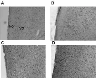
Figure 5. Coronal sections of the rat brain showing the DRN (proxi-mal, relative to the aqueduct [Aq]) where Fos-immunoreactive cells
were quantiied in the (A) control group (no social instigation and no
aggressive behavior), (B) social instigation and aggressive behavior group, (C) no social instigation and aggressive behavior group, and (D) social instigation and aggressive behavior group.
of aggression such as social instigation. Some studies have shown an increase in Fos expression in the medial amygdala, bed nucleus of the stria terminalis, paraventricular nucleus of the hypothalamus, and periaqueductal gray after maternal aggression (Gammie & Nelson, 2001). Parental male mice that take care of offspring exhibit an increase in Fos expression in the medial preoptic area, ventromedial hypothalamus, and medial amygdala (Trainor, Finy, & Nelson, 2008), and these same regions also express Fos in the context of maternal aggression (Gammie, 2005). Fos is expressed for only short periods of time (15-90 min) after exposure to a stimulus (Morgan, Cohen, Hempstead, & Curran, 1987; Morgan & Curran, 1991; Gammie & Nelson, 2001; Knyshevski, Connor, Harrison, Ricci, & Melloni, 2005). However, some studies have shown that Fos expression can persist, representing cellular activation in response to chronic physiological (Fenelon, Poulain, & Theodosis, 1993; Miyata, Nakashima, & Kiyohara, 1994) and behavioral (Fenelon et al., 1993; Matsuda, Peng, Yoshimura, Wen, Fukuda, & Sakanaka, 1996) stimuli.
Experiments conducted with male rats found that, compared to LO and VO, regions of the prefrontal cortex cause an increase in neuronal activation after aggressive encounters (Halasz et al., 2006), and this activation was particularly higher in male mice previously selected for high rates of aggression (Haller et al., 2006). Thus, the increased expression of Fos in the VO PFC in the present study is similar to previous studies.
The manifestations of abnormal aggressive behavior are related to or caused by decreased prefrontal cortex activity (Damasio, Grabowski, Frank, Galaburda, & Damasio, 1994; Volkow et al., 1995; Golden, Jackson, Peterson-Rhone, & Gontkovsky, 1996; Critchley et al., 2000; Davidson, Putnam, & Larson, 2000; Hawkins & Trobst, 2000; Bassarath, 2001; Best, Williams, & Coccaro, 2002; Veit et al., 2002; Blair, 2004; Halász et al., 2006). In contrast to these results, however, the present study found that social instigation followed by aggressive behavior increased neuronal activation in the VO PFC, and other studies have also shown similar results (Halász et al., 2006; Haller et al., 2006). Halász et al. (2006) argued that this discrepancy with
regard to PFC deiciency in humans who exhibit violent
behaviors are not found in rodents and can be explained by the differences between the acute and chronic functioning of the PFC. For example, acute activation as observed in the present study and chronic dysfunction in violent persons may affect aggressive behavior through independent mechanisms. Prefrontal cortex
deiciencies have been shown to alter moral judgments
in humans, and behavioral disturbances have been suggested to occur through this route (Price, Daffner, Stowe, & Mesulam, 1990; Nichelli, Grafman, Pietrini, Clark, Lee, & Miletich, 1995; Greene, Nystrom, Engell, Darley, & Cohen, 2004; Luo, Nakic, Wheatley, Richell,
Martin, & Blair, 2006). Thus, prefrontal deicits lead to violence via deiciencies in moral judgments (Halász
et al., 2006). In contrast, acute activation of the PFC
No social instigation + aggressive behavior ( =5)n
Social instigation + aggressive behavior ( =5)n
Frequency
30
25
20
15
10
5
0
* Lateral attack
30
25
20
15
10
5
0
In summary, the present results demonstrated
that lactating rats subjected to social instigation and
aggressive behavior on PPD 5 exhibited an increase in Fos expression in the VO PFC in the presence of their pups, but no changes in Fos expression were found in the DRN. Future experiments that analyze Fos immunohistochemistry in other brain regions in lactating
rats subjected to social instigation and aggressive
behavior may further elucidate the modulation and expression of maternal aggression.
References
Agrati, D., Zuluaga, M. J., Fernández-Guasti, A., Meikle, A., & Ferreira, A. (2008). Maternal condition reduces fear behaviors but not the endocrine response to an emotional threat in virgin female rats. Hormones and Behavior, 53(1), 232-240.
Bassarath, L. (2001). Neuroimaging studies of antisocial behavior.
Canadian Journal of Psychiatry, 46, 728-732.
Best, M., Williams, J. M., & Coccaro, E. F. (2002). Evidence for a dysfunctional prefrontal circuit in patients with an impulsive aggressive disorder. Proceedings of the National Academy of Sciences of the United States of America, 99, 8448-8453.
Blair, R. J. (2001). Neurocognitive models of aggression, the antisocial personality disorders, and psychopathy. Journal of Neurology, Neurosurgery and Psychiatry, 71, 727-731.
Blair, J. R. (2004). The roles of orbital frontal cortex in the modulation of antisocial behavior. Brain and Cognition, 55, 198-208. Bridges, R. (1996). Biochemical basis of paternal behavior in the
rat. In J. S. Rosenblatt, & C. T. Snowdon (Eds.), Parental care:
evolution, mechanisms, and adaptive signiicance (pp. 215-242). San Diego: Academic Press.
Camozzato, T. S., Winkelmann-Duarte, E. C., Padilha, C. B., Miguel, S. P., Bonzanini, L., Anselmo-Franci, J. A., Fernandes, M. C., & Lucion, A. B. (2009). Neonatal handling reduces the number of cells in the medial preoptic area of female rats. Brain Research,
247, 92-99.
Cardinal, R. M., Winstanley, C. A., Robbins, T. W., & Everitt, B. J. (2004). Limbic corticostriatal systems and delayed reinforcement.
Annals of the New York Academy of Sciences, 1021, 33-50. Cezario, A. F., Ribeiro-Barbosa, E. R., Baldo, M. V., & Canteras, N.
S. (2008). Hypothalamic sites responding to predator threats: The role of the dorsal premammillary nucleus in unconditioned and conditioned antipredatory defensive behavior. European Journal of Neuroscience, 28, 1003-1015.
Consiglio, A. R., Borsoi, A., Pereira, G., & Lucion, A. B. (2005).
Effects of oxytocin microinjected into the central amygdaloid
nucleus and bed nucleus of the stria terminalis on maternal aggressive behavior in rats. Physiology and Behavior, 85, 354-362. Consiglio, A. R., & Bridges, R. S. (2009). Circulating prolactin,
MPOA prolactin receptor expression and maternal aggression in lactating rats. Behavioural Brain Research, 197, 97-102.
Critchley, H. D., Simmons, A., Daly, E. M., Russell, A., van Amelsvoort, T., Robertson, D. M., Glover, A., & Murphy, D. G. (2000). Prefrontal and medial temporal correlates of repetitive violence to self and others. Biological Psychiatry, 47, 928-934. Damasio, H., Grabowski, T., Frank, R., Galaburda, A. M., & Damasio,
A. R. (1994). The return of Phineas Gage: Clues about the brain from the skull of a famous patient. Science, 264, 1102-1105.
Erskine, M. S., Barield, R. J., & Goldman, B. D. (1978). Intraspeciic ighting during late pregnancy and lactation in rats and effects of
litter removal. Behavioral Biology, 23, 206-218.
Erskine, M. S., Barield, R. J., & Goldman, B. D. (1980). Postpartum
aggression in rats: I. Effects of hypophysectomy. Journal of Comparative and Physiological Psychology, 94, 484-494. Factor, E. M., Mayer, A. D., & Rosenblatt, J. S. (1993). Peripeduncular
nucleus lesions in the rat: I. Effects on maternal aggression, lactation, and maternal behavior during pre- and postpartum periods. Behavioral Neuroscience, 107, 166-185.
Fenelon, V. S., Poulain, D. A., & Theodosis, D. T. (1993). Oxytocin neuron activation and Fos expression: A quantitative immunocytochemical analysis of the effect of lactation, parturition, osmotic and cardiovascular stimulation. Neuroscience, 53, 77-89. Ferreira, A., Dahlof, L. G., & Hansen, S. (1987). Olfactory mechanisms
in the control of maternal aggression, appetite, and fearfulness: Effects of lesions to olfactory receptors, mediodorsal thalamic nucleus, and insular prefrontal cortex. Behavioral Neuroscience,
101, 709-717; see also p. 746.
Ferreira, A., Pereira, M., Agrati, D., Uriarte, N., & Fernández-Guasti, A. (2002). Role of maternal behavior on aggression, fear and anxiety. Physiology and Behavior, 77, 197-204.
Fish, E. W., Faccidomo, S., & Miczek, K. A. (1999). Aggression heightened by alcohol or social instigation in mice: reduction by the 5-HT1B receptor agonist CP-94,253. Psychopharmacology,
146, 391-399.
Gammie, S. C., & Nelson, R. J. (2001). cFOS and pCREB activation and maternal aggression in mice. Brain Research, 898, 232-241. Gammie, S. C. (2005). Current models and future directions for
understanding the neural circuitries of maternal behaviors in rodents.
Behavioral and Cognitive Neuroscience Reviews, 4, 119-135. Giovenardi, M., Padoin, M. J, Cadore, L .P., & Lucion, A. B. (1998).
Hypothalamic paraventricular nucleus modulates maternal aggression in rats: Effects of ibotenic acid lesion and oxytocin antisense. Physiology and Behavior, 63, 351-359.
Giovenardi, M., Consiglio, A. R., Barros, H. T. M., & Lucion, A. B. (2000). Pup age and aggressive behavior in lactating rats. Brazilian Journal of Medical and Biological Research, 33, 1083-1088. Golden, C. J., Jackson, M. L., Peterson-Rohne, A., & Gontkovsky,
S. T. (1996). Neuropsychological correlates of violence and aggression: A review of the clinical literature. Aggression and Violent Behavior, 1, 3-25.
Greene, J. D., Nystrom, L. E., Engell, A. D., Darley, J .M., & Cohen,
J .D. (2004). The neural bases of cognitive conlict and control in moral judgment. Neuron, 44, 389-400.
Halász, J., Tóth, M., Kalló, I., Liposits, Z., & Haller, J. (2006). The activation of prefrontal cortical neurons in aggression: A double labeling study. Behavioural Brain Research, 175, 166-175. Haller, J., Tóth, M., Halász, J., & De Boer, S. F. (2006). Patterns of
violent aggression-induced brain c-fos expression in male mice selected for aggressiveness. Physiology and Behavior, 88, 173-182.
Hawkins, K. A., & Trobst, K. K. (2000). Frontal lobe dysfunction and
aggression: Conceptual issues and research indings. Aggression and Violent Behavior, 5, 147-157.
Insel, T. R. (1986). Postpartum increases in brain oxytocin binding.
Neuroendocrinology, 44, 515-518.
Kheramin, S., Body, S., Herrera, F. M., Bradshaw, C. M., Szabadi, E., Deakin, J. F., & Anderson, I. M. (2005). The effect of orbital prefrontal cortex lesions on performance on a progressive ratio schedule: implications for models of inter-temporal choice.
Kinsley, C. H., & Bridges, R. S. (1990). Morphine treatment and reproductive condition alter olfactory preferences for pup and adult male odors in female rats. Developmental Psychobiology, 23, 331-347.
Knyshevski, I., Connor, D. F., Harrison, R. J., Ricci, L. A., & Melloni, R. H., Jr. (2005). Persistent activation of select forebrain regions in aggressive, adolescent cocaine-treated hamsters. Behavioural Brain Research, 159, 277-286.
Lonstein, J. S., & Gammie, S. C. (2002). Sensory, hormonal, and neural control of maternal aggression in laboratory rodents.
Neuroscience and Biobehavioral Reviews, 26, 869-888.
Luo, Q., Nakic, M., Wheatley, T., Richell, R., Martin, A., & Blair, R. J. (2006). The neural basis of implicit moral attitude: An IAT study using event-related fMRI. Neuroimage, 30, 1449-1457.
Madruga, C. S., Xavier, L. L., Achaval, M., Sanvitto, G. L., & Lucion, A. B. (2006). Early handling, but not maternal separation, decreases emotional responses in two paradigms of fear without changes in mesolimbic dopamine. Behavioural Brain Research, 166, 241-246. Matsuda, S., Peng, H., Yoshimura, H., Wen, T. C., Fukuda, T., &
Sakanaka, M. (1996). Persistent c-fos expression in the brains of mice with chronic social stress. Neuroscience Research, 26, 157-170. Miyata, S., Nakashima, T., & Kiyohara, T. (1994). Expression of c-fos
immunoreactivity in the hypothalamic magnocellular neurons during chronic osmotic stimulations. Neuroscience Letters, 175, 63-66.
Morgan, J. I., Cohen, D. R., Hempstead, J. L., & Curran, T. (1987). Mapping patterns of c-fos expression in the central nervous system after seizure. Science, 237, 192-197.
Morgan, J. I., & Curran, T. (1991). Stimulus-transcription coupling in the nervous system: Involvement of the inducible proto-oncogenes
fos and jun. Annual Review of Neuroscience, 14, 421-451. Nelson, R. J., & Trainor, B. C. (2007). Neural mechanisms of
aggression. Nature Reviews Neuroscience, 8(7), 536-546. Nichelli, P., Grafman, J., Pietrini, P., Clark, K., Lee, K. Y., & Miletich,
R. (1995). Where the brain appreciates the moral of a story.
Neuroreport, 6, 2309-2313.
Olivier, B., & Young, L. J. (2002). Animal models of aggression. In K. L. Davis, D. Charney, J. T. Coyle, & C. Nemeroff (Eds.), Neuropsychopharmacology: The ifth generation of progress (pp. 1699-1708). Philadelphia: Lippincott Williams and Wilkins. Paxinos, G., & Watson, C. (1998). The rat brain in stereotaxic
coordinates, 4th edition. San Diego: Academic Press.
Potegal, M. (1991). Attack priming and satiation in female golden hamsters: Tests of some alternatives to the aggression arousal interpretation. Aggressive Behavior, 17, 327-335.
Price, B. H., Daffner, K. R., Stowe, R. M., & Mesulam, M. M. (1990). The compartmental learning disabilities of early frontal lobe damage. Brain, 113, 1383-1393.
Rosenblatt, J. S. (1975). Selective retrieving by maternal and nonmaternal female rats. Journal of Comparative and Physiological Psychology, 88, 678-686.
Russel, J. A., & Leng, G. (1998). Sex, parturition and motherhood without oxytocin? Journal of Endocrinology, 157, 343-359. Séguin, J. R. (2004). Neurocognitive elements of antisocial behavior:
Relevance of an orbitofrontal cortex account. Brain and Cognition,
55, 185-197.
Svare, B. (1990). Maternal aggression: hormonal, genetic, and developmental determinants. In N. A. Krasnegor, & R. S. Bridges (Eds.), Mammalian parenting: Biochemical, neurobiological, and behavioral determinants (pp. 118-132). New York: Oxford University Press.
Trainor, B. C., Finy, M. S., & Nelson, R. J. (2008). Paternal aggression in a biparental mouse: Parallels with maternal aggression.
Hormones and Behavior, 53, 200-207.
Veiga, C. P., Miczek, K. A., Lucion, A. B., & De Almeida, R. M.
M. (2007). Effect of 5-HT1B receptor agonists injected into the
prefrontal cortex on maternal aggression in rats. Brazilian Journal of Medical and Biological Research, 40, 825-830.
Veiga, C. P., Miczek, K. A., Lucion, A. B., & De Almeida, R. M. M. (2011). Social instigation and aggression in postpartum female rats: Role of 5-HT1A and 5-HT1B receptors in the dorsal raphé nucleus and prefrontal cortex. Psychopharmacology, 213, 475-487. Veit, R., Flor, H., Erb, M., Hermann, C., Lotze, M., Grodd, W., &
Birbaumer, N. (2002). Brain circuits involved in emotional learning in antisocial behavior and social phobia in humans. Neuroscience Letters, 328, 233-236.
Volkow, N. D., Tancredi, L. R., Grant, C., Gillespie, H., Valentine, A., Mullani, N., Wang, G. J., & Hollister, L. (1995). Brain glucose metabolism in violent psychiatric patients: A preliminary study.
Psychiatry Research, 61, 243-253.

![Figure 5. Coronal sections of the rat brain showing the DRN (proxi- (proxi-mal, relative to the aqueduct [Aq]) where Fos-immunoreactive cells were quantiied in the (A) control group (no social instigation and no aggressive behavior), (B) social instigati](https://thumb-eu.123doks.com/thumbv2/123dok_br/19011706.467684/5.892.102.436.452.737/coronal-sections-relative-immunoreactive-quantiied-instigation-aggressive-instigati.webp)
