Treatment time of ultrasound therapy interferes
with the organization of collagen fibers in rat tendons
Thiago S. Farcic1,2,3, Cristiano S. Baldan1,2,4, Carla G. Cattapan1,
Nivaldo A. Parizotto5, Silvia M. A. João1, Raquel A. Casarotto1
ABSTRACT | Background: The application time of therapeutic ultrasound is an infrequently studied dosimetric variable that affects tissue repair. Objectives: The aim of this study was to evaluate the effects of different treatment times of
therapeutic ultrasound (US) on the organization of collagen ibers in the tendons of rats. Method: Forty Wistar rats were
selected (300±45 g), and the rats were divided into ive groups (n=8 for each group): Control, without tenotomy or any
treatment; tenotomy group, with tenotomy and without treatment; US groups (US1, US2, and US3), subjected to tenotomy
and treated with US for one, two, or three minutes per area of the transducer, respectively. The animals were sacriiced on the 12th post-operative day, and the tendons were surgically removed for analyses of the collagen iber organization
by means of birefringence analysis. Results: The collagen ibers exhibited better aggregation and organization in the
US3 group compared with the tenotomy group (p<0.05). Conclusions: The indings suggest that US applied for three minutes per treated area improves the organization of collagen ibers during rat tendon repair.
Keywords: therapeutic ultrasound; Achilles tendon; physical therapy; collagen; wound healing.
HOW TO CITE THIS ARTICLE
Farcic TS, Baldan CS, CattapanCG, ParizottoNA, JoãoSMA, Casarotto RA. Treatment time of ultrasound therapy interferes with the organization of collagen ibers in rat tendons. Braz J Phys Ther. 2013 May-June; 17(3):263-271. http://dx.doi.org/10.1590/ S1413-35552012005000090
1 Physical Therapy Department, Occupational Therapy and Fonoaudiology, Universidade de São Paulo (USP), São Paulo, SP, Brazil 2 Physical Therapy Course, Health Science Institute, Universidade Paulista (UNIP), São Paulo, SP, Brazil
3 Physical Therapy Course, Mario Schenberg College, São Paulo, SP, Brazil
4 Physical Therapy Course, Health College, Universidade Metodista de São Paulo (UMESP), São Paulo, SP, Brazil 5 Physical Therapy Department, Universidade Federal de São Carlos (UFSCar), São Carlos, SP, Brazil
Received: 07/06/2012 Revised: 10/23/2012 Accepted: 11/26/2012
a r t i c l e
Introduction
The healing process of tendon injuries can take months, although the use of electrotherapeutic resources may help accelerate recovery and prevent functional complications that might otherwise delay the rehabilitation process1. The high incidence of
these injuries justifies more studies to improve tendon repair by reducing recovery time and the time to return to functional activities2. Researchers have
studied non-pharmacological treatment modalities to accelerate tissue repair, including therapeutic ultrasound (US) and low-intensity laser therapy1,3-6.
US therapy has been used to treat musculoskeletal injuries, particularly in tissues with a high percentage of collagen fibers4,7-12. Harvey et al.13 described
the following physiological responses involved in soft tissue repair when the tissue is submitted
to US: acceleration of inlammatory responses by
promoting the release of histamines, macrophages, and monocytes; increases in cellular metabolism and collagen synthesis; and decreases in edema and pain14-17.
Studies regarding the efficacy of US therapy demonstrate that there are no precise guidelines for its parameters, particularly with respect to the dose-response of the treatments18-21. Most professionals justify their use of US therapy with their clinical
experience18,22-24.
The dose-response of therapeutic US is inluenced
by many variables, including frequencies, intensities, irradiation times, application modes, type and coupling techniques, and early post-injury interventions25-28. A systematic review on the effectiveness of US therapy in musculoskeletal injuries found no evidence of its effectiveness. Warden and McMeeken19 concluded that US is often used by physical therapists in sport rehabilitation, but there is no evidence to support the
dosimetry used. Robertson18 noted that a therapeutic window ranging from 0.16 to 0.5 W/cm² in pulsed
area, and the size of the treated areas, make comparisons impossible. These considerations
are corroborated by the indings of Speed29, who supported the exclusion of US therapy due to a lack
of evidence.
The US application time inluences the amount
of energy applied to the tissue, which is calculated
by the following formula: Energy (Joules) = Power
(Watts) * Time (Seconds)30. The treatment time is
an infrequently studied variable in US therapy. The relationship between the time of application and the treated area has been described by Oakley31. Oakley31
proposed that each area of 11/
2 times the size of the
transducer should be treated for one to two minutes. Furthermore, for each area adjacent to the transducer, an additional one to 11/
2 minutes of treatment was
advised, although these relationships were not grounded or justified. Hoogland32 recommended
a maximum treatment time of 15 minutes and at least 1 minute for each treated area. Conversely,
Olsson et al.24 concluded that there was no deinitive
recommended treatment time for US application.
Experimental studies on tendon repair using
ultrasound follow the recommendation of 1-2 minutes per transducer treated6,33,34. The rationale for this treatment is based on previous studies using the same application time or following the recommendations of Oakley31 and Hoogland32, who
proposed this time empirically. There are no studies in cell culture, animal tendons or human tendons concerning different US application times. Thus, there is no evidence to justify the choice of this dosimetric parameter for tendon healing. The effects of irradiation time as a function of frequency and depth of penetration has been evaluated in human muscle tissue by Draper et al.25. There is a good basis
for US application in individuals with little adipose tissue overlying the muscle. The effect of treatment time on tendon healing needs to be studied to improve
the eficiency of ultrasound application.
Therefore, the aim of this study was to evaluate the effects of different US therapy treatment times
on the organization of collagen ibers in rat tendons.
Method
This study was approved by the ethical research board of the Faculty of Medicine of the Universidade
de São Paulo (USP), São Paulo, SP, Brazil (nº 065/11 04/13/2011). The surgical procedures followed the ethical guidelines for animal experiments of the Council for the International Organization of Medical
Sciences, the standards of the Brazilian Society of
Laboratory Animal Sciences, and the current national
legislation on procedures for the scientiic use of animals in research (Federal Law 11,794, October 9th, 2008).
Animals
Forty male Wistar rats were used in this study
(weight: 300±45 g, age: 90 days). The animals were
kept in a controlled environment at a temperature of
25°C and a light/dark cycle of 12 hours and were
provided with a balanced diet for rodents and water
ad libitum. The rats were randomly divided into ive
groups of eight rats. The number of animals needed to
achieve statistical signiicance was based on previous
studies35,36.
The animals were divided into the following groups: control group, without surgery and US therapy; tenotomy group, with tenotomy of the Achilles tendon and without US therapy; US1 group, with tenotomy of the Achilles tendon and US treatment for one minute per transducer area, totaling two minutes of treatment; US2 group, with tenotomy of the Achilles tendon and US treatment for two minutes per area of the transducer, totaling four minutes of treatment; and the US3 group, with tenotomy of the Achilles tendons and US treatment for three minutes per area of the transducer, totaling
six minutes of treatment.
Experimental model
The model for the total tenotomy of the middle portion of the Achilles tendon (transverse incision of the dissected tendon) was based
upon experiments performed by Cunha et al.14,
Reddy et al.37, Koeke et al.38, and Carrinho et al.39.
Surgical technique
The animals were weighed and anesthetized with veterinarian tiletamine hydrochloride and
zolazepam hydrochloride (Zoletil 50 - VIRBAC®) at
a dose of 25 mg/kg of body mass via intraperitoneal
injections40. The posterior areas of the right tibia,
corresponding to the location of the Achilles tendon, were epilated. Afterwards, asepsis was achieved with
alcohol 70%, and the skin and the panniculus carnosus
were cut longitudinally over the site corresponding to the middle third of the Achilles tendon with a scalpel
blade, exposing the tendon to obtain a cross-section22
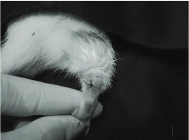
(Figure 1). After sectioning the tendon, the skin incision was completely sutured in the central area
using 4.0 nylon thread (Figure 2). Post-operatively,
of Ceftriaxone in the contralateral hind limb for antibiotic prophylaxis and were returned to sterilized
cages without immobilization14,37-39,41. The right Achilles tendon was submitted to US in the US1, US2 and US3 groups.
Treatment protocols
The Sonacel BIOSET® US emitter was used. The
design of its transducer, which has an area of 0.5 cm2, was modiied to it the posterior portion of
the rats’ hind limbs and the size of the lesions. The equipment was calibrated with an acoustic
scale (GT210 Glutymax®) and an oscilloscope
(Intermetro®).
The animals were submitted to the irst application
of US therapy 24 hours after the surgery. The ultrasonic irradiation employed the following parameters: 1 MHz frequency, pulsed mode
with 20% of the duty cycle (2 ms emission/8 ms interval), 100 Hz repetition frequency, 0.5 W/cm² of
intensity (spatial average time average - SATA), and
0.5 cm² ERA and was performed once per day. The treated area was 1 cm² in size. The animals were sacriiced after the 10th treatment session on the
12th post-operative day. The irradiations occurred
consecutively at one day intervals after the ifth
treatment day3,4,18,38,42.
During the US applications, the transducer was placed perpendicular to the treated area using movement techniques. Water-soluble gel was used as a coupling agent to better conduct the waves and facilitate movement over the rat’s skin27,43. The animals were also stabilized with a standard retainer (Figure 3)14,38.
Preparation and analysis of the histological slides
The animals were sacriiced using a CO2 chamber, and the damaged tendon areas were surgically removed. For the quantitative assessment of tendon repairs, the removed tendons were placed
in a 10% formalin solution for 24 hours and were
subsequently dehydrated with four alcohol baths
of one hour each at dilutions of 50%, 70%, 80%, and 100%. Subsequently, the samples were washed in xylene for one hour and embedded in parafin at 58°C. The blocks were longitudinally cut into serial sections using a microtome (LAICA®) with a standard
thickness of 7 µm. The sections were mounted on glass slides without staining and identiied by a
blinded evaluator.
The uneven penetration of light through an object is a measure of birefringence. This measure assesses the density and organization of the analyzed material and is used to evaluate the organization and structure
of the collagen ibers. The purpose of this procedure
Figure 1. Longitudinal incision and exposure of the Achilles
tendon on the posterior of the rat’s hind limb.
Figure 2. Surgical incision sutures with 4.0 nylon surgical thread.
Figure 3. Constraint and irradiation of the Achilles tendon on the
was to analyze the organization, the aggregation states, and the alignment of the tendon collagen
ibers by measuring their birefringence14,20,39. For the birefringence analyses, the slides for each group were
immersed in distilled water (refractive index η=1.333) for 30 minutes. The optical retard (OR) was measured using a polarized light microscope (LEICA®) with a
10X/0.22 objective, pol. 0.9 condenser, Senarmont λ/4 compensator, and monochromatic light (λ=546 nm) obtained by an interference ilter (LEICA®) of the
LAMAV (Vitreous materials laboratory, Department
of material Engineering of UFSCar). These types of
analyses have been used to quantitatively measure
the degrees of organization of the collagen ibers
in several studies14,20,38. The resulting measures, in degrees, were converted into nanometers (nm) by multiplying the values by 3.03. The total
birefringence of the collagen ibers was measured
after soaking the tendons in distilled water. To
perform the measurements along the axis of the tendon, the longitudinal axis of the collagen ibers
was oriented 45° from the direction of the propagation
of the transmission light. In this position, the collagen ibers have the highest OR. Measurements were taken at ive different points on the central areas of
the tendons, which corresponded to the areas of the injuries14,20,38,39. The birefringence data were collected by two previously trained, independent, and blinded assessors.
Statistical analyses
The data distributions were tested using the Kolmogorov-Smirnov test. Subsequently, the data were analyzed using the Kruskal-Wallis test, followed by Dunn’s post-hoc test to investigate
differences between the groups with a signiicance
level of α<0.05.
Results
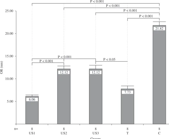
Kruskall-Wallis tests revealed statistically
signiicant differences among the groups with respect to the OR of the ive different areas of each injury
region (p<0.0001). Dunn’s multiple post-hoc tests demonstrated statistically significant differences
when comparing the OR data between the following groups: US1xUS2, US1xUS3, US1xC, US2xC, US3xT, and CxT (Figure 4).
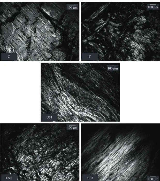
Qualitative histological analyses using polarized
light microscopy allow the deinition of normality
standards regarding the organization of the collagen
ibers (Figure 5). When comparing the images in
Figure 5, it is possible to observe that the animals of the US3 group demonstrated better organization of
the collagen ibers when compared to the other treated groups (US1 and US2). It is also possible to observe that the collagen ibers of the animals in the US1 and
tenotomy groups were not organized.
Discussion
The aim of this study was to evaluate the effects of different US application times on tendon repair in tenotomized rats, and the results suggested that application times of less than three minutes were
insuficient to promote adequate organization of the collagen ibers. The literature also does not provide clear deinitions regarding US therapy treatment
times. US doses are the most frequently studied
Figure 5. Images related to the qualitative observations of the birefringence analyses of the tenotomized rat tendons. Specimens were
variables15,30, and there is evidence that lower doses
are more effective for tissue repair44.
The dose standards employed in the present study were those most often used in US therapy studies, and the treatment times were the studied variables. The
intensity (0.5 W/cm²) was based upon the research of Carvalho et al.4, Cunha et al.14, Reddy et al.37, Koeke et al.38,and Silva et al.45. The form of
ultrasonic pulsed emission was chosen based on
research conducted by Carvalho et al.4, Cunha et al.14,
Frasson et al.17,Blume et al.22, Koeke at al.38, and Belanger et al.46. The frequency of the equipment was justiied by the experiments by Carvalho et al.4,
Piedade et al.11, Cunha et al.14, and Koeke et al.38. The treatment duration is known to depend upon the area of the injury. Oakley31 recommended that
the treatment time be related to the treated area, with one or two minutes for each area that is 11/
2 times
the size of the transducer. According to Oakley31,
subsequent treatment times can be increased from 1 to 11/
2 minutes. This subsequent treatment
time is based upon empirical studies or clinical
observations. There are no clinical or experimental studies that support these indings, and this lack
of information concerning treatment should be considered an issue for further study. The results of the present study do not corroborate the results of Oakley31 regarding the application times needed
to promote better alignment of collagen ibers in rat
tendons.
The application time is an important variable to be considered in US therapy dosimetry and determines the amount of energy applied to the treated
tissue. Alexander et al.30, in a systematic review of
the use of US in various shoulder pathologies, noted that studies that employed the average energy of 4.2 J per treatment session were clinically effective,
whereas those that applied an average of 2.019 J were
not successful. The duration of application may have
inluenced these data.
The components of the extracellular matrix of the
connective tissues present in tendons, particularly
the collagen ibers, exhibit viscoelastic properties
that depend on the amount of time the tissue is
exposed to deformations or strains to keep their
morphofunctional characteristics47. There is a direct relationship between the alignment of collagen ibers of the extracellular matrix and the biomechanical properties of tendon: when the collagen ibers exhibit better organization, the lexibility and strength of
these tendons also improve48.
Thus, three minutes of US application could have
suficient biomechanical effects, compression and/or
tension to promote the mechanisms of cellular and
matrix extracellular signaling for the realignment of the collagen ibers49.
In this study, the OR in the tendon scar tissues of rats demonstrated statistically signiicant differences when 18 J of energy (group US3) was applied to the
affected area. However, when other magnitudes of
energy, namely, 6 J (US1 group) or 12 J (group US2),
were deposited on the treated area, no differences
were observed regarding the OR between the experimental and control groups.
The most commonly employed application time
is ive minutes, and the US is generally applied to
an area twice the size of the transducer, resulting in an application time of 2.5 minutes per treated area6,38. The size of the treated area is a variable that has not been described in studies related to US therapy and tendon repair. Wood et al.6, Cunha et al.14,and
Koeke et al.38 applied US therapy to an area of 1 cm², but these data were not described in the articles and were instead personally provided by the authors. The present study demonstrated that longer application times are needed to promote better alignment of
the collagen ibers. One hypothesis to explain the
differences between the results of this and previous studies is that the control of the treated areas may not have been accurately measured, making it impossible
to compare indings. This variable must be considered when designing the experiment so that comparisons
between studies can be made.
The relevance of studying and developing techniques to treat tendon injuries relates to the long recovery times and the resulting functional disabilities of tendon injuries, which may take weeks or even months to fully recover1,3,50. The tensile
strength of the tendons is promoted by the alignment
of their collagen ibers and by the types and amount of ibers. Thus, this study used an experimental model already deined in the literature, described by Enwemeka and Reddy1 and reproduced by other
authors14,37-39,41, that exhibits easy reproducibility, lower costs, and high levels of reliability.
The levels of collagen iber organization in the injured area are measured according to the OR in
the polarized light microscope, which provides quantitative data. The brightness of the birefringence image reveals the level of aggregation of the collagen fibers14. During the process of tissue repair, the
OR values tend to decrease39, corroborating the results of the present study, in which the US1 group
demonstrated the lowest OR values, followed by the
The indings of the present study suggest that
three minutes of application per area of the US transducer results in positive effects on the collagen
iber organization in rat tendons during the tissue repair process, producing signiicant changes in the
collagen optical retards. Shorter application times
produced no signiicant changes in the optical delays
when compared to the tenotomy group.
The transposition of the experimental indings to
the clinical application of therapeutic ultrasound on tendon healing should be evaluated with respect to the difference between the application times of 2 and 3 minutes. Further studies are needed to assess the difference between these treatment times with regard to human healing.
References
1. Enwemeka CS, Reddy K. The biological effects of laser
therapy and other physical modalities on connective tissue repair processes. J Laser Ther. 2000;12:22-30. http:// dx.doi.org/10.5978/islsm.12.22
2. Tumilty S. Achilles tendon ruptures: rising incidence in New Zealand follows international trends.
Phys Ther Rev 2007;2(1):59-65. http://dx.doi. org/10.1179/108331907X174998
3. Saini NS, Roy KS, Bansal PS, Singh B, Simran PS. A preliminary study on the effect of ultrasound
therapy on the healing of surgically severed achilles
tendons in ive dogs. J Vet Med A Physiol Pathol Clin Med. 2002;49(6):321-8. PMid:12227476. http://dx.doi. org/10.1046/j.1439-0442.2002.00441.x
4. Carvalho PT, Silva IS, Reis FA, Belchior AC, Aydos RD, Facco GG, et al. Histological study of tendon healing in
malnourished Wistar rats treated with ultrasound therapy.
Acta Cir Bras. 2006;21(Suppl 4):13-7. http://dx.doi. org/10.1590/S0102-86502006001000004
5. Neves MA, Pinildi CE, Wood VT, Gobbato RC, Da Silva FM, Parizotto NA, et al. Different Power Settings of LLLT on the Repair of the Calcaneal Tendon. Photomed Laser Surg. 2011;29(10):663-8. PMid:21668375. http://dx.doi. org/10.1089/pho.2010.2919
6. Wood VT, Pinfildi CE, Neves MA, Parizoto NA, Hochman B, Ferreira LM. Collagen changes and
realignment induced by level laser therapy and low-intensity ultrasound in the calcaneal tendon. Lasers Surg
Med. 2010;42(6):559-65. PMid:20662033. http://dx.doi. org/10.1002/lsm.20932
7. Johns LD. Nonthermal effects of therapeutic ultrasound: the frequency resonance hypothesis. J Athl
Train. 2002;37(3):293-9. PMid:16558674 PMCid:164359. 8. Chipchase LS, Trinkle D. Therapeutic ultrasound:
clinician usage and perception of eficacy. Hong Kong Physiother J. 2003;21(1):5-14. http://dx.doi.org/10.1016/ S1013-7025(09)70034-X
9. Larsen A, Kristensen G, Thorlacius-Ussing O, Oxlund
H. The influence of ultrasound on the mechanical
properties of healing tendons in rabbits. Acta
Orthop. 2005;76(2):225-30. PMid:16097548. http:// dx.doi.org/10.1080/00016470510030616
10. Wong RA, Schumann B, Townsend R, Phelps CA.
A survey of therapeutic ultrasound use by physical
therapists who are orthopaedic certiied specialists. Phys Ther. 2007;87(8):986-94. PMid:17553923. http://dx.doi. org/10.2522/ptj.20050392
11. Piedade MC, Galhardo MS, Battlehner CN, Ferreira MA, Caldini EG, De Toledo OM. Effect of ultrasound therapy
on the repair of gastrocnemius muscle injury in rats.
Ultrasonics. 2008;48(5):403-11. PMid:18384832. http:// dx.doi.org/10.1016/j.ultras.2008.01.009
12. Romano CV, Barbieri CH, Mazzer N, Volpon JB, Shimano AC, Roncaglia FB. O ultra-som terapêutico não aumentou as propriedades mecânicas de tendões flexores após reparo. Acta Ortop Bras. 2010;18(1):10-4. http://dx.doi. org/10.1590/S1413-78522010000100001
13. H a r v e y W , D y s o n M , P o n d J B , G r a h a m e R. The stimulation of protein synthesis in human fibroblasts by therapeutic ultrasound. Rheumatol Rehabil. 1975;14(4):237. PMid:1198016. http://dx.doi. org/10.1093/rheumatology/14.4.237
14. Cunha A, Parizotto NA, Vidal BC. The effect of
therapeutic ultrasound on repair of the achilles tendon (tendo calcaneus) of the rat. Ultrasound Med
Biol. 2001;27(12):1691-6. http://dx.doi.org/10.1016/ S0301-5629(01)00477-X
15. Ng CO, Ng GY, See EK, Leung MC. Therapeutic
ultrasound improves strength of achilles tendon repair in
rats. Ultrasound Med Biol. 2003;29(10):1501-6. http:// dx.doi.org/10.1016/S0301-5629(03)01018-4
16. Monte-Raso VV, Barbieri CH, Mazzer N, Fazan VPS.
Os efeitos do ultra-som terapeutico nas lesões por esmagamento do nervo ciatico de ratos: analise funcional
da marcha. Rev Bras Fisioter. 2006;10(1):113-9. http:// dx.doi.org/10.1590/S1413-35552006000100015 17. Frasson NF, Taciro C, Parizotto NA. Análise
nanoestrutural da ação do ultra-som terapêutico sobre
o processo de regeneração do tendão de ratos. Fisioter
Pesqui. 2009;16(3):198-204. http://dx.doi.org/10.1590/ S1809-29502009000300002
18. Robertson VJ. Dosage and treatment response in randomized clinical trials of therapeutic ultrasound. Phys
Ther Sport. 2002;3(3):124-33.
19. Warden SJ, McMeeken JM. Ultrasound usage and dosage in sports physiotherapy. Ultrasound Med
Biol. 2002;28(8):1075-80. http://dx.doi.org/10.1016/ S0301-5629(02)00552-5
20. Vidal BC. Image analysis of tendon helical superstructure
using interference and polarized light microscopy.
Micron. 2003;34(8):423-32. http://dx.doi.org/10.1016/ S0968-4328(03)00039-8
22. Blume K, Matsuo E, Lopes MS, Lopes LG. Dosimetria
proposta para o tratamento por ultra-som: uma revisão de
literatura. Fisioter Mov. 2005;18(3):55-64.
23. Franco AD, Pereira LE, Groschitz M, Aimbiré F, Martins RA, Carvalho RA. Análise do efeito do ultra-som no edema inlamatório agudo: estudo experimental. Fisioter Mov. 2005;18(2):19-24.
24. Olsson DC, Martins VMV, Pippi NL, Mazzanti A, Tognoli GK. Ultra-som terapêutico na cicatrização tecidual. Ciênc Rural. 2008;38(4):1199-207. http://dx.doi.org/10.1590/ S0103-84782008000400051
25. Draper DO, Castel JC, Castel D. Rate of temperature
increase in human muscle during 1 MHz and 3
MHz continuous ultrasound. J Orthop Sports Phys Ther. 1995;22(4):142-50. PMid:8535471.
26. Van der Windt DA, Van der Heijden GJ, Van den Berg SG, Ter Riet G, De Winter AF, Bouter LM. Ultrasound
therapy for musculoskeletal disorders: a systematic review.
Pain. 1999;81(3):257-71. http://dx.doi.org/10.1016/ S0304-3959(99)00016-0
27. Casarotto RA, Adamowski JC, Fallopa F, Bacanelli F. Coupling agents in therapeutic ultrasound: acoustic and thermal behavior. Arch Phys Med Rehabil. 2004;Jan;85(1):162-5. http://dx.doi.org/10.1016/ S0003-9993(03)00293-4
28. Fu SC, Shum WT, Hung LK, Wong MW, Qin L, Chan KM.
Low-intensity pulsed ultrasound on tendon healing: a study of the effect of treatment duration and treatment initiation.
Am J Sports Med. 2008;36(9):1742-9. PMid:18645043. http://dx.doi.org/10.1177/0363546508318193
29. Speed CA. Therapeutic ultrasound in soft tissue lesions. Rheumatology. 2001;40(12):1331-6. PMid:11752501. http://dx.doi.org/10.1093/rheumatology/40.12.1331
30. Alexander LD, Gilman DR, Brown DR, Brown JL, Houghton PE. Exposure to low amounts of ultrasound
energy does not improve soft tissue shoulder pathology:
a systematic review. Phys Ther. 2010;90(1):14-25. PMid:19910457. http://dx.doi.org/10.2522/ptj.20080272
31. Oakley EM. Application of continuous beam ultrasound
at therapeutic levels. Physiotherapy. 1978;64(6):169-72. PMid:674399.
32. Hoogland R. Terapia Ultrasónica [manual]. Enraf Nonius; 1986.
33. Aiyegbusi AI, Duru FI, Akinbo SR. The Morphology
of the Healing Tendon: A Comparison of the Effects of Intrasound Therapy and Therapeutic Pulsed Ultrasound. Connect Tissue Res. 2012;53(6):478-84. PMid:22574701. http://dx.doi.org/10.3109/03008207.2012.690793
34. Ng GY.Comparing therapeutic ultrasound with microamperage stimulation therapy for improving the
strength of Achilles tendon repair. Connect Tissue Res. 2011 Jun;52(3):178-82. PMid:20672987. http:// dx.doi.org/10.3109/03008207.2010.500752
35. Eckelman WC, Kilbourn MR, Joyal JL, Labiris R, Valliant JF. Justifying the number of animals for each experiment. Nucl Med Biol. 2007;34(3):229-32. PMid:17383571. http://dx.doi.org/10.1016/j.nucmedbio.2007.01.005 36. Damy SB, Camargo RS, Chammas R, Figueiredo
LFPd. Aspectos fundamentais da experimentação
animal - aplicações em cirurgia experimental. Rev Assoc Med Bras. 2010;56(1):103-11. PMid:20339795. http:// dx.doi.org/10.1590/S0104-42302010000100024 37. Reddy GK, Stehno-Bittel L, Enwemeka CS. Laser
photostimulation accelerates wound healing in diabetic rats.
Wound Repair Regen. 2001;(3):248-55. PMid:11472621. http://dx.doi.org/10.1046/j.1524-475x.2001.00248.x 38. Koeke PU, Parizotto NA, Carrinho PM, Salate AC.
Comparative study of the efficacy of the topical
application of hydrocortisone, therapeutic ultrasound and phonophoresis on the tissue repair process in rat tendons.
Ultrasound Med Biol. 2005;31(3):345-50. PMid:15749557. http://dx.doi.org/10.1016/j.ultrasmedbio.2004.12.005 39. Carrinho PM, Renno AC, Koeke P, Salate
AC, Parizotto NA, Vidal BC. Comparative study using 685-nm and 830-nm lasers in the tissue repair of tenotomized tendons in the mouse. Photomed Laser Surg. 2006;24(6):754-8. PMid:17199477. http://dx.doi. org/10.1089/pho.2006.24.754
40. Baldan C, Pasqual AM, Schiavinato AM, Casarotto RA. Dose-dependência do laser de baixa intensidade (670 nm)
na viabilidade de retalhos cutâneos randômicos em ratos.
J Health Sci Inst. 2010;28(4):359-62.
41. Arruda ERB, Rodrigues NC, Taciro C, Parizotto NA. Influência de diferentes comprimentos de onda da laserterapia de baixa intensidade na regeneração tendínea do rato após tenotomia. Rev Bras Fisioter. 2007;11(4):283-8. http://dx.doi.org/10.1590/S1413-35552007000400007
42. Bélanger A. Therapeutic eletrophysical agents: evidence
behind practice. 2nd ed. Philadelphia. Lippincott Williams
& Wilkins; 2010.
43. Draper DO, Edvalson CG, Knight KL, Eggett
D, Shurtz J. Temperature increases in the human achilles tendon during ultrasound treatments with commercial ultrasound gel and full-thickness and
half-thickness gel pads. J Athl Train. 2010;45(4):333-7. PMid:20617906 PMCid:2902025. http://dx.doi. org/10.4085/1062-6050-45.4.333
44. Ng GY, Fung DT. The effect of therapeutic ultrasound
intensity on the ultrastructural morphology of tendon
repair. Ultrasound Med Biol. 2007;33(11):1750-4. PMid:17630092007;33(11):1750-4. http://dx.doi.org/10.1016/j. ultrasmedbio.2007.05.019
45. Silva JMN, Carvalho JP, Moura Júnior MJ, Arisawa EA, Martin AA, Sá HP, et al. Estudo da ação do ultrassom terapêutico em modelo experimental de tendinite em ratos Wistar. Cons Saúde. 2010;9(4):625-32. http://dx.doi. org/10.5585/conssaude.v9i4.2380
46. Belanger A. Therapeutic eletrophysical agents: evidence
behind practice. 2nd ed. Philadelphia. Lippincott Williams
& Wilkins; 2010.
47. Culaw EM, Clark CH, Merrilees MJ. Connective tissues: matrix composition and its relevance to physical therapy. Phys Ther. 1999;79(3):308-19.
48. Aparecida de Aro A, Vidal BC, Pimentel ER.
Biochemical and anisotropical properties of tendons.
49. Chiquet M. Regulation of extracellular matrix gene expression by mechanical stress. Matrix Biol. 1999;18(5):417-26. http://dx.doi.org/10.1016/ S0945-053X(99)00039-6
50. Soma CA, Mandelbaum BR. Repair of acute Achilles tendon ruptures. Orthop Clin North Am. 1995;26(2):239-47. PMid:7724190.
Correspondence
Thiago Saikali Farcic Av. Yojiro Takaoka, 3500
CEP 06500-000, Santana de Parnaiba, SP, Brasil