Detection of highly and minimally leukotoxic
Actinobacillus
actinomycetemcomitans
strains in patients with
periodontal disease
Detecção de cepas de
Actinobacillus actinomycetemcomitans
de
máxima e mínima leucotoxicidade em pacientes com
doença periodontal
Sheila Cavalca Cortelli*
Antonio Olavo Cardoso Jorge** José Roberto Cortelli***
Shawn Francis Jordan**** Violet Ibyola Haraszthy*****
ABSTRACT:This study examined the prevalence of highly and minimally leukotoxicActinobacillus actinomycetemco-mitansin patients with periodontal disease. Pooled subgingival plaque samples from 136 patients with some form of periodontal disease were examined. Subjects were between 14 and 76 years of age. Clinical examinations included pe-riodontal pocket depth (PD), plaque index (PI) and bleeding index (BI). The obtained plaque samples were examined for the presence of highly or minimally leukotoxicA.actinomycetemcomitansstrains by the polymerase chain reaction (PCR). Chi-square and logistic regression were performed to evaluate the results. Forty-seven subjects were diagnosed with gingivitis, 70 with chronic periodontitis and 19 with aggressive periodontitis. According to chi-square there was no significant correlation detected between PD (c2= 0.73), PI (c2= 0.35), BI (c2= 0.09) and the presence of the highly le-ukotoxicA.actinomycetemcomitans.The highly leukotoxicA.actinomycetemcomitansstrains were correlated with subjects that were 28 years of age and younger (c2= 7.41). There was a significant correlation between highly leukoto-xicA.actinomycetemcomitansand aggressive periodontitis (c2= 22.06). This study of a Brazilian cohort confirms the strong association between highly leukotoxicA.actinomycetemcomitansstrains and the presence of aggressive perio-dontitis.
DESCRIPTORS:Actinobacillus actinomycetemcomitans; Leukotoxin; Gingivitis; Periodontitis; Polymerase chain reac-tion.
RESUMO:O presente estudo avaliou a prevalência deActinobacillus actinomycetemcomitansde máxima e mínima leu-cotoxicidade em indivíduos com doença periodontal. Foram analisadas amostras de placa bacteriana subgengival de 136 indivíduos, entre 14 e 76 anos de idade, com algum tipo de doença periodontal. Os parâmetros clínicos avaliados incluíram profundidade de sondagem (PS), índice de placa bacteriana (IPB) e índice de sangramento gengival (ISG). A presença de amostras deA. actinomycetemcomitansde máxima e mínima leucotoxicidade foi avaliada por reação em cadeia da polimerase (PCR). Os resultados foram analisados empregando-se teste qui-quadrado e análise de regressão logística. Quarenta e sete indivíduos apresentaram gengivite, 70 indivíduos periodontite crônica e 19 indivíduos perio-dontite agressiva. O teste qui-quadrado não demonstrou correlação significativa entre PS (c2= 0,73), IPB (c2= 0,35), ISG (c2= 0,09) e a presença de amostras de máxima leucotoxicidade.A. actinomycetemcomitansde máxima leucotoxi-cidade apresentou associação com indivíduos com idade inferior a 28 anos (c2= 22,06). Os resultados observados nes-sa população brasileira confirmam a forte associação existente entre amostras deA. actinomycetemcomitansde máxi-ma leucotoxicidade e periodontite agressiva.
DESCRITORES:Actinobacillus actinomycetemcomitans; Leucotoxina; Gengivite; Periodontite; Reação em cadeia da po-limerase.
*Assistant Professor, Microbiology and Periodontics; ***Assistant Professor, Periodontics – University of Taubaté. **Full Professor, Microbioloy and Immunology, State University of São Paulo;
INTRODUCTION
The presence of periodontal pathogens can be frequently detected in healthy subjects. This indi-cates that not all humans are equally susceptible and/or that there is variation in virulence and pathogenic potential (Tonetti e Mombelli23, 1999; Zadeh et al.26, 1999). Bacteria associated with periodontal disease have a number of virulence factors such as endo- and exotoxins, that can di-rectly damage host tissues, and immune evasion strategies which shield the bacteria from host de-fenses.
Every strain ofA. actinomycetemcomitans studi-ed to date is resistant to serum-mstudi-ediatstudi-ed killing. They are not killed by unseparated serum and any serum component by implication, which would in-clude the membrane attack complex, platelet-deri-vedb-lysin, acute phase proteins and lysozyme. To clear an infection by a serum-resistant organism necessarily involves host phagocytes (Zadeh et al.26, 1999). However,
A. actinomycetemcomitans has been shown to be capable of inhibiting poly-morphonuclear leukocytes chemotaxis (Van Dyke et al.24, 1982; Fives-Taylor
et al.7, 1996). Further-more, phagocytosis inhibition is associated withA. actinomycetemcomitansFc binding proteins.A. ac-tinomycetemcomitanscan also inhibit the leukocy-tes fusion with lysosomes (Fives-Taylor et al.8, 1999).
A. actinomycetemcomitans produce a leuko-toxin (ltx) that is an RTX (repeats in leuko-toxin) leuko-toxin. The RTX family members share similar gene orga-nization and are expressed from an operon con-sisting of four genes that are designated C (ltxC), A (ltxA), B (ltxB) and D (ltxD) in transcriptional order. The structural gene is designated as the gene A and the three remaining genes flanking the A gene are required to activate and transport the toxin (Lallyet al.12, 1989). Leukotoxins are cell-specific. The toxin binds to neutrophils, monocytes and a subset of lymphocytes (Zadeh et al.26, 1999). Cell lysis might be induced by the rapid formation of highly conductive ion channels that lead to mem-brane depolarization, loss of intracellular K+, os-motic swelling, and subsequently cell death (Slots20, 1994; Lally
et al.12, 1996; Karakelian et al.11, 1998; Fives-Taylor
et al.8, 1999). The expres-sion of leukotoxins is tightly regulated in many strains, although some strains produce high levels of leukotoxins (Brogan et al.5, 1994; Mombelli
et al.15, 1999; Paju
et al.17, 2000). The promoter
re-gions of high producers have a 530 bp deletion that may be involved in repressing leukotoxin ex-pression (Brogan et al.4, 1994). Leukotoxins may protect A. actinomycetemcomitans against phagocytosis by human polymorphonuclear leu-kocytes (Johansson et al.10, 2000). Patients with aggressive periodontitis were shown to have a sub-stantially higher prevalence of highly leukotoxic strains than chronic periodontitis individuals (Zambonet al.28, 1996; Haraszthy
et al.9, 2000). The aim of the present study was to examine the prevalence of highly and minimally leukotoxic Actinobacillus actinomycetemcomitans in patients with periodontal disease.
MATERIALS AND METHODS
Subject population
A total of 136 subjects, males and females, aged from 14 to 76 years with gingivitis (47), chronic (70) and aggressive (19) periodontitis were enrolled in this study. Individuals were previously classi-fied as gingivitis, chronic or aggressive perio-dontitis patients, based upon clinical and radio-graphic findings (periodontal probing depth - PD, loss of clinical attachment and periapical radio-graphs) and on the diagnostic criteria presented in the World Workshop of Periodontology 1999 (AAP2, 1999). Clinical examinations also included Plaque Index - PI and Bleeding Index - BI (Ainamo, Bay1, 1975).
Subjects were excluded if any of the following conditions were detected: use of systemic antibi-otic in the previous 6 months and periodontal therapy undertaken less than 12 months before the study. All subject participation was preceded by approval of all protocols by the appropriate in-stitutional review board and the obtainment of a signed consent from participants.
Microbial Sampling and Polymerase Chain
Reaction
microfuge (Eppendorf) and DNA was isolated from the samples (Instagene Purification Matrix, Bio Rad, Hercules, USA).
PCR was performed using a thermocycler (Perkin Elmer Cetus) with Taq polymerase (Amershan, Arlington Heights, USA). Using previ-ously published sequences (Broganet al.5, 1994), 2 oligonucleotide primers were used to the A. actinomycetemcomitansleukotoxin promoter regi-on:
•A - 5’-GCAGGATCCATATTAAATCTCCTTGT-3’; •B - 5’-GCGGTCGACAACCTGATAACAGTATT-3’.
They were used to amplify either a 492 base pair product characteristic of highly leukotoxic A. actinomycetemcomitansor a 1022 base pair prod-uct characteristic of minimally leukotoxic A. actinomycetemcomitans. PCR was performed using standard conditions. The reaction mixture con-sisted of 100 ml of 10 mM Tris-HCl pH 8.3 (Amershan, Arlington Heights, USA), 50 mM KCl (Amershan, Arlington Heights, USA), 1.5 mM MgCl2(Amershan, Arlington Heights, USA), 100mM each of dATP, dCTP, dGTP, dTTP (Amershan, Arlington Heights, USA), 0.001% (w/v) gelatin, 100 ng of each primer (Invitrogen/Gibco, San Diego, USA), 0.5 UTaqpolymerase, and 100 ng of genomic DNA. The thermocycle profile was 25 cy-cles of 30 seconds at 95ºC, 30 seconds at 55ºC, and 30 seconds at 72ºC. The amplification prod-ucts were analyzed by electrophoresis in 2% sub-marine agarose gels (Sigma, Dorset, UK) and visu-alized by UV illumination (Bio Rad, Hercules, USA) following ethidium bromide (Amershan, Arlington Heights, USA) staining. These agarose gels were compared with both positive (JP2 and 652 strains) and negative controls.
Statistical analysis
Chi-square and logistic regression were per-formed to evaluate the results. The level of signifi-cance considered was set at p < 0.05. The relation between variants and periodontal diagnosis was established by logistic regression. Age, gender, smoking, PD, PI, BI, PCR detection and highly or minimally leukotoxic A. actinomycetemcomitans were considered in separated or multiple analyses. The probability of aggressive periodontitis – P(AP) – was calculated as follows:
RESULTS
Graph 1 shows the distribution of highly and minimally leukotoxic A. actinomycetemcomitans. The major prevalence of highly leukotoxic A. actinomycetemcomitans was observed in aggres-sive periodontitis subjects followed by chronic periodontitis and gingivitis (c2= 22.06). No statisti-cally significant difference was observed between gingivitis and chronic periodontitis related to highly leukotoxic A. actinomycetemcomitans (c2 = 0.17). In contrast, highly leukotoxic
A. actinomycetemcomitans was correlated with sub-jects 28 years of age and younger (c2= 7.41).
Minimally leukotoxicA. actinomycetemcomitans showed no correlation with periodontal status (c2= 0.91). No association between PD (c2= 0.73), PI (c2= 0.35) and BI (c2= 0.09) and highly and mi-nimally leukotoxicA. actinomycetemcomitanswas detected.
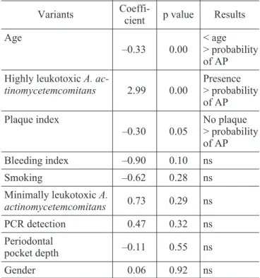
Separated logistic regression analyses showed correlation between periodontal status and age, highly leukotoxicA. actinomycetemcomitansand PI (Table 1). Multiple logistic regression analyses sho-wed correlation between periodontal disease, age and PD (Table 2).
The probability of aggressive periodontitis was calculated. Graph 2 shows the results. The mini-mum and maximini-mum values of PD found among all subjects included in the present study were ap-plied in multiple logistic regression. Age was also considered for analyses.
GRAPH 1 -Highly and minimally leukotoxic Actinobacil-lus actinomycetemcomitansaccording to periodontal sta-tus. G - gingivitis (n = 47); CP - chronic periodontitis (n = 70); AP - aggressive periodontitis (n = 19).
P(AP)
exp (5.58 2 age 4 periodontal pocke =
+ - +
1
DISCUSSION
A. actinomycetemcomitans is an important pathogen in some forms of periodontitis. Aggres-sive periodontitis is the most notorious disease as-sociated withA. actinomycetemcomitans.The prev-alence of this pathogen is nearly 90% in aggressive periodontitis and 30-50% in severe chronic periodontitis. Furthermore, one of the most impor-tant virulence factors ofA. actinomycetemcomitans is leukotoxin (Lally et al.12, 1996; Karakelian et al.11,1998; Fives-Tayloret al.8, 1999; Zadehet al.26, 1999). In the present study, the periodontalstatus was associated with highly leukotoxic A. actinomycetemcomitans. There was significant cor-relation between highly leukotoxic strain and
ag-gressive periodontitis (c2= 22.06) (Graph 1). Addi-tionally, we observed that the highly leukotoxicA. actinomycetemcomitansstrain was correlated with subjects 28 years of age and younger (c2= 7.41). These data agree with the results obtained by Ávila-Campos, Velásquez-Meléndez3 (2002) who observed thatA.actinomycetemcomitanswas asso-ciated with disease in fifty Brazilian patients with clinical and radiographic evidence of alveolar bone loss confined to the molar and incisor teeth. Nakagawaet al.16, 2001, analyzed the serumal and salivary levels of antibodies against the leukotoxin produced by A. actinomycetemcomitans in Brazil-ian patients with aggressive periodontitis and in healthy controls. The authors found significantly higher serumal levels of IgG in patients with ag-gressive periodontitis, when they were compared with the healthy controls. However, Lima et al.13, 2001, isolated a high leukotoxin producer from a healthy subject and moderate producers from periodontal disease patients.
On the other hand, no relation was observed be-tween plaque index (c2 = 0.35), bleeding index (c2= 0.09) and highly and minimally leukotoxic
A. actinomycetemcomitans. These data are in accor-dance with other studies which strongly showed that there is more probability to detect A. actinomycetemcomitans when clinical signs of in-flammation were evident (Mombelli et al.14, 1995; Zambon et al.27, 1994; Tanner
et al.22
, 1996). Par-ticularly in chronic periodontitis subjects, A. actinomycetemcomitanswas weakly related to the bleeding index, while there was no correlation be-tween the organism and any clinical index in ag-gressive periodontitis subjects (Bontaet al.41985; Renvertet al.181997; Shiloah e Patters19, 1997).
When periodontal pocket depth is considered, different authors have relatedA. actinomycetemco-mitansto deeper pockets (Slotset al.21, 1980; Wolff et al.25, 1993; Zambon27, 1994; Tanner et al.22, 1996). In the present study no correlation was ob-served between periodontal pocket depth and highly and minimally leukotoxicA. actinomycetem-comitans. However, multiple logistic regression analyses showed that periodontal pocket depth was related to highly leukotoxicA. actinomycetem-comitans(p < 0.05).
Separated logistic regression was applied to analyze the relation between risk factors and peri-odontalstatus(Table 1). Age and PD were strongly associated with aggressive periodontitis (p < 0.05) according to multiple logistic regression (Table 2). TABLE 1 -Estimate coefficient, p value and statistically
significance established by logistic regression related to isolated analyses of each considered variant.
Variants
Coeffi-cient p value Results
Age
–0.33 0.00
< age > probability of AP
Highly leukotoxicA.
ac-tinomycetemcomitans 2.99 0.00
Presence > probability of AP
Plaque index
–0.30 0.05
No plaque > probability of AP
Bleeding index –0.90 0.10 ns
Smoking –0.62 0.28 ns
Minimally leukotoxicA.
actinomycetemcomitans 0.73 0.29 ns
PCR detection 0.47 0.32 ns
Periodontal
pocket depth –0.11 0.55 ns
Gender 0.06 0.92 ns
AP - aggressive periodontitis; PCR detection - presence ofA. actinomycetemcomitans; PCR - polymerase chain reaction; ns - not significant.
TABLE 2 -Results of multiple logistic analyses.
Variant Age Periodontal pocket depth
Coefficient –0.42 0.74
Different ages and values of PD found in the present study originated a diagram of probabilities for aggressive periodontitis. Subjects 20 years of age or younger and PD between 9 and 10 mm showed 100% of probability of aggressive peri-odontitis. On the other hand, subjects 40 years of age or older, even showing similar PD (9 to 10 mm) had probability close to zero to exhibit aggressive periodontitis (Graph 2). The present research showed an association between age, PD and ag-gressive periodontitis.
A. actinomycetemcomitans is implicated in ag-gressive periodontitis and it is generally conside-red a major etiologic microorganism in this form of disease. However, the role of the pathogen in chro-nic periodontitis is more difficult to make clear sin-ce A. actinomycetemcomitans is found in a much more microbiologically complex and diverse plaque ecology compared toA. actinomycetemcomitans in-fections in aggressive periodontitis (Zambon27, 1994). The relationships between bacteria in chro-nic periodontitis are frequent. For example,P. gin-givalis and P. intermedia produce proteases that can decrease the toxicity ofA. actinomycetemcomi-tans. Thus, in some patientsA. actinomycetemco-mitanscan exhibit a complementary role in develo-ping periodontal disease (Chen, Slots6, 1999).
CONCLUSIONS
The major prevalence of highly leukotoxicA. ac-tinomycetemcomitans was observed in aggressive periodontitis. Minimally leukotoxic strains showed no correlation with periodontalstatus. This study of a Brazilian cohort confirms the strong associati-on between highly leukotoxicA. actinomycetemco-mitansstrains and the presence of aggressive peri-odontitis.
ACKNOWLEDGEMENTS
This work was supported in part by University of Taubaté (UNITAU) and Paulista State University (UNESP). The authors are grateful to Doctor Jo-seph J. Zambon for their technical assistance at the State University of New York at Buffalo.
REFERENCES
1. Ainamo J, Bay I. Problems and proposals for recording gin-givitis and plaque. Int Dent J 1975;25:229-35.
2. American Academy of Periodontology. Ann Periodontol 1999;4:18-9.
3. Ávila-Campos MJ, Velásquez-Meléndez G. Prevalence of putative periodontopathogens from periodontal patients and healthy subjects in São Paulo, SP, Brazil. Rev Inst Med Trop S Paulo 2002;44:1-5.
4. Bonta Y, Zambon JJ, Genco RJ, Neiders ME. Rapid identi-fication of periodontal pathogens in subgingival plaque: comparison of indirect immunofluorescence microscopy with bacterial culture for detection ofActinobacillus acti-nomycetemcomitans. J Dent Res 1985;64:793-8.
5. Brogan JM, Lally ET, Poulsen K, Killian M, Demuth DR. Regulation of Actinobacillus actinomycetemcomitans
leukotoxin expression: analysis of the promoter regions of
leukotoxic and minimally leukotoxic strains. Infect Immun 1994;62:501-8.
6. Chen C, Slots J. Microbiological tests for Actinobacillus actinomycetemcomitans and Porphyromonas gingivalis. Periodontol 2000 1999;20:53-64.
7. Fives-Taylor PM, Meyer DH, Mintz KP.Virulence factors of the periodontopathogen Actinobacillus actinomycetem-comitans.J Periodontol 1996;67:291-7.
8. Fives-Taylor PM, Meyer DH, Mintz KP, Brissette C. Viru-lence factors of Actinobacillus actinomycetemcomitans. Periodontol 2000 1999;20:136-67.
9. Haraszthy VI, Hariharan G, Tinoco EMB, Cortelli JR, Lally ET, Davis E,et al. Evidence for the role of highly leucotoxic
Actinobacillus actinomycetemcomitansin the pathogenesis of localized juvenile and other forms of early onset periodontitis. J Periodontol 2000;71:912-22.
10. Johansson A, Sandstrom G, Claesson R, Hanstrom L, Kalfas S. Anaerobic neutrophil-dependent killing of
Actinobacillus actinomycetemcomitans in relation to the bacterial leukotoxicity. Eur J Oral Sci 2000;108:136-46. 11. Karakelian D, Lear JD, Lally ET, Tanaka JC.
Characteriza-tion of Actinobacillus actinomycetemcomitans leukotoxin pore formation in HL60 cells. Biochem Biophys Acta 1998;1406:175-87.
12. Lally ET, Kieba IR, Golub EE, Lear JD, Tanaka JC. Struc-ture/function aspects of Actinobacillus actinomycetem-comitansleukotoxin. J Periodontol 1996;67:298-308. 13. Lima FL, Farias FF, Campos PC, Totola AH, Tavares CAP,
Costa JE, et al. Leukotoxic activity of Actinobacillus actinomycetemcomitansisolated from human and non-hu-man primates. Braz J Microbiol 2001; 32: 250-6.
14. Mombelli A, Rutar A, Lang NP. Correlation of the periodon-talstatus6 years after puberty with clinical and microbio-logical conditions during puberty. J Clin Periodontol 1995;2:300-5.
15. Mombelli A, Gmur R, Lang NP, Corbert E, Frey J.
Actinobacillus actinomycetemcomitans in Chinese adults: serotype distribution and analysis of the leukotoxin gene promoter locus. J Clin Periodontol 1999;26:505-10. 16. Nakagawa RI, Guazeli-Amin V, Hidalgo MM, Trevisan Jr W,
Itano EN. Anticorpos antileucotoxina contraActinobacillus actinomycetemcomitans em amostras de soro e saliva de pacientes com periodontite juvenil localizada. Pesqui Odontol Bras 2001;15:5-11.
17. Paju S, Carlson P, Jousimies-Somer H, Asikainen S. Heter-ogeneity ofActinobacillus actinomycetemcomitans strains in various human infections and relationships between serotype, genotype, and antimicrobial susceptibility. J Clin Microbiol 2000;38:79-84.
18. Renvert S, Dahlén G, Snyder B. Clinical and microbiologi-cal effects of subgingival antimicrobial irrigation with citric acid as evaluated by an enzyme immunoassay and culture analysis. J Periodontol 1997;68:346-52.
19. Shiloah J, Patters MR, Dean JW, Bland P, Toledo G. The survival rate of Actinobacillus actinomycetemcomitans, Porphyromonas gingivalis,andBacteroides forsythus, fol-lowing 4 randomized treatment modalities. J Periodontol 1997;68:720-8.
20. Slots J. A. actinomycetemcomitans. In: Nisengard RJ, Newman MG. Oral Microbiology and Immunology. 2nded. Philadelphia: Saunders; 1994. P.218-27.
21. Slots J, Reynolds HS, Genco RJ. Actinobacillus actinomycetemcomitansin human periodontal disease: a cross sectional microbiological investigation. Infect Immun 1980;29:1013-20.
22. Tanner A, Kent R, Maiden MFJ, Taubman MA. Clinical, mi-crobiological and immunological profile of healthy, gingivi-tis and putative active periodontal subjects. J Periodontal Res 1996;31:195-204.
23. Tonetti MS; Mombelli A. Early onset periodontitis. Ann Periodontol 1999;4:39-52.
24. Van Dyke TE, Bartholomew E, Genco RJ, Slots J, Levine MJ. Inhibition of neutrophil chemotaxis by soluble bacte-rial products. J Periodontol 1982;53:502-8.
25. Wolff LF, Aeppli M, Pihlstrom B, Anderson L, Stoltenberg J, Osborn J,et al. Natural distribution of 5 bacteria associ-ated with periodontal disease. J Clin Periodontol 1993;20:699-70.
26. Zadeh HH, Nichols FC, Miyasaki KT. The role of the cell-mediated immune response to Actinobacilus actinomycetemcomitans and Porphyromonas gingivalis in periodontitis. Periodontol 2000 1999;20:239-88.
27. Zambon JJ.Actinobacillus actinomycetemcomitansin adult periodontitis. J Periodontol 1994;65:892-3.
28. Zambon JJ, Haraszthy VI, Hariharan G, Rally ET, Demuth DR. The microbiology of early-onset periodontitis: associa-tion of highly leukotoxic Actinobacillus actinomycetem-comitans strains with localized juvenile periodontitis. J Periodontol 1996;67:282-90.