Comparison between two experimental protocols to promote
osteoporosis in the maxilla and proximal tibia of female rats
Comparação entre dois protocolos experimentais para promover
osteoporose no osso maxilar e na tíbia proximal de ratas
Juliana Mazzonetto Teófilo*
Ana Carolina Bernardes Azevedo** Sérgio Olavo Petenusci***
Renata Mazaro****
Teresa Lúcia Lamano-Carvalho***
ABSTRACT:The effects of two experimental protocols (ovariectomy associated or not with a low calcium diet) used to
promote osteoporosis in the rat maxilla and proximal tibia were compared 5 and 11 weeks after surgery. Female Wistar rats were ovariectomized or sham-operated. Half of the ovariectomized rats were fed a low Ca++diet (ovx*) and the re-maining ovariectomized (ovx) and sham animals received a standard chow. At sacrifice, the proximal metaphysis was excised from the tibia and the molars were extracted from the hemi-maxilla. Dry (60°C/overnight) and ash (700°C/14 h) weights were measured and the ashes were used for Ca++measurement by means of a colorimetric method. After 5 weeks, ovx caused no alteration while ovx* decreased proximal metaphysis (17%) and maxilla (35%) bone mass. After 11 weeks, ovx caused a 14% bone mass reduction in the proximal metaphysis but not in the maxilla, while ovx* caused a comparable bone mass reduction (30%) in both bone segments. Calcium concentration was not al-tered in any experimental condition. The results show that estrogen deficiency is insufficient to cause maxillary osteo-porosis in rats over an 11-week period and a long-term ovariectomy is needed to exert deleterious effect on proximal metaphysis bone mass. When a low Ca++diet is associated with estrogen deficiency, however, a relatively precocious harmful effect is observed, twice as pronounced in the maxilla than in the proximal metaphysis. On a long-term basis, ovariectomy associated with a low Ca++diet seems to be equally injurious to both proximal metaphysis and maxilla.
DESCRIPTORS:Osteoporosis; Tibia; Maxilla; Ovariectomy.
RESUMO:Comparou-se o efeito de dois protocolos experimentais (ovariectomia associada ou não à dieta pobre em
Ca++) utilizados para promover osteoporose em maxila e metáfise proximal de ratas, nos períodos de 5 e 11 semanas pós-cirurgia. Ratas Wistar foram ovariectomizadas ou submetidas à cirurgia simulada. Metade das ratas ovariectomi-zadas recebeu dieta pobre em Ca++(ovx*) e as demais (ovx), assim como as que sofreram falsa cirurgia, receberam dieta comercial. Foram coletados o osso maxilar (após extração dos molares) e a metáfise proximal da tíbia para medidas do peso seco (60ºC/12 h) e da cinza óssea (700ºC/14 h), utilizada para dosagem de Ca++(método colorimétrico). Cinco se-manas após a cirurgia, não se observaram alterações nos parâmetros investigados no grupo ovx, enquanto no grupo ovx* houve redução da massa óssea da metáfise proximal (17%) e da maxila (35%). Após 11 semanas, o grupo ovx apresentou 14% de redução da massa óssea da metáfise proximal, mas não da maxila, enquanto no grupo ovx* obser-vou-se diminuição de 30% em ambos os segmentos ósseos. A concentração de Ca++na cinza não se alterou em nenhu-ma condição experimental. Os resultados mostram que apenas a deficiência de estrógeno não é suficiente para provo-car osteoporose maxilar em ratas num período de até 11 semanas, mas que nesse período já se observa seu efeito deletério na massa da metáfise proximal. Quando se associa a ovariectomia à dieta pobre em Ca++, observa-se diminu-ição da massa óssea após 5 semanas, 2 vezes maior na maxila do que na metáfise proximal da tíbia.
DESCRITORES:Osteoporose; Tíbia; Maxila; Ovariectomia.
*Doctorate Student, Oral Rehabilitation Area, School of Dentistry of Ribeirão Preto; **Undergraduate Student, School of Dentistry of Ribeirão Preto (on CNPq/PIBIC Scholarship); ***Full Professors, Department of Morphology, Stomatology and Physiology, School of Dentistry of Ribeirão Preto; ****Doctorate Student, Physiology Area, School of Medicine of Ribeirão Preto – University of São Paulo.
INTRODUCTION
Ovariectomized rats have been widely used as an animal model to simulate human post-menopausal accelerated bone loss. Besides
estro-gen deficiency, a decrease in intestinal calcium (Ca++) absorption also occurs with aging and may
contribute to the accompanying bone loss, which results in osteoporosis when bone mass falls to a level at which it is more susceptible to fracture12
Thus, a low Ca++ diet accompanied or not by
ovariectomy has been sometimes utilized as an ex-perimental protocol to simulate human osteoporosis7,12,14-16,19
.
The literature shows that the effects of estrogen deficiency on bone characteristics such as size, mass and density are site-dependent, with the cancellous appendicular (proximal femur and tibia) and axial (vertebra) bones being by far those most investigated for osteopenia due to a higher incidence of spontaneous fractures observed at these skeletal sites. There is comparatively little information about the incidence of increased alve-olar bone loss in estrogen-deficient women8,11,13,20,21
and the animal studies conducted to investigate whether alveolar bone may also be influenced by factors causing systemic osteoporosis have pro-duced conflicting results2,5,7,9,10,14,18,19,22,23,25.
Considering that contradictory results may arise from different experimental designs, the pur-pose of the present study was to compare the ef-fects of two protocols (ovariectomy associated or not with a low calcium diet) used to promote osteo-porosis in the maxillar alveolar bone and proximal tibia after shorter (5 weeks) and longer (11 weeks) periods of treatment.
MATERIAL AND METHODS
Female Wistar rats (209.7 ± 4.2 g initial body weight) were bilaterally ovariectomized (ovx, n = 40) or sham-operated (sham, n = 20) under 2,2,2, tribromoethanol anesthesia (Aldrich, Mil-waukee, USA; intraperitoneal injection of 25 mg/100 g body weight). A single intramuscular injection of a polyvalent veterinary antibiotic (Pentabiótico Veterinário, Wyeth, São Bernardo do Campo, SP, Brazil; 0.2 ml/rat) was administered immediately after surgery. Half of the ovx rats re-ceived a low calcium (0.1% Ca++
) and phosphorus (0.5% P) diet (Rhoster Ind. Com., Vargem Grande Paulista, SP, Brazil) from the day of surgery to sac-rifice, while the remaining ovx animals as well as those from the sham group were fed a standard laboratory chow (Nuvilab, Curitiba, PR, Brasil) containing 1.4% Ca++ and 0.8% P. The animals
were housed in a climate-controlled environment (temperature 23± 2ºC, light cycle with 12 h light beginning at 6:00 a.m.) and received a solid diet and tap waterad libitum.
The rats were killed with an intraperitoneal overdose of sodium pentobarbital 5 or 11 weeks following surgery (n = 10 per group), their left tibia
and hemi-maxilla were removed, freed of soft tis-sues and stored at -20ºC until the measurements were performed. The molars were extracted from the left hemi-maxilla and the corresponding region of the alveolar process (from the mesial face of the first molar to the distal face of the third molar) was also resected for analysis. The bone segments were maintained overnight at 60ºC for the determina-tion of dry weight (organic + mineral contents) and then ashed at 700ºC for 14 h. After weighing, the ashes (mineral content) were used for measure-ment of Ca++ by a colorimetric method
(Spectro-photometer B380, Micronal, São Paulo, Brasil) us-ing specific commercial kits (Labtest Sistemas Diagnósticos Ltda., Belo Horizonte, MG, Brazil).
Statistical analysis
Differences between groups were analyzed by the non-parametric Kruskal-Wallis test.
RESULTS
The results (Table 1, summarized in Table 2) showed that estrogen deficiency resulting from 5 weeks of bilateral ovariectomy was ineffective in promoting any significant alteration in either the proximal metaphysis or maxillary bone mass (esti-mated by dry and ash weights in addition to total Ca++amount). During the same period, a low Ca++
diet associated with ovariectomy resulted in a sig-nificantly decreased bone mass in the proximal metaphysis (17%) and maxilla (30%-40%).
Following a longer post-ovariectomy period (11 weeks), estrogen deficiency resulted in a slight bone mass reduction in the proximal metaphysis (14% decrease in the dry weight, while ash weight and Ca++
content tended to show a non significant decrease). These changes were not observed in the maxilla.
A low Ca++ diet provided to ovariectomized rats
for 11 weeks caused an apparently comparable bone mass reduction in both the proximal metaphysis (a decrease of 23% and 27% in dry and ash weights, and of 32% in Ca++
amount) and maxilla (a decrease of 31% and 28% in dry and ash weights, and of 32% in Ca++
amount). Calcium con-centration in the proximal metaphysis and maxilla of ovariectomized rats (ovx and ovx* groups) did not differ significantly from that of sham animals.
DISCUSSION
os-teoporosis, this correlation still lacks confirma-tion. In edentulous women with osteoporotic frac-tures, symptomatic osteoporosis seems to be a se-vere risk factor for residual ridge reduction of the maxilla but not of the mandible24
. An association between skeletal osteoporosis and decreased man-dibular bone density has been suggested by some investigators but not recognized by others18. A
crit-ical review shows that techncrit-ical difficulties besides inadequate experimental designs have made it dif-ficult to draw conclusions about this topic4
. Thus, an adequate animal model of oral bone osteoporo-sis, to be used to investigate the implications of clinical procedures such as tooth extraction, im-plants, bone grafting, ridge augmentation, in
addi-tion to pathological processes such as periodontal disease and the efficacy of therapeutic agents against oral bone loss, would be advantageous.
Ovariectomized animals have been helpful in providing an insight regarding human post-menopausal osteoporosis because both share many characteristics including an increased rate of bone turnover with resorption exceeding forma-tion. However, the deleterious effect of ovariectomy on oral bones remains unconvincing. Elovicet al.2
examined the long-term effect (up to 200 days) of ovariectomy on the rat mandible. Since the experi-ment included both adult and old rats, the effect of aging in addition to ovariectomy could also be identified. The authors concluded that estrogen depletion contributes to oral bone loss, an effect that may be accentuated by aging. The effect of ag-ing and ovariectomy was also investigated on the rat mandibular condyle and no significant alter-ation in bone mineral density was found by dual-energy X-ray absorptiometry up to 60 days post-ovariectomy, probably because the thickness of cortical bone obscured any possible change in trabecular bone. However, estrogen deficiency seemed to cause a significantly large marrow area, allowing the authors to speculate that osteoporotic changes may occur in the mandibular condyle22.
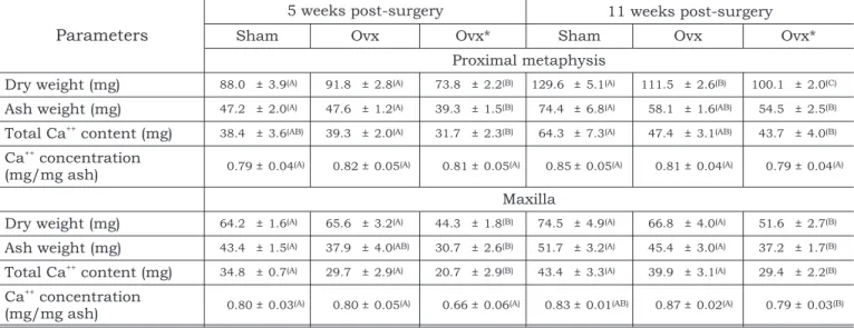
A histometric analysis performed 2 months post-ovariectomy showed no significant change in TABLE 1 -Proximal metaphysis and maxilla from sham-operated rats (sham), ovariectomized rats receiving a standard laboratory chow (ovx) and ovariectomized rats receiving a low Ca++diet (ovx*). The results are expressed as mean±SEM
(standard error of the mean). For each post-surgery period and each bone parameter, different letters denote statistically significant differences between the experimental groups (Kruskal-Wallis test, A¹B¹C,a= 0.05).
Parameters
5 weeks post-surgery 11 weeks post-surgery
Sham Ovx Ovx* Sham Ovx Ovx*
Proximal metaphysis
Dry weight (mg) 88.0 ± 3.9(A) 91.8 ± 2.8(A) 73.8 ± 2.2(B) 129.6 ± 5.1(A) 111.5 ± 2.6(B) 100.1 ± 2.0(C)
Ash weight (mg) 47.2 ± 2.0(A) 47.6 ± 1.2(A) 39.3 ± 1.5(B) 74.4 ± 6.8(A) 58.1 ± 1.6(AB) 54.5 ± 2.5(B)
Total Ca++
content (mg) 38.4 ± 3.6(AB) 39.3 ± 2.0(A) 31.7 ± 2.3(B) 64.3 ± 7.3(A) 47.4 ± 3.1(AB) 43.7 ± 4.0(B)
Ca++
concentration
(mg/mg ash) 0.79 ± 0.04(A) 0.82 ± 0.05(A) 0.81 ± 0.05(A) 0.85 ± 0.05(A) 0.81 ± 0.04(A) 0.79 ± 0.04(A)
Maxilla
Dry weight (mg) 64.2 ± 1.6(A) 65.6 ± 3.2(A) 44.3 ± 1.8(B) 74.5 ± 4.9(A) 66.8 ± 4.0(A) 51.6 ± 2.7(B)
Ash weight (mg) 43.4 ± 1.5(A) 37.9 ± 4.0(AB) 30.7 ± 2.6(B) 51.7 ± 3.2(A) 45.4 ± 3.0(A) 37.2 ± 1.7(B)
Total Ca++
content (mg) 34.8 ± 0.7(A) 29.7 ± 2.9(A) 20.7 ± 2.9(B) 43.4 ± 3.3(A) 39.9 ± 3.1(A) 29.4 ± 2.2(B)
Ca++
concentration
(mg/mg ash) 0.80 ± 0.03(A) 0.80 ± 0.05(A) 0.66 ± 0.06(A) 0.83 ± 0.01(AB) 0.87 ± 0.02(A) 0.79 ± 0.03(B)
TABLE 2 -Bone mass decrease (estimated by mean per-cent reduction of dry and ash weights and total Ca++
amount) in the proximal metaphysis and maxilla of ovariectomized rats receiving a standard laboratory chow (ovx) and ovariectomized rats receiving a low Ca++
diet (ovx*), as compared to sham-operated animals.
Bone
5 weeks post-surgery
11 weeks post-surgery
Ovx Ovx* Ovx Ovx*
Proximal
metaphysis » ¯17% ¯14% ¯28%
Maxilla » ¯35% » ¯30%
the amount of compact or trabecular bone in the mandible and only a 10-25% increase in bone po-rosity in the maxilla25
. The authors concluded that rats, due to their peculiar masticatory habits plac-ing large loads on oral bones, are not a suitable ex-perimental model for studying oral bone loss re-lated to skeletal osteoporosis and that, to worsen oral osteopenia, it would be mandatory to combine ovariectomy with a mechanical unloading, i.e. af-ter molar extraction. In fact, ovariectomy-induced estrogen depletion has been shown to affect bone healing/remodeling after molar extraction by in-creasing bone resorption and reducing bone for-mation, an effect observed earlier in the maxilla9,10,17,18than in the mandible3,4. Compared to
the distal femur, the changes in the edentulous mandible of ovariectomized rats take longer, possi-bly due to a larger proportion of trabecular bone composing the femur, while the edentulous man-dible contains primarily cortical bone4
.
In this respect, literature data have shown that the maxillary cortical bone shell, like the mandibu-lar one, is not markedly influenced by short- or long-term estrogen depletion18
. The reason why trabecular bone is lost faster than cortical bone is that trabecular bone turnover is greater than corti-cal turnover due to the much greater number of bone cells and larger surface area in the former6
. The present results support literature data showing that the metaphysis of a long bone is more affected by estrogen deficiency than the oral bone and that ovariectomy alone is not effective in af-fecting maxillary bone mass, at least over an 11-week period.
Additionally, a low Ca++diet has been used as a
protocol for oral osteoporosis in female rats. A Ca++
diet reduced to 6% of the control one, administered for 16 weeks, caused practically the same rate of reduction in the cancellous bone of the proximal tibia, first tail vertebra and mandible15
. The au-thors emphasized that a common denominator of all bone segments investigated was that they con-tained cancellous bone readily available for the maintenance of calcium homeostasis.
A combined ovariectomy and low Ca++diet is an
experimental design seldom used to investigate os-teoporosis in oral bones. Moriyaet al.14
compared by radiographic and visual inspection the bone mineral density and bone loss in rat femur, tibia, maxilla and mandible following ovariectomy and/or a low (0.005%) Ca++
diet administered for 4
weeks. Although bone mineral density was de-creased in all bones by a low Ca++diet associated or
not with ovariectomy (but not by ovariectomy alone), no significant alteration was detected re-garding alveolar bone loss (evaluated by the dis-tance from the cemento-enamel junction to the bone crest at the center of the molars mesial root). Contrarily, a significant increase in both bone for-mation and resorption, resulting in a decreased bone volume, were detected by histomorphological analysis applied to the cortical maxillary bone and to the cancellous bone of mandible and proximal tibia, from 12 to 32 weeks post-ovariectomy asso-ciated to a low (0.02%) Ca++
diet19
.
The present results show that feeding ovariectomized rats a low (0.1%) Ca++
diet caused a bone mass reduction which was slight in the proxi-mal metaphysis but more pronounced in the maxilla, as early as during the 5thweek
post-sur-gery, although on a long-term basis the treatment seemed equally injurious to both bones. It has been demonstrated that dietary Ca++
deficiency seems to induce bone loss in both cortical and cancellous bone whereas bone loss due to estrogen deficiency is mainly confined to cancellous bone16
. Moreover, rats fed a diet containing more than 1% Ca++
(as is the case for most standard chows) have reduced bone sensitivity to ovariectomy, whereas in ovariectomized animals fed a low Ca++ diet the
decrease in Ca++
absorption due to ovariectomy be-comes significant and bone loss is enhanced12
. In the present study, Ca++concentration in the
proximal metaphysis and maxilla of ovariecto-mized rats receiving or not a low Ca++
diet did not differ significantly from that of sham-operated ani-mals. It has been shown that ovariectomy in rats, like the postmenopausal period in women, results in loss of bone matrix with no alteration in bone matrix mineralization6
. Osteoporosis has been de-fined as a generalized, progressive diminution in bone mass, causing weakness of skeletal strength, even though the ratio of mineral to organic ele-ments is unchanged in the remaining normal bone1
. A low bone mass accompanied by trabecular disruption and cortical porosity is seen in osteoporosis, in contrast to an equally low bone mass with disturbances in mineralization ob-served in osteomalacia4
CONCLUSION
In conclusion, the animal model of a low Ca++
diet administered to rats with ovariectomy-in-duced estrogen deficiency described here proved to be an effective protocol for maxillary osteoporosis, even during a short-term experimental period, promising to be useful in future investigations re-garding the implications of clinical procedures, pathological processes and the efficacy of thera-peutic agents against oral bone loss.
ACKNOWLEDGEMENTS
The authors thank Dr. Augusto Cesar C. Spadaro for providing the facilities for Ca++
mea-surement and Ana Cristina M. Polizelo, Adriana M. G. Silva and Edna A. S. Moraes for technical assis-tance. The research was supported by CNPq (520128/96-6) and FAPESP (99/08858-5).
REFERENCES
1. Berkow R, Fletcher AJ. The Merck manual of diagnosis and therapy (1987)apudJahangiri L, Kim A, Nishimura I. Ef-fect of ovariectomy on the local residual ridge remodeling. J Prosthet Dent 1997;77:435-43.
2. Elovic RP, Hipp JA, Hayes WC. Ovariectomy decreases the bone area fraction of the rat mandible. Calc Tissue Internat 1995a;56:305-10.
3. Elovic RP, Hipp JA, Hayes WC. Maxillary molar extraction causes increased bone loss in the mandible of ovariectomized rats. J Bone Miner Res 1995b;10:1087-93. 4. Elsubeihi ES, Heersch JMN. Effects of postmenopausal
os-teoporosis in the mandible. In: Zarb G, Lekholm U, Albrektsson T, Tenenbaum H. Aging, osteoporosis and dental implants. London: Quintessence Books; 2002. p. 207-15.
5. Gilles JA, Carner DL, Dallas MR, Holt SC, Bonewald LF. Oral bone loss is increased in ovariectomized rats. J Endod 1997;23:419-22.
6. Grynpas MD. The concept of bone quality in osteoporosis. In: Zarb G, Lekholm U, Albrektsson T, Tenenbaum H. Aging, osteoporosis and dental implants. London: Quintes-sence Books; 2002. p. 25-34.
7. Hara T, Sato T, Oka M, Mori S, Shirai H. Effects of ovariectomy and/or dietary calcium deficiency on bone dy-namics in the rat hard palate, mandible and proximal tibia. Arch Oral Biol 2001;46:443-51.
8. Horner K, Devlin H, Alsop CW, Hodgkinson IM, Adams JE. Mandibular bone mineral density as a predictor of skeletal osteoporosis. Br J Radiol 1996;69:1019-25.
9. Hsieh YD, Devlin H, McCord F. The effect of ovariectomy on the healing tooth socket of the rat. Arch Oral Biol 1995;40:529-31.
10. Jahangiri L, Kim A, Nishimura I. Effect of ovariectomy on the local residual ridge remodeling. J Prosthet Dent 1997;77:435-43.
11. Jeffcoat MK, Chestnuti CH. Systemic osteoporosis and oral bone loss: evidence shows increased risk factors. J Am Dent Assoc 1993;124:49-56.
12. Kalu DN. The ovariectomized rat model of postmenopausal bone loss. Bone and Mineral 1991;15:175-92.
13. Kribbs PJ. Comparison of mandibular bone in normal and osteoporotic women. J Prosthet Dent 1990;63:218-22.
14. Moriya Y, Ito K, Murai S. Effects of experimental osteoporo-sis on alveolar bone loss in rats. J Oral Sci 1998;40:171-5. 15. Rosenquist JB, Lundgren S. Sensitivity to a low calcium
diet in different bones: an experimental study in the adult rat. Scand J Dent Res 1992;100:327-9.
16. Shen V, Birchman R, Xu R, Lindsay R, Dempester DW. Short-term changes in histomorphometric and biochemi-cal turnover markers and bone mineral density in estro-gen- and/or dietary calcium-deficient rats. Bone 1995;16:149-56.
17. Shimizu M, Sasaki T, Ishihara A, Furuya R, Kawawa T. Bone wound healing after maxillary molar extraction in ovariectomized aged rats. J Electron Microsc 1995; 47:517-26.
18. Shimizu M, Furuya R, Kawawa T, Sasaki T. Bone wound healing after maxillary molar extraction in ovariectomized aged rats: quantitative backscattered electron image anal-ysis. Anat Rec 2000;259:76-85.
19. Shirai H, Sato T, Oka M, Hara T, Mori S. Effect of calcium supplementation on bone dynamics of the maxilla, mandi-ble and proximal tibia in experimental osteoporosis. J Oral Rehabil 2002;29:287-94.
20. Taguchi A, Tanimoto K, Suei Y, Otani K, Wada T. Oral signs as indicators of possible osteoporosis in elderly women. Oral Surg Oral Med Oral Pathol Oral Radiol Endod 1995;80:612-6.
21. Taguchi A, Tanimoto K, Suei Y, Ohama K,; Wada T. Rela-tionship between the mandibular and lumbar vertebral bone mineral density at different postmenopausal stages. Dentomaxillofac Radiol 1996;25:130-5.
22. Tanaka M, Ejiri S, Kohno S, Ozawa H. The effect of aging and ovariectomy on mandibular condyle in rats. J Prosthet Dent 1998;79:685-90.
23. Tanaka M, Ejiri S, Toyooka E, Kohno S, Ozawa H. Effects of ovariectomy on trabecular structures of rat alveolar bone. J Periodontol Res 2002;37:161-5.
24. von Wowern N, Kollerup G. Symptomatic osteoporosis: a risk factor for residual ridge reduction of the jaws. J Prosthet Dent 1992;67:656-60.
25. Zaffe D, Paganelli C, Cocchi D. Induction and pharmaco-logical treatment of oral osteopenia in rats. Minerva Stomatol 1999;48:45-62.