Mailing Address: Rui André Quadros Bebiano da Providência e Costa •
Couraça de Lisboa nº22 - 3000-434 – Coimbra – Portugal E-mail: [email protected]
Manuscript received May 22, 2009; revised manuscript received July 11, 2009; accepted August 06, 2009.
Hereditary Hemorrhagic Telangiectasia: Rare Cause of Pulmonary
Hypertension?
Rui Providência, Maria do Carmo Cachulo, Gisela Veríssimo Costa, Joana Silva, Carlos Graça Lemos, A.M.
Leitão-Marques
Coimbra’s Hospital Center, Cardiology Department, Coimbra - Portugal
:
A 73-year-old woman was admitted to the emergency room with predominantly right-sided heart failure and anemia. Following clinical and imagiological evaluation, a diagnosis of pulmonary hypertension (PH) associated with Hereditary Hemorrhagic Telangiectasia (HHT) was confirmed. The initial response to bosentan plus sildenafil was good, including improvement in functional class and reduction of edema, allowing her to be discharged. Unfortunately, the patient died, due to her underlying condition, before the effects of the combination treatment could be fully assessed. PH should be considered in patients with HTT and screening for pulmonary hypertension should be performed in these patients and their relatives.
Key Words
Telangiectasia, hereditary hemorrhagic; hypertension, pulmonary; epistaxis; vascular diseases.
Introduction
Rendu-Osler-Weber syndrome, also known as hereditary hemorrhagic telangiectasia (HHT), was first described by Osler, Rendu and Hanes in the 19th century1-4. It is a dominant
autosomal disease, with a prevalence of <5 in 8,0005, and it
is caused by mutations in the genes codifying for transforming growth factor β (TGF-β) signaling receptors, endoglin and activin receptor-like kinase type-1 (ALK-1)1.
The syndrome is characterized by telangiectasias affecting the skin and mucous membranes, most frequently the hands, feet, lips, tongue and conjunctiva. Spontaneous and repeated epistaxis is often present, as well as gastrointestinal hemorrhages causing ferropenic anemia. Diagnostic imaging techniques have also revealed pulmonary (30% of patients), hepatic (<30%) or brain (10-20%) arteriovenous anomalies6,7.
Shovinet al8diagnostic definition of HHT requires the presence
of at least three of the following: epistaxis, telangiectasias, visceral vascular anomalies, or a family history of the disease8.
Some HHT patients also present pulmonary complications
such as pulmonary hypertension. These are more common in patients with ALK-1 mutations9 – also associated with other
inherited forms of primary pulmonary hypertension – although the precise prevalence is not known.
Here we describe the case of an elderly woman with HTT and severe pulmonary hypertension.
Case report
A 73-year-old woman presented twice at the Emergency Room with mainly right-sided heart failure. She also had edema of the lower limbs, Godet sign, severe dyspnea after brief exercise (New York Heart Association [NYHA] class III) and orthopnea when placed at a 45º angle. Her medical history included chronic anemia (due to gastro-duodenal angiodysplasias), frequent and abundant epistaxis over a period of more than 20 years, atrial fibrillation, arterial hypertension and venous disease of the lower limbs. She had had a total hysterectomy five years previously due to the presence of myomas. A previous endoscopic study had also indicated friable angiodysplasias in the gastric body and duodenum, for which the patient had received argon plasma coagulation therapy. There was no record of pulmonary parenchymatous disease, sleep apnea or use of any anorexigenic substance.
Signs of HHT (i.e. angiodysplasias in the tongue, oral mucous membrane and limbs) had also been present in her father, as well as in a paternal aunt and cousin. During physical examination, the patient was conscious, oriented and cooperative. She was eupneic and showed no visible signs of respiratory distress syndrome. Pulse oximetry showed O2 saturation levels of 96%. On cardiopulmonary auscultation, breath sounds were symmetric, with some inspiratory basal rales heard on both sides. A systolic murmur was detected in the pulmonary valve region. No abdominal alterations were apparent. Her skin and mucous membranes were hydrated, but pale. Telangiectasias were visible in her tongue, oral mucous membrane and skin (face and arms).
An electrocardiogram showed atrial fibrillation with controlled ventricular response and non-specific alterations of anterolateral repolarization. Cardiac remodeling was evident in a posterior-anterior (PA) thoracic radiogram, and transthoracic echocardiography. Doppler ultrasonography showed moderate to severe tricuspid regurgitation, which made it possible to estimate the systolic pressure of the pulmonary artery (SPPA) at 76 mmHg (56 gradient VD/AD plus 20mmHg for total absence of collapse of the inferior vena cava). The presence of congenital cardiac shunt was excluded following a transesophageal echocardiogram. Pulmonary angio-CT showed dilatation of the pulmonary
e34
After careful analysis of all clinical, analytical and imaging data, a diagnosis of pulmonary hypertension associated with HTT was suggested. It was proposed that hepatovenous shunts led to hyperflow conditions in the pulmonary circulation, which, in turn, resulted in the development of pulmonary hypertension.
Subsequent catheterization indicated average pressure in the pulmonary artery of 56 mmHg (74/40/56), increased pulmonary vascular resistance (5.1 Wood units), with normal pulmonary capillary pressure and elevated cardiac index – (5.3 L.min-1.m-2). The coronariography showed the coronary arteries
to be undamaged, but with an exaggerated blush. Initially, the patient was treated with intraduodenal digoxin (0.25 mg), enalapril maleate (5 mg), daflon (500 mg), furosemide (2 mg/kg), ranitidine (300 mg) and an iron–folate supplement; however, her condition did not improve. Following the diagnosis of pulmonary hypertension, she commenced therapy with bosentan plus sildenafil. This brought about a gradual improvement in her condition.
During her stay in hospital the patient acquired nosocomial pneumonia due to Klebsiella pnemoniae infection, which was treated with piperacillin + tazobactam for two weeks and presented several episodes of epistaxis and hematemesis, necessitating an upper endoscopy, resumption of argon therapy, a number of transfusions (total: 14 units of red cells) and further treatment with intravenous esomeprazole and sucralfate, oral folate, iron and vitamin B12.
After 65 days, the patient’s heart condition had clearly improved (to NYHA class II); she had no edema in the lower limbs, and could walk small distances, with assistance, so she was discharged. One week later, she was again admitted to the emergency room with melena and anemia. She was given a transfusion of 2 units of red cells and argon plasma therapy to treat gastroduodenal angiodysplasias. A week
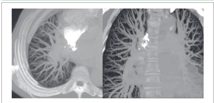
Figure 1 – A – Pulmonary angio-CT image, axial plane, showing pulmonary hypervascularization, affecting the whole lung, even the most peripheral segments. B - coronal plane: Note the pulmonary trunk and dilatation of the main branches.
Figure 2 – Abdominal angio-CT images conirming the previous hepatic
indings with the presence of arteriovenous istula.
trunk and its main branches, with abundant vasculature up to the periphery of the pulmonary fields, but no thrombi or arteriovenous fistulae (Figure 1). Laboratory tests showed values of <0.3mg/dL and 2941.3pg/mL for C-reactive protein and N-terminal prohormone brain natriuretic peptide, respectively. Malformations of the hepatic arterial and venous systems (increased caliber of vessels, with tortuous aspect and turbulent and rapid flow rates) were suggested by Doppler ultrasonography and confirmed by the abdominal angio-CT (Figure 2).
Arq Bras Cardiol 2009; 94(3) : e34-e36 Providencia et al HHT as rare cause of pulmonary hypertension
e35
later she was admitted again with dyspnea due to bilateral pneumonia (caused by a non-identified micro-organism) and severe anemia, for which she received two units of red cells and intravenous antibiotics. She was discharged when her hemoglobin levels increased from 5.2 g/dL to 9.2g/dL, but died just a few days later, due to an uncontrollable upper gastrointestinal hemorrhage.
Discussion and conclusions
This patient had well-developed vascular disease affecting multiple organ sites (liver, lungs and gastrointestinal tract). Despite a good response to combination therapy with bosentan and sildenafil for pulmonary hypertension, she eventually died as a consequence of her underlying disease, making it difficult to assess the hemodynamic functional evolution of the patient during treatment. There are very few reports of pulmonary hypertension associated with HHT in the literature; the rate of development of pulmonary hypertension in these patients is unknown and little is known regarding optimum treatment.In a previous case study,Schlag et al10 reported that the number
of episodes of epistaxis could increase with prostanoids, whereas a good response to bosentan was observed.
The reason why some patients with ALK-1 mutations develop HHT only, whereas others develop only idiopathic
pulmonary hypertension and still others develop both conditions is not known. In order to diagnose pulmonary hypertension associated with HHT, one must carefully consider the patient’s history and confirm the signs and symptoms of the condition using a number of different diagnostic imaging procedures. As HHT has a dominant autosomal transmission mechanism, we recommend that relatives of patients with HHT undergo periodic transthoracic Doppler echocardiography (every 3 years), in order to assess the systolic pressure of the pulmonary artery.
Potential Conflict of Interest
No potential conflict of interest relevant to this article was reported.
Sources of Funding
There were no external funding sources for this study.
Study Association
This study is not associated with any post-graduation program.
1 Rendu H. Epistaxis repetees chez un sujet porteur de petits angiomes cutanes et muquez. Gaz des Hôpitaux. 1896; 13: 1322-3.
2 Osler W. On a family form of recurring epistaxis, associated with multiple telangiectases of the skin and mucous membranes. Bull Johns Hopkins Hosp. 1901; 12: 333-7.
3 Weber F. Multiple hereditary developmental angiomata (telangiectases) of the skin and mucous membranes associated with recurring hemorrhages. Lancet. 1907; 2: 160-2.
4 Hanes FM. Multiple hereditary telangiectases causing hemorrhage (hereditary hemorrhagic telangiectasia). Bull Johns Hopkins Hosp. 1909; 20: 63-73. 5 Fernández LA, Sanz-Rodriguez F, Blanco FJ, Bernabéu C, Botella LM.
Hereditary hemorrhagic telangiectasia, a vascular dysplasia affecting the TGF-β signaling pathway. Clin Med Res. 2006; 4 (1): 66-78.
6 Begbie ME, Wallace GMF, Shovlin CL. Hereditary haemorragic telangiectasia
(Osler-Weber-Rendu syndrome): a view from the 21st century. Postgrad Med J. 2003; 79: 18-24.
7 de Melo JC, de Araújo AP, Monteiro ES, Rongel EB, Felipe H, Ferreira MC, et al. Pulmonary arteriovenous fistula: a case report and review of the literature. Arq Bras Cardiol. 1989; 53 (1): 43-8.
8 Shovlin CL, Guttmacher AE, Buscarini E, Faughnan ME, Hyland RH, Westermann CJ, et al. Diagnostic criteria for hereditary haemorrhagic telangiectasia (Rendu-Osler-Weber syndrome). Am J Genet. 2000; 91 (1): 66-7.
9 Abdalla SA, Gallione CJ, Barst RJ, Horn EM, Knowles JA, Marchuk DA, et al. Primary pulmonary hypertension in families with hereditary haemorrhagic telangiectasia. Eur Respir J. 2004; 23: 373-7.
10 Schlag K, Opitz C, Wensel R, Felix S, Ewert R. Pulmonary hypertension in hereditary haemorrhagic telangiectasia (Rendu-Osler-Weber disease). Progression over 10 years. Dtsch Med Wochenschr. 2005; 130 (23): 1434-7.
References
Arq Bras Cardiol 2009; 94(3) : e34-e36 Providencia et al
HHT as rare cause of pulmonary hypertension