Abstract
Background: Cardiogenic shock and acute pulmonary edema are the major causes of death of patients with scorpion
envenomation, whose pathophysiological mechanism remains controversial.
Objective: To investigate the correlation between myocardial perfusion abnormalities and left ventricular contractile function in victims of scorpion envenomation.
Methods: Fifteen patients underwent ECG-gated myocardial perfusion scintigraphy (gated SPECT) within 72 hours of,
and 15 days after scorpion envenomation. Images were analyzed by means of a semi-quantitative visual perfusion score (0 = normal, 4 = absent) and motion score (0 = normal, 4 = akinesia), using the 17-segment model. Summed perfusion (SPS) and summed motion (SMS) scores were calculated for each patient. Ejection fraction (LVEF) was calculated by a commercially available software.
Results: At baseline, 12 out of the 15 patients presented abnormal myocardial perfusion and contractility. Mean values of SPS, SMS and LVEF were 12.5 ± 7.3, 17.0 ± 12.8, and 44.6 ± 16.0%, respectively. A positive correlation between SPS and SMS (r = 0.68; p = 0.005) and negative correlation between SPS and LVEF (r = -0.75; p = 0.0021) were found. The follow-up studies showed recovery of global contractility (LVEF of 68.9 ± 9.5, p = 0.0002), segmental contractility (SMS of 2.6 ± 3.1, p = 0.0009) and perfusion (SPS of 3.7 ± 3.3, p = 0.0003). Improvement of LVEF correlated positively with improvement of SPS (r = 0.72; p = 0.0035).
Conclusions: Myocardial perfusion abnormalities are common in scorpion envenomation and correlate topographically
with the contractile dysfunction. Recovery of contractility correlates with reversibility of perfusion defects. These findings suggest the participation of myocardial perfusion abnormalities in the pathophysiology of this form of acute ventricular failure. (Arq Bras Cardiol 2010; 94(4):418-425)
Key Words: Scorpions; Radionuclide Imaging; Perfusion; Ventricular Dysfunction; Pulmonary Edema.
Mailing address: Marcus Vinicius Simões •
Rua Humaitá 461, apartamento 24 - Santa Cruz - 14020-680 - Ribeirão Preto, SP - Brazil
E-mail: simoesmv@yahoo.com
Manuscript received October 05, 2008; revised manuscript received June 25, 2009; accepted July 09, 2009.
Assessment of Myocardial Perfusion and Function in Victims of
Scorpion Envenomation Using Gated-SPECT
Alexandre Baldini de Figueiredo, Palmira Cupo, Antônio O. Pintya, Fábio Caligaris, José A. Marin-Neto, Sylvia
E.Hering, Marcus Vinicius Simões
Hospital das Clínicas da Faculdade de Medicina de Ribeirão Preto - USP - São Paulo, SP - Brazil
echocardiographic and hemodynamic alterations consistent with cardiac damage, contractile dysfunction, and acute left ventricular failure accompanying this clinical syndrome2-9.
Although the venom seems to have some direct effect on myocardial fibers, most of the authors agree that the determining factor for cardiac dysfunction is the effect of the high concentrations of catecholamines10. In addition to the
hemodynamic overload caused by increased blood pressure and venous return, excessive catecholaminergic stimulation is also known to lead to myocardial damage.
Another possible mechanism that is frequently suggested to be responsible for the cardiac alterations seen in scorpion envenomation is myocardial ischemia. In fact, the electrocardiogram of victims of severe scorpion envenomation frequently shows changes consistent with myocardial ischemia: ST-segment elevation and depression, and electrically inactive zones, commonly associated with elevated levels of cardiac enzymes. Myocardial ischemia in scorpion envenomation may result from overstimulation of alpha-adrenergic
Introduction
Scorpions account for one of the oldest public health problems that humankind has ever faced. In Brazil, scorpion envenomation is common, even considering that mild cases are underreported. According to the National Health Foundation (Fundação Nacional de Saúde - FUNASA), 24,826 cases were reported between January 1990 and December 1993, with 143 deaths1.
receptors, which would lead to microvascular constriction of the coronary circulation.
The participation of myocardial ischemia in the pathophysiology of scorpion envenomation in humans has been suggested in few clinical studies11-13. In a previous report,
we demonstrated the presence of severe transient alterations of the myocardial perfusion in victims of severe scorpion envenomation14. However, assessment of the ventricular function
and myocardial perfusion were performed using different - and not simultaneous - imaging methods: echocardiography and myocardial perfusion SPECT, respectively. The objective of the present study was to thoroughly analyze the topographic correlation between perfusion and left ventricular function abnormalities, including the follow-up of the temporal profile, by analyzing those parameters simultaneously using gated-SPECT myocardial perfusion imaging.
Methods
Inclusion criteria
Patients of both genders, with no age limit, who were previously healthy and victims of scorpion envenomation clinically classified as moderate or severe (by the presence of systemic symptoms of sympathetic or parasympathetic hyperactivity or cardiopulmonary manifestations that translated into systemic action of the venom) were prospectively selected for inclusion in this study. The trial was analyzed and approved by the Ethics Committee of the Ribeirão Preto School of Medicine, University of Sao Paulo, and a written informed consent was obtained from the persons responsible for the patients.
Exclusion criteria
Patients who experienced clinical instability that made impossible their removal to the Service of Nuclear Medicine in order to undergo scintigraphy studies in a timely fashion, pregnant women and nursing women were excluded from the study.
Study protocol
All patients received scorpion venom antiserum preceded by intravenous corticosteroids and antihistaminic drugs to prevent or minimize immediate hypersensitivity reactions (hydrocortisone, dexchlorpheniramine and ranitidine), and supportive care including nasal oxygen cannula, diuretics and inotropic agents in the cases for which it was deemed necessary. The following tests were performed at admission and then serially in all patients included in the study: complete blood count, renal function, electrolytes, blood glucose, CK-MB, electrocardiogram, chest radiography.
Myocardial perfusion study - gated SPECT
All patients included in the study underwent myocardial perfusion scintigraphy with administration of 99mTc-SESTAMIBI at
rest within the first 72 hours following scorpion envenomation. The tests were repeated within one to two weeks of the onset of symptoms. In two patients who remained with severe perfusion abnormalities (summed perfusion score equal to or
higher than 8) the tests were repeated later in order to follow up the course of the abnormalities found.
All tests were performed in a DST-Sopha Medical Vision digital gamma camera equipped with two rectangular middle-field detectors coupled to low-energy high-resolution collimators with energy acceptance window of 20% centered at 140 Kev (photopeak of 99m-Tc).
The emission tomography images were acquired one hour after intravenous administration of 14.8 MBq/kg (0,4mCi/kg) of 99mTc - SESTAMIBI at rest, using the acquisition protocol
with a 180º scanning angle, patient in the supine position, step-and-shoot mode circular orbit, from the right anterior oblique view to left posterior oblique view, with 32 projections, 60 seconds per projection, and a 64x64 matrix. Images were acquired in synchrony with ECG (gated SPECT) for the analysis of global and segmental left ventricular contractile function using acquisition with 8 frames per cardiac cycle,
with a 50% acceptance window around the mean value of
the RR interval.
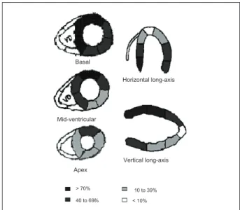
Myocardial perfusion images were processed in a dedicated computer using commercially available software programs (Sopha Medical Vision). After manually setting up the spatial reorientation axes and calculating the tomographic views in the three orthogonal planes, the images were submitted to analysis. Two independent observers carried out a semi-quantitative analysis (visual analysis scores) of each image as regards perfusion and contractile function of the different myocardial segments; occasional disagreements were settled by consensus. For this analysis, the left ventricular wall was subdivided into 17 segments15 (Figure 1),
and a semi-quantitative visual perfusion score (0 = normal, 1 = slightly low uptake, 2 = moderately low uptake, 3 = severely low uptake, 4 = no uptake) and motion score (0 = normal, 1 = mild hypokinesia, 2 = moderate hypokinesia, 3 = severe hypokinesia and 4 = akinesia) was attributed to each segment.
Left ventricular ejection fraction was calculated automatically using “The Cedars-Sinai Quantitative Gated SPECT” (QGS) software16.
Statistical analysis
Variables were expressed as mean and standard deviation of the mean.
For the analysis of the topographic correlation between perfusion and segmental motion abnormalities, the chi-square test of heterogeneity of frequency distribution was used. For the analysis of the correlation between the intensity of segmental abnormalities of perfusion and motion, the Cohen’s kappa coefficient was calculated.
To verify the significance of the difference between the means of the variables obtained at baseline and in the follow-up, the paired t-test was used when the sample distribution of the variables was normal; otherwise, the Wilcoxon test was used.
The Kolmogorov-Smirnoff test was used to test whether the sample distribution of the variables was normal.
Results
Clinical and laboratory aspects
Fifteen patients were included in the study (n = 15); nine
were males, and the mean age was 7.6 ± 4 years. The majority of the patients were admitted to the Emergency Department within the first three hours after the sting, ranging from
one to 6.5 hours. All patients presented with some sign of
envenomation in addition to local pain, the most frequent of which being vomiting, diaphoresis, and alteration of the level
of consciousness. Of the 15 cases, 12 were clinically classified
as severe; seven developed acute pulmonary edema, and six required the use of vasoactive amines. Some of the clinical and laboratory characteristics of these patients are shown in Table 1. Six patients presented significantly increased serum CK-MB levels (above twofold the normal limits).
Electrocardiographic abnormalities
All patients (except for no. 7) presented significant electrocardiographic abnormalities at some time during the follow-up. The abnormalities most frequently found, in addition to sinus tachycardia, were T-wave and ST-segment abnormalities (elevation and/or depression). In four cases, a transient “electrically inactive zone” was also observed, that disappeared within a few days.
Assessment of myocardial perfusion
Of the 15 patients included in the study, 12 (80%)
presented significant alterations of myocardial perfusion (summed perfusion score > 4), all of them with scorpion envenomation clinically classified as severe. Only one patient with moderate envenomation presented a normal perfusion study. The number of segments involved per patient was 7.9
± 4.2 and the summed perfusion score was 12.5 ± 7.3. The distribution of the perfusion defects did not respect the limits or follow the distribution of the main coronary territories (Figure 2). A high frequency of involvement of the basal portions of the ventricular walls (particularly the anterior and septal walls) was observed, and this frequency decreased progressively in the mid portions and especially the apical region. The frequency distribution of perfusion defects by myocardial segment is shown in Figure 3.
Assessment of Left Ventricular Function
Reduced left ventricular ejection fraction was frequently observed (10 patients), with a mean of 44.6% ± 16 %. Ejection fraction could not be assessed in only one patient due to technical problems related to the very small size of the ventricular cavity.
In all patients who presented decreased global left ventricular systolic performance, this was associated with significant abnormalities of the segmental wall motion. As happens for perfusion defects, the left ventricular basal and mid-segments were those whose motion was more severely affected; the apical segment was the less severely affected (Figure 4).
A significant negative correlation was observed between the summed perfusion score and left ventricular ejection fraction
Basal
Horizontal long-axis VENTRICULAR SEGMENTS
Mid-ventricular
Basal anterior
1. 10. Mid-inferior
Basal anteroseptal
2. 11. Mid-inferolateral
Basal inferoseptal
3. 12. Mid-anterolateral
Basal inferior
4. 13. Apical anterior
Basal inferolateral
5. 14. Apical septal
Basal anterolateral
6. 15. Apical inferior
Mid-anterior
7. 16. Apical lateral
Mid-anteroseptal
8. 17. Apex
Mid-inferoseptal 9.
Figure 1 - Left ventricular wall segmentation used in the analysis of perfusion and motion.
Table 1 - Demographic, clinical and laboratory aspects of the patients
studied at admission in the Emergency Unit
Mean ± SD Minimum Maximum
Age (years) 7.6 ± 4.3 1 15
HR (bpm) 107 ± 40.8 50 175
Systolic BP (mmHg) 131.9 ± 22.1 90 163
Diastolic BP (mmHg) 92.4 ± 22.1 50 127
∆t sting/ perfusion
study (hours) 30.9 ± 17.5 8 68
Glucose (mg/dl) 301.9 ± 59.8 188 400
Potassium (mEq/l) 3.1 ± 0.3 2.75 3.7
CKMB (U/l) 77.1 ± 61.8 11 251
Creatinine (mg/dl) 0.87 ± 0.2 0.7 1.1
(r = -0.75; p = 0.0021 – Pearson’s correlation analysis). Also,
a positive correlation was observed between the summed perfusion score and the intensity of motion abnormalities as
assessed by the summed motion score (r = 0.68; p = 0.0050
- Pearson’s correlation analysis).
Figure 2 - Illustrative example of baseline and follow-up myocardial perfusion studies of a patient with severe scorpion envenomation. Short-axis, vertical long-axis, and horizontal long-axis tomographic views are represented; in each box, the upper images correspond to the baseline study and the lower images, to the follow-up study. In the baseline study, severe perfusion defect can be observed in the basal and mid-segments of the anterior, inferior and septal left ventricular walls. Full recovery of perfusion abnormalities in the follow-up study.
of 120), motion abnormalities were concomitantly found in
101. On the other hand, among the 135 segments with normal
perfusion, 113 also had normal motion. The overall agreement
was 84% (chi square = 114.50; p < 0.0001).
Also, a positive correlation was observed between the intensity and extension of myocardial perfusion defects, as quantified by the summed perfusion score, and peak
CK-MB levels (r = 0.61; p = 0.0152). Additionally, a significant
association was observed between the occurrence of electrocardiographic abnormalities and the presence of perfusion defects (p = 0.0440 - Fisher’s exact test). Of the 12 patients with perfusion defects in the baseline study, eight (67%) presented significant ischemic alterations on the electrocardiogram: ST-segment elevation or depression of at least 1 mm, or pathological Q-wave in at least two contiguous leads. On the other hand, the three patients who did not present significant perfusion defects in the baseline studies had only abnormalities in T-wave morphology and duration.
Late assessment
During the clinical course, almost all patients presented significant improvement of the myocardial perfusion pattern, with significant reduction of the summed perfusion score (3.7
± 3.3) in relation to the acute phase study (p = 0.0003, paired
t-test p = 0.003, Figure 5).
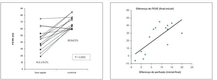
Improvement of perfusion was accompanied by recovery of the global and segmental left ventricular systolic performance. In the follow-up study, a reduction in the summed motion score (2.6 ± 3.1, p = 0.0009, paired t-test) and significant increase in left ventricular ejection fraction (68.9 ± 9.5%, p =
0.0002, paired t-test, Figure 6) were observed in comparison to the acute phase study.
A positive correlation was observed between the recovery
of LVEF and reduction of perfusion scores (r = 0.65; p =
0.008, Figure 7), thus showing that patients with higher degrees of recovery of ventricular function had concomitant improvement of perfusion defects.
Discussion
Our results show that, in patients with moderate to severe scorpion envenomation, left ventricular systolic dysfunction is closely related to myocardial perfusion defects. The association between perfusion and function abnormalities may be demonstrated both in the agreement of the topographic distribution of the segmental involvement and in the positive correlation between the individual values of severity of the global perfusion defect, as estimated by the summed perfusion scores, and the intensity of left ventricular systolic dysfunction, as assessed by the reduction of ejection fraction.
The sequential observation of the behavior of perfusion and function abnormalities provides important additional support to the correlation between these two defects. Recovery of the global and segmental ventricular dysfunction and improvement of the perfusion defects occurred concomitantly, and this was demonstrated by the positive correlation of the difference of perfusion scores between the acute and late phases and the improvement of left ventricular ejection fraction in the same period.
Figure 3 - Graphic representation of percentage frequency distribution of perfusion abnormalities by segment.
Basal
Horizontal long-axis
Vertical long-axis Mid-ventricular
Apex
> 70%
Indirect evidence supporting the role of myocardial ischemia in the genesis of ventricular systolic dysfunction could be found in the positive correlation between the intensity of perfusion defects in the acute phase of scorpion envenomation, the level of cardiac enzyme (CK-MB) elevation, and the magnitude of electrocardiographic abnormalities.
Taken together, these results strongly suggest that myocardial ischemia plays a relevant role in the pathophysiology of acute transient heart failure associated with scorpion envenomation.
In the present study, the assessment of global and segmental ventricular function was performed by obtaining ECG-gated myocardial perfusion images, which permitted simultaneous assessment of myocardial perfusion and function, thus providing greater power of observation of the topographic correlation between the abnormalities found. To the best of our knowledge, this is the first investigation to use this method to study the correlation between these functional parameters in victims of scorpion envenomation. The literature has repeatedly demonstrated the accuracy and reproducibility of the quantification of left ventricular ejection fraction using gated-SPECT in relation to several other types of assessment methods (echocardiography, radionuclide ventriculography, contrast ventriculography)16-18.
Alteration of myocardial perfusion after scorpion envenomation in humans was first described by Geron et al6
who reported perfusion defects in the basal segments of the anterior, septal, inferior and posterolateral walls in resting 201Tl
scintigraphy (planar images) in a 14-year-old female patient with severe scorpion envenomation, acute pulmonary edema and ventricular dysfunction.
In Bahloul et al’s study13, six patients with signs of left
ventricular dysfunction following scorpion envenomation underwent 201Tl myocardial perfusion scintigraphy within 12
to 17 hours of the event. Myocardial perfusion defects were observed in all patients. The tests were repeated in only two
patients after six and 15 days, and showed partial recovery
of the abnormalities initially found.
The results obtained in the present study corroborate some of Bahoul et al13 initial observations and add other
important information, especially regarding recovery of perfusion and function abnormalities in the short term after envenomation.
Persistence of perfusion defects
Although both global and segmental left ventricular contractile function had been considered normal in the follow-up studies of all patients, this did not occur in regard to myocardial perfusion, since four patients still persisted with significant perfusion deficits in the follow-up (summed score greater than 4). Persistent perfusion defects in the follow-up study may represent residual functional abnormality of the microcirculation with reduced myocardial flow at rest that is still subject to normalization in later studies. On the other hand, it is also important to consider that several patients in this case series presented significant elevation of cardiac enzyme levels that translated into tissue damage. This result suggests that mild perfusion defects in follow-up images may reflect
Figure 4 - Graphic representation of percentage frequency distribution of wall motion abnormalities by segment.
Basal
Horizontal long-axis
Vertical long-axis Mid-ventricular
Apex
> 70%
40 to 69% < 10% 10 to 39%
Figure 5 - Time proile of the summed perfusion score between the baseline assessment and follow-up assessment. Paired t-test (p = 0.0003).
0 5 10 15 20 25
f a s e inic ia l s e guim e nto
12,5±7,3
3,7±3,3 P = 0,0003
the presence of small islands of myocardial fibrosis which are not sufficient to cause noticeable impairment of the global or segmental left ventricular contractile function, but, in some cases, are evidenced in studies known to be sensitive, as is the case of perfusion scintigraphy. We could hypothesize that these initially subclinical structural changes may be accompanied by ventricular remodeling and impaired ventricular function in the long term.
The hypothesis that scorpion envenomation may cause late impairment of the left ventricular function was first proposed by Sundararaman et al19. In a case-control study
of scorpion envenomation as a risk factor for the subsequent development of idiopathic dilated cardiomyopathy. The results of Sundararaman’s study could suggest that, despite the apparent recovery of myocardial function after scorpion envenomation, some degree of subclinical structural cardiac damage would persist, and this could, after a few years, serve as the substrate for its progression, as well as for cardiac remodeling and development of symptomatic ventricular dysfunction. Thus, the finding of late persistent perfusion defects in our case series could be an indirect support to this hypothesis.
We believe that the elucidation of the pathophysiological meaning of the persistent perfusion defects observed in the follow-up studies would require the performance of a new investigation especially designed for this purpose, that is, with later serial assessments of perfusion and left ventricular function.
Pathophysiological mechanisms
The analysis of our results shows that the myocardial perfusion defects found in patients with scorpion envenomation could hardly be explained by spasm of subepicardial coronary branches. The preferential involvement of left ventricular basal and mid-segments and the tendency to spare the apical region does not follow the distribution of the main territories of the coronary vessels, which favors the hypothesis of microvascular constriction. In fact, several pathophysiological arguments exist that support the idea that the hyperadrenergic state, which is a prominent aspect of scorpion envenomation, could cause microcirculatory spasm and ischemia in different regions and organs.
Previous studies in experimental models demonstrated the correlation between adrenergic hyperstimulation and induction of microcirculation constriction and ischemia in different vascular territories20. Induction of myocardial
ischemia by excessive catecholamine stimulation could occur as a result of either an intense increase in myocardial oxygen consumption (by means of the significant hemodynamic
overload caused by increased blood pressure, tachycardia, increased peripheral vascular resistance and venous return) or by the combination of coronary spasm and/or microcirculation vasoconstriction20,21.
Evidences of induction of microcirculatory spasm caused by adrenergic hyperstimulation in scorpion envenomation have been published by Zeghal et al22 who used an experimental
model of scorpion envenomation in anesthetized rats. In this study, venom administration induced a 30 to 40-fold increase in plasma catecholamine levels and was associated with a dose-dependent increase of blood pressure, as well as of total peripheral, muscular, and renal vascular resistances. These effects were almost entirely suppressed when the rats were pre-treated with phentolamine (an alpha-adrenergic blocker). Our results show that in scorpion envenomation there is significant systemic and regional vasoconstriction, very likely due to alpha-adrenergic hyperstimulation.
Myocardial ischemia as a pathophysiological mechanism seems particularly relevant when the manifestations of severe scorpion envenomation are considered. In this context, the presence of electrocardiographic alterations consistent with myocardial ischemia and damage, as well as the elevation of serum enzyme levels indicative of lesion of cardiac fibers have drawn the attention of many investigators2,3,9. In the present
study, these abnormalities were also found, thus corroborating previous observations. Electrocardiographic abnormalities were common in our case series, and in some cases they were similar to those found in acute myocardial infarction. Significant elevations of enzyme levels (CK-MB) were observed
in eight out of the 15 patients (values of at least 50 U/l, twofold
above normal levels); other five presented borderline values
(between 25 and 50 U/l).
Data presented above support the hypothesis that regional wall motion dysfunction in severe scorpion envenomation is caused by spasm of the coronary microcirculation triggered by excessive local release of catecholamines from cardiac sympathetic nerve fibers which, in turn, results from the action of neurotoxins of the scorpion venom.
Figure 7 - Scatter plot showing the correlation between improvement of perfusion and improvement of LVEF. Pearson’s correlation index (r = 0.65; p = 0.008)
-10 0 10 20 30 40 50 60
-5 0 5 10 15 20 25
Diferença de perfusão (inicial-final) Diferença de FEVE (final-inicial)
Figure 6 - Time proile of left ventricular ejection fraction between the baseline assessment and follow-up assessment. Paired t-test (p = 0.0002).
0 10 20 30 40 50 60 70 80 90
fase aguda controle
44,6 ±16,0%
68,9±9,5%
Whether catecholamines act locally, inducing microvascular constriction through simple stimulation of alpha receptors at the coronary arteries, and thus leadingto hypoperfusion, ischemia and myocardial stunning, or whether they act indirectly through a complex neurohormonal interaction in which other neuropeptides and cytokines participate, as was suggested by Bahloul et al23 and by Nouira et al24, are
questions that still lack a definitive answer. The results of the present study strongly support the first hypothesis as the major determinant of all this cascade of events, although it is likely that there is also the adjuvant participation of several of the other factors mentioned above.
Study limitations
One of the limitations of gated SPECT quantification of left ventricular ejection fraction that may have influenced the final results of the present investigation is the size of the ventricular cavity. Most of the algorithms are known to tend to underestimate ventricular volumes and overestimate ejection fraction values in small hearts. This phenomenon originates from the low spatial resolution of cardiac radionuclide images that result in an almost complete obliteration of the ventricular cavity at the end-systole in patients with reduced ventricular size. Since the majority of the patients included in this study were children, this certainly influenced the determination of the contractile function. Thus, the left ventricular ejection fraction values that, as a whole, were already reduced in the acute phase
of scorpion envenomation, could be actually overestimated. The comparative analysis of ejection fraction between studies of the acute phase and follow-up is not affected by this effect, since it occurs in both phases similarly.
The results of the present study corroborate the hypothesis that myocardial perfusion abnormalities participate in the pathogenesis of acute heart failure that is frequently found in patients with severe scorpion envenomation.
Acknowledgements
Marcus V. Simões is supported by CNPq (process PQ Nº309082/2007-2)
Potential Conflict of Interest
No potential conflict of interest relevant to this article was reported.
Sources of Funding
There were no external funding sources for this study.
Study Association
This article is part of the thesis of master submitted by Alexandre Baldini de Figueiredo, from Faculdade de Medicina de Ribeirão Preto, Universidade de São Paulo.
References
11. Silveira N P, Moraes-Santos T, Azevedo AD, Freire-Maia L. Effects of Tityus serrulatus scorpion venom and one of its purified toxins (toxin γ) on the isolated guinea pig heart. Comp Biochem Physiol C. 1991; 98 (2-3): 329-36.
12. Margulis G, Sofer S, Zalstein E, Zucker N, Ilia R, Gueron M. Abnormal coronary perfusion in experimental scorpion envenomation. Toxicon. 1994;
32 (12): 1675-8.
13. Bahloul M, Hamida CB, Chtourou K, Ksibi H, Dammak H, Kallel H, et al. Evidence of myocardial ischaemia in severe scorpion envenomation myocardial perfusion scintigraphy study. Intensive Care Med. 2004; 30 (3): 461-7.
14. Cupo P, Figueiredo AB, Pazin Filho A, Pintya AO, Tavares Junior GA, Caligaris F, et al. Acute left ventricular dysfunction of severe scorpion envenomation is related to myocardial perfusion disturbance. Int J Cardiol. 2007; 116 (1): 98-106.
15. Hansen CL, Goldstein RA, Akinboboye DD, Berman DS, Botvinick EH,
Churchwell KB, et al. Myocardial perfusion and function: single photon emission computed tomography. J Nucl Cardiol. 2007; 14 (6): e39-60.
16. Germano G, Kiat H, Kavanagh PB, Moriel M, Mazzanti M, Su HT, et al. Automatic quantification of ejection fraction from gated myocardial perfusion
SPECT. J Nucl Med. 1995; 36 (11): 2138-47.
17. Nichols K, Depuey EG, Rozanski A. Automation of gated tomographic left
ventricular fraction. J Nucl Cardiol. 1996; 3 (6 Pt 1): 475-82.
18. Nichols K, Depuey EG, Rozanski A, Salensky H, Friedman MI. Image enhancement of severely hipoperfused myocardia for computation of tomographic ejection fraction. J Nucl Med. 1997; 38 (9): 1411-7.
19. Sundararaman T, Olithselvan M, Sethuraman KR, Narayan KA. Scorpion envenomation as a risk factor for development of dilated cardiomyopathy. J
Assoc Physicians India. 1999; 47 (11): 1047-50.
1. Fundação Nacional de Saúde (FUNASA). Manual de diagnóstico de tratamento de acidentes por animais peçonhentos. Brasília: Fundação Nacional de Saúde; 2001.
2. Hering SE, Jurca M, Vichi F, Azevedo-Marques MM, Cupo P. “Reversible cardiomyopathy ” in patients with severe scorpion envenoming by Tityus serrulatus: evolution of enzymatic, electrocardiographic and echocardiographic alterations. Ann Trop Paediatr. 1993;13 (2): 173-82.
3. Amaral CFS, Lopes JA, Magalhães RA, Rezende NA. Electrocardiographic, enzymatic and echocardiographic evidence of myocardial damage after Tityus serrulatus scorpion poisoning. Am J Cardiol. 1991; 67 (7): 655-7.
4. Amaral CFS, Rezende NA, Freire-Maia L. Acute pulmonary edema after Tityus serrulatus sting in children. Am J Cardiol. 1993; 71 (2): 242-5.
5. Gueron M, Margulis G, Sofer S. Echocardiographic and radionuclide
angiographic observations following scorpion envenomation by Leiurus quinquestriatus. Toxicon. 1990; 28 (9): 1005-9.
6. Geron M, Margulis G, Ilia R, Sofer S. The management of scorpion envenomation 1993 [letter]. Toxicon. 1993; 31 (9): 1071-83.
7. Karnad DR. Hemodynamic patterns in patients with scorpion envenomation.
Heart. 1998; 79 (5): 485-9.
8. Elatrous S, Nouira S, Besbes-Ouanes L, Bussarsar M, Boukef R, Marghali S, et al.
Dobutamine in severe scorpion envenomation. Chest. 1999;116 (3): 748-53.
9. Cupo P, Hering SE. Cardiac troponin I release after severe scorpion envenoming by Tityus serrulatus. Toxicon. 2002; 40 (6): 823-30.
10. Behonick GS, Novak MJ, Nealley EW, Baskin SI. Toxicology update: the cardiotoxicity of the oxidative stress metabolites of catecholamines
20. Simons M, Downing SE. Coronary vasoconstriction and catecholamine
cardiomyopathy. Am Heart J. 1985; 109 (2): 297-304.
21. Rona G. Catecholamine cardiotoxicity. J Mol Cell Cardiol. 1985; 17 (4): 291-306.
22. Zeghal K, Sahnoun Z, Guinot M, Richer C, Giudicelli JF. Characterization and mechanisms of the cardiovascular and haemodynamic alterations induced
by scorpion venom in rats. Fundam Clin Pharmacol. 2000; 14 (4): 351-61.
23. Bahloul M, Kallel H, Rekik N, Ben Hamida C, Chelly H, Bouaziz M. Cardiovascular dysfunction following severe scorpion envenomation:
mechanisms and physiopathology. Presse Med. 2005; 34 (2 Pt 1): 115-20.
24. Nouira S, Elatrous S, Besbes L, Boukef R, Devaux C, Aubrey N, et al. Neurohormonal activation in severe scorpion envenomation: correlation
with hemodynamics and circulation toxin. Toxicol Appl Pharmacol. 2005;