Neurofibromatoses: part 1
–
diagnosis and
differential diagnosis
Das neurofibromatoses: parte 1
–
diagnóstico e diagnóstico diferencial
Luiz Oswaldo Carneiro Rodrigues1, Pollyanna Barros Batista1, Eny Maria Goloni-Bertollo2, Danielle de Souza-Costa1, Lucas Eliam6, Miguel Eliam7, Karin Soares Gonçalves Cunha3, Luiz Guilherme Darrigo-Junior4, José Roberto Lopes Ferraz-Filho2, Mauro Geller5, Ingrid F. Gianordoli-Nascimento1, Luciana Gonçalves Madeira1, Leandro Fernandes Malloy-Diniz1, Hérika Martins Mendes1, Débora Marques de Miranda1, Erika Cristina Pavarino2, Luciana Baptista-Pereira1, Nilton A. Rezende1, Luíza de
Oliveira Rodrigues1, Carla Menezes da Silva1, Juliana Ferreira de Souza1, Márcio Leandro Ribeiro de Souza1, Aline Stangherlin1, Eugênia Ribeiro Valadares1, Paula Vieira Teixeira Vidigal1
ABSTRACT
Neurofibromatoses (NF) are a group of genetic multiple tumor growing predisposition diseases: neurofibromatosis type 1 (NF1), neurofibromatosis type 2 (NF2) and schwannomatosis (SCH), which have in common the neural origin of tumors and cutaneous signs. They affect nearly 80 thousand of Brazilians. In recent years, the increased scientific knowledge on NF has allowed better clinical management and reduced complication morbidity, resulting in higher quality of life for NF patients. In most cases, neurology, psychiatry, dermatology, clinical geneticists, oncology and internal medicine specialists are able to make the differential diagnosis between NF and other diseases and to identify major NF complications. Nevertheless, due to its great variability in phenotype expression, progressive course, multiple organs involvement and unpredictable natural evolution, NF often requires the support of neurofibromatoses specialists for proper treatment and genetic counseling. This Part 1 offers step-by-step guidelines for NF differential diagnosis. Part 2 will present the NF clinical management.
Keywords:neurofibromatoses, neurofibromatosis type 1, neurofibromatosis type 2, schwannomatosis.
RESUMO
Neurofibromatoses (NF) constituem um grupo de doenças genéticas com predisposição ao crescimento de múltiplos tumores: tipo 1 (NF1), tipo 2 (NF2) e schwannomatose (SCH). Estas doenças têm em comum a origem neural dos tumores e os sinais cutâneos. Afetam cerca de 80 mil brasileiros. O maior conhecimento científico sobre as NF tem permitido melhor manejo clínico, redução da morbidade das complicações e melhor qualidade de vida. Na maioria dos casos, os especialistas em neurologia, dermatologia, genética clínica, oncologia e medicina interna estão capacitados a realizar o diagnóstico diferencial e identificar suas principais complicações. Devido à sua variabilidade fenotípica, curso progressivo, multiplicidade de órgãos acometidos e evolução imprevisível, as NF frequentemente necessitam de especialistas em NF para o acompanhamento. A Parte 1 deste texto oferece orientações para o diagnóstico de cada tipo de NF e discute os diagnósticos diferenciais com outras doenças. A Parte 2 oferecerá orientações em relação ao manejo clínico das NF.
Palavras-chave:neurofibromatoses, neurofibromatose tipo 1, neurofibromatose tipo 2, schwannomatose.
Individuals with neurofibromatoses (NF) usually present to neurologists with one or more of the following clinical find-ings: cognitive and developmental deficits, macrocephaly,
deafness, blindness, neuropathic pain, seizures or benign/ malign tumors of the central nervous system (CNS). Also, NF affected persons may be referred to psychiatrists due
1Universidade Federal de Minas Gerais, Belo Horizonte MG, Brazil;
2Universidade de São Paulo, Faculdade de Medicina de São José do Rio Preto, Sao Jose do Rio Preto, Brazil; 3Universidade Federal Fluminense, Faculdade de Medicina, Niteroi RJ, Brazil;
4Universidade de São Paulo, Faculdade de Medicina de Ribeirão Preto, Ribeirão Preto, Brazil; 5Universidade Federal do Rio de Janeiro, Faculdade de Medicina, Rio de Janeiro RJ, Brazil; 6Radiologia Anchieta, Brasilia DF, Brazil;
7Instituto de Neurologia de Goiânia, Goiania GO, Brazil.
Correspondence:Luiz Oswaldo Carneiro Rodrigues; Centro de Referência em Neurofibromatoses, Hospital das Clínicas, Faculdade de Medicina da Universidade Federal de Minas Gerais; Alameda Álvaro Celso, 55; 30150-260 Belo Horizonte MG - Brasil; E-mail: [email protected]
Financial support:CNPq, Capes and Fundação de Amparo à Pesquisa do Estado de Minas Gerais.
Conflict of interest:There is no conflict of interest to declare.
Received 08 November 2013; Received in final form 18 November 2013; Accepted 25 November 2013. DOI:10.1590/0004-282X20130241
to behavior disorders, including attention deficit/hyperactiv-ity disorder (ADHD) and autism and/or learning difficulties. There are three distinct diseases under NF denomination: neurofibromatosis type 1 (NF1), neurofibromatosis type 2 (NF2) and schwannomatosis (SCH). The NF share the gen-etic autossomal dominant predisposition to multiple tumor growth and the neural origin of tumors and cutaneous sig-nals. They are progressive, present a great heterogeneity in phenotype expression and have unpredictable natural course. The NF differ profoundly in age of first manifesta-tions, symptoms, evolution and prognosis1.
NF diseases affect nearly 80 thousands of Brazilian indi-viduals2and if NF is suspected, it is necessary to distinguish
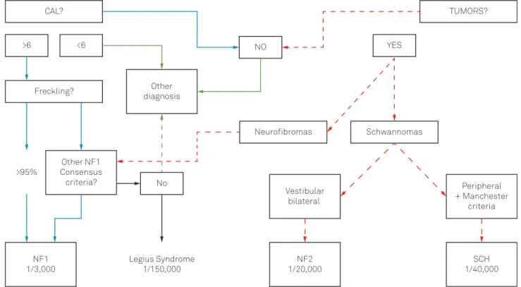
NF1 from NF2 and SCH. Figure 1 depicts a stepwise dia-gnosis pathway to distinguish the three NF diseases. In most cases, the differential diagnosis among the NF diseases is made with certainty. Nevertheless, special NF patients pre-sent overlap with some other diseases because of: a) cafe au lait lesions (CAL); b) localized hyper growth syndromes; c) neurofibromas-like tumors; and d) multiple endocrine neoplasia. These particular situations should deserve atten-tion from the NF specialist.
The present Part 1 text offers step-by-step guidelines for differential diagnosis among the neurofibromatosis types
and other diseases. A further Part 2 will present the NF clin-ical management. Basic information for patients can be found in www.amanf.org.br.
STEP BY STEP DIAGNOSIS OF NF1, NF2 AND SCH
Figure 1 depicts a practical step-by-step guide to differ-entiate NF1 from NF2 and SCH. The first two questions to be answered are: are there cafe au lait lesions (CAL)? Are there tumors?
STEP 1- ARE THERE CAFÉ AU LAIT LESIONS?
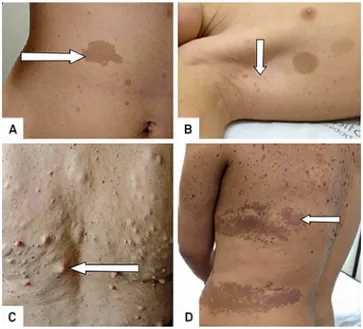
Yes - CAL are usually multiple and present or develop at birth or in early infancy and are observed in 99% of people with NF1 by age of 3 years. They are macules with uniform ovoid shape and smooth outline, varying of light to dark brown color (Figure 2A). Its size varies from 5 mm (infancy) to 30 mm (adulthood), but they can be bigger than 20 cm and involve an entire anatomic region4. The CAL size is
pro-portionate to body growth and they are randomly distribu-ted, although scalp, palms, and soles are spared5. Many
Legius Syndrome 1/150,000
No
YES
TUMORS?
>95%
Freckling?
Neurofibromas Schwannomas Other
diagnosis
Vestibular bilateral
NF2 1/20,000 NF1
1/3,000
SCH 1/40,000
Peripheral + Manchester
criteria Other NF1
Consensus criteria?
>6 <6 NO
CAL?
Figure 1.Practical flowchart to differentiate NF diseases: neurofibromatosis type 1 (NF1), type 2 (NF2) and schwannomatosis
other hyperpigmented skin lesions are commonly misdiag-nosed as CAL, such as the lesion in Figure 2D.
Six or more typical CAL are a strong sign (95%) of NF1. NF1 is the most common condition associated with multiple CAL and patients with a higher number of CAL are more likely to develop NF1, but CAL can also be seen in other syn-dromes (Table 5). On the other hand, the presence of more than three CAL is detected in only 0.3% of children with no evidence of genetic disorders6.
STEP 2–ARE THERE AXILLARY AND INGUINAL
FRECKLING?
Yes- Axillary and/or inguinal freckling are
hyperpigmen-ted spot macules (1-3mm) and the second most common feature in NF1 (Figure 2B). Their appearance is similar to solar-induced freckling, but in NF1 they occur typically in areas with minimal to none sun exposure. Freckling gen-erally appears between 3 and 5 years of age in the axillae and/or inguinal region. Other sites include neck and breasts, around the lips, and even the trunk in adults, but in these sites they are not a diagnostic criterion6.
Until recent years, the presence of CAL associated with freckling was enough to the NF1 diagnosis. Nevertheless, the discovery of the Legius syndrome (LEGIUS), which has in common with NF1 the CAL, the freckling, macrocephaly and cognitive deficits, made required a third criterion to make the NF1 diagnosis certain7. LEGIUS could be
consid-ered the fourth NF disease3.
STEP 3 –ARE THERE OTHER NF1 CONSENSUS
CRITERIA?
The diagnostic criteria for NF1 have been established by a National Institute of Health consensus of experts in 1987. A person is thought to have NF1 if they have two or more of the Table 1 criteria.
STEP 4–ESTABLISHED NF1 DIAGNOSIS (PREVIOUS
VON RECKLINGHAUSEN’S DISEASE)
NF1 is the most prevalent human monogenetic disease (incidence of 1:2,500 live births)14, it is an autossomal
dom-inant disorder caused by hereditary or new mutations in the neurofibromin protein codification gene, located in Chromosome 17 (locus 17q11.2), which results in neural and other tissues dysplasia and increased tumor formation of neural origin15.
Figure 2.Cutaneous lesions (arrows): Multiple cafe au lait lesions (CAL) (A); freckling and two CAL (B); Discrete neurofi-bromas (C); Pigmented skin lesions (epidermal nevus) mis-diagnosed as CAL and freckling in a non-NF1 patient (D). Pictures made with patient’s permission at theCentro de Referência em Neurofibromatoses.
Table 1.National Institute of Health NIH consensus criteria8 and the approximate Brazilian prevalence of the criteria9-11, which are similar to international literature, except for tumors (recent review prevalence presented in bold parenthesis12). The descriptions of cafe au lait (CAL), freckling, neurofibro-mas, optical glioma (OPG) and osseous dysplasia are presented in the text. Lisch Nodules are harmartomas in the iris, which are specific of Neurofibromatosis (NF1). Many features of NF1 increase in frequency with age. At age 1, only 50% of patients fulfill the NIH criteria, but at age 8, almost all patients have fulfilled the diagnostic criteria13.
Criteria Description Prevalence CAL Six or more CAL.5 mm in the larger
diameter in prepubertal individuals and.15 mm in the larger diameter
in post pubertal individuals
95%
Freckling Two or more freckles in the axillary or inguinal regions 87%
Neurofibromas Two or more of any type
(cutaneous, subcutaneous, spinal) (40 - 60%)75%
Or one plexiform 35%(60%)
Glioma Optical nerve or optical pathway tumor (astrocytoma) diagnosed by
nuclear magnetic resonance
6% (15-20%)
Lisch nodules Two or more nodular harmartomas in the iris, diagnosed by the
ophthalmologist
78%
Osseous
dysplasia A distinctive osseous lesion such assphenoid dysplasia or thinning of long bone cortex with or without
pseudoarthrosis
5%
Familial history
A first-degree relative (parent, sibling, or offspring) with established
NF1 by the above criteria
STEP 5 –ARE THERE TUMORS?
Most NF cases are associated with some CAL; therefore, its absence suggests other diseases. Nevertheless, consider-ing that some special NF patients do not have CAL, the next step is to search for tumors.
The common types of tumors associated with NF1 are neurofibromas (nfbs), plexiform neurofibromas (PNF), malignant peripheral nerve sheath tumors (MPNST) and optic glioma (OPG) (see below). The presence of two or more of these tumors is strongly suggestive of NF1 and other NIH criteria must be investigated (STEP 4). If no other criterion is found, this situation deserves a NF specialist support. On the other side, the presence of two or more schwannoma tumors (see below) is suggestive of both NF2 and SCH.
NF1 ASSOCIATED TUMORS
Although a specialized physician usually recognizes the different types of neurofibromas, the diagnostic certainty needs further histological studies. Below, we present a brief description of NF1 commonest tumors2,16and detailed
histo-logical information can be found elsewhere17.
NEUROFIBROMAS (WHO GRADE I)
Neurofibromas, the hallmark tumors of NF1, are benign tumors of the peripheral nerve sheath. They exhibit extens-ive cellular heterogeneity (Schwann cells, perineural cells, mast cells, fibroblasts and axons in an extracellular matrix) and it can be classified into two major types: localized and PNF.
Localized neurofibromas are the most prevalent type
of tumor in NF1. They tend to develop from skin sensory nerves and usually present as a cutaneous and/or sub-cutaneous tumor that remains associated with a single nerve ending. The nfbs are more commonly found on the skin, but other sites can also be affected, such as the spinal roots, heart, stomach, larynx, bladder, bowels and oral mucosa.
Plexiform neurofibromas are almost always associated
with NF1 and they are classified as benign peripheral nerve sheath tumors that involve multiple nerve fascicles or large branches of a major nerve. PNF are the first and foremost source of morbidity in NF1, due to their tendency to grow to large sizes and their capacity to cause significant deform-ity and compression of adjacent structures. Some may be present at birth, but most of them usually become apparent during the first two years of life and, if not present by then, rarely develop after adolescence. The growth rate associated
with these neoplasms is unpredictable, with periods of rapid growth followed by periods of relative inactivity.
MALIGNANT PERIPHERAL NERVE SHEATH TUMOR (WHO GRADE II, III OR IV)
MPNST commonly derive from pre-existing PNF. The NF1 patient lifetime risk of developing MPNST is 8-13% (being the double in the NF1 microdeletion subtype). Because MPNST develop from malignant progression of pre-existing PNF, the risk of developing MPNST increases to almost 50% in patients with NF1 and PNF18. MPNST
occurrence in childhood and adolescence is uncommon. Incidence is higher among adults in the third to sixth decade of life. MPNST should be suspected when the patient devel-ops persistent pain with no other explanation and/or unex-plained neurological deficit associated with changes in texture and enlargement of the PNF. Histological differenti-ation between a PNF with atypical features and a low grade MPNST is difficult, since these lesions actually represent a histological continuum and it is possible to find lesions with areas of atypical features adjacent to areas of frank MPNST.
GLIOMAS
Gliomas are the predominant CNS tumor type associated with NF1 and they may occur in all parts of the nervous sys-tem, with a preference for the optic pathways, brainstem and cerebellum. Frequent NF1-associated gliomas include optic pathway gliomas (OPG), other pilocytic astrocytomas, dif-fuse astrocytomas and glioblastomas.
OPG (WHO GRADE 1)
The majority of OPG in NF1 patients are pilocytic astro-cytomas that are located within the optic nerve. Macroscopically most pilocytic astrocytomas are a relatively circumscribed, slowly growing soft-grey mass. Intra or para-tumoural cyst formation is common. Optic nerve tumors also often show collar-like involvement of the subarachnoid space. Histopathologically, this astrocytic tumor of low to moderate cellularity exhibits an often biphasic pattern with varying proportions of compacted bipolar cells with Rosenthal fibers and loose-textured multipolar cells with microcysts and granular bodies/hyaline droplets. Rare mitosis, hyperchromatic and pleomorphic nuclei, glomeru-loide vascular proliferation, infarct-like necrosis and infiltra-tion of leptomeninges are compatible with the diagnosis of pilocytic astrocytoma and are not signs of malignancy19.
DIFFUSE ASTROCYTOMAS (WHO GRADE II)
That is a diffusely infiltrating astrocytoma that typically affects young adults and is characterized by a high degree of cellular differentiation and slow growth; the tumor occurs throughout the CNS but is preferentially supratentorial and has an intrinsic tendency for malignant progression to ana-plastic astrocytoma and, ultimately, glioblastoma.
GLIOBLASTOMA (WHO GRADE IV)
The most malignant neoplasm with predominant astrocy-tic differentiation; histological features include nuclear atypia, pleomorphic cells, mitotic activity, vascular thrombosis, micro-vascular proliferation and necrosis. It typically affects adults and is preferentially located in the cerebral hemispheres. Most glioblastomas manifest rapidly de novo, without recog-nizable precursor lesions (primary glioblastoma). Secondary glioblastomas develop slowly from diffuse WHO grade II astro-cytoma or anaplastic astroastro-cytoma (WHO grade III). Due to their invasive nature, glioblastomas cannot be completely removed and, despite progress in radio/chemotherapy, less than half of patients survive more than one year, with older age being the most significant adverse prognostic factor.
OTHER NF1 ASSOCIATED TUMORS
NF1 patients present with a higher risk of developing other tumors such as:
SARCOMAS
Gastrointestinal stromal tumor (GIST)
NEUROENDOCRINE/NEUROECTODERMAL TUMOURS
Pheochromocytomas Carcinoid tumor
Medullar thyroid carcinoma C-cell hyperplasia
HEMATOPOIETIC TUMORS
Juvenile chronic myeloid leukemia Juvenile xanthogranuloma Acute lymphocytic leukemia Non Hodgkin lymphoma
STEP 6–ARE THERE BILATERAL VESTIBULAR
SCHWANNOMAS (BVS)?
Yes–The presence of BVS is a hallmark of NF2. NF2 is an
autossomal dominant disorder caused by hereditary or new mutations in the merlin protein codification gene located in chromosome 22 (locus 22q12.2). NF2 presents with BVS, multiple meningiomas, juvenile subcapsular cataract and peripheral schwannomas and other neural tumors.
SCHWANNOMAS (WHO GRADE I)
Schwannomas are benign true nerve sheath neoplasms composed entirely of Schwann cell proliferation, well cir-cumscribed, nodular or ovoid, usually encapsulated dermal, subcutaneous and central nerves tumors2.
The prevalence of NF2 vary from 1:25,000-33,000 indivi-duals20and it presents with deafness and/or tinnitus (50%),
balance dysfunction (5%), focal weakness or sensory change (18%), seizure (5%), ocular symptoms (7%) and genetic or radiographic diagnosis of an asymptomatic family member (10%), during the second decade or early adulthood21.
Nevertheless, some special cases of NF2 are less evident. The Manchester (modified NIH) diagnostic criteria for NF2 have been expanded to include patients with no family his-tory who have multiple schwannomas and or meningiomas, but who have not yet developed bilateral vestibular nerve tumors22. The diagnosis is best confirmed using high quality
MRI of the brain.
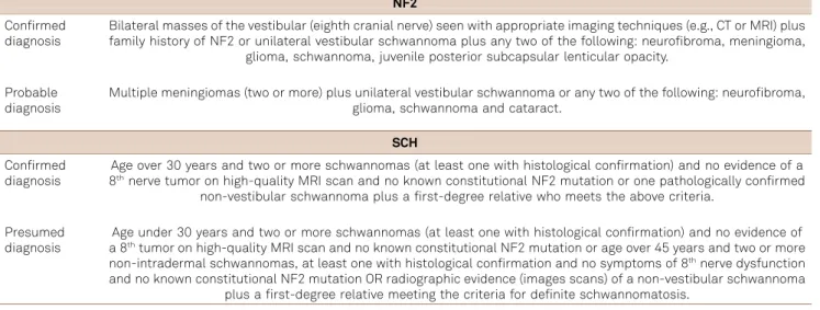
If there are two or more painful schwannomas, the pre-sent step is to complete the diagnosis of SCH with the fol-lowing criteria23. Table 2 presents the consensus criteria
for NF2 and SCH diagnosis.
SCH typically becomes symptomatic during the third decade of life and presents a prevalence of 1:40,000 indivi-duals. The molecular biology of SCH has not been well estab-lished yet and a candidate gene for SCH, called INI1, was identified, but the NF2 gene and possibly other genes may also be involved. Unlike NF1 and NF2, SCH does not have a clear pattern of inheritance.
The Table 3 summarizes the main clinical differential characteristics of NF1, NF2 and SCH.
NF1 VARIATIONS
There are genetically related (allelic) disorders, where apparently pathogenic NF1 mutations have been demon-strated in a few individuals or families who do not have NF1 according to the NIH Diagnostic Criteria4. They have
MOSAICISM OR SEGMENTAL NF
Occasionally NF1, NF2 (rarely) and schwannomatosis (often) occur in mosaic forms. Mosaicism in NF1 is observed more frequently and results from somatic mutations. Early
somatic mutations cause generalized disease, clinically indistinguishable from no mosaic forms. Later somatic mutation gives rise to localized disease often described as segmental. In individuals with mosaic or localized manifesta-tions of NF1 (segmental NF1), disease features are limited to
Table 2.Neurofibromatosis type 2 (NF2) and schwannomatosis (SCH) Manchester consensus diagnosis criteria22,23. NF2
Confirmed
diagnosis Bilateral masses of the vestibular (eighth cranial nerve) seen with appropriate imaging techniques (e.g., CT or MRI) plusfamily history of NF2 or unilateral vestibular schwannoma plus any two of the following: neurofibroma, meningioma, glioma, schwannoma, juvenile posterior subcapsular lenticular opacity.
Probable diagnosis
Multiple meningiomas (two or more) plus unilateral vestibular schwannoma or any two of the following: neurofibroma, glioma, schwannoma and cataract.
SCH Confirmed
diagnosis 8Age over 30 years and two or more schwannomas (at least one with histological confirmation) and no evidence of athnerve tumor on high-quality MRI scan and no known constitutional NF2 mutation or one pathologically confirmed
non-vestibular schwannoma plus a first-degree relative who meets the above criteria.
Presumed
diagnosis a 8Age under 30 years and two or more schwannomas (at least one with histological confirmation) and no evidence ofthtumor on high-quality MRI scan and no known constitutional NF2 mutation or age over 45 years and two or more
non-intradermal schwannomas, at least one with histological confirmation and no symptoms of 8thnerve dysfunction
and no known constitutional NF2 mutation OR radiographic evidence (images scans) of a non-vestibular schwannoma plus a first-degree relative meeting the criteria for definite schwannomatosis.
Table 3.Clinical differential characteristics of neurofibromatosis (NF1, NF2) and schwannomatosis (SCH).
Features NF1 NF2 SCH
First signs and symptoms Infancy Beginning of adulthood Adulthood.30 years
Typical findings Multiple CAL and neurofibromas Deafness and balance dysfunction Pain
Ocular findings Lisch Nodules Juvenile posterior subcapsular cataract None
Growth Macrocephaly, short stature,
cognitive deficits
Normal Normal
Tumors Neurofibromas: (cutaneous, plexiform
and intraneural - 60%) Schwannomas: bilateral vestibular(.95%), other cranial (24-51%), cutaneous (59-68%), peripheral
nerve (42%)
Schwannomas: peripheral and painful
Gliomas: benign (optic pathways 15-20%) and malignant (0,8%) Pheochromocytomas (0,1-13%)
Meningiomas: intracranial (50%) Ependymomas (33-53%)
Mesothelioma
Prevalence 1:2,500 a 1:7,800 1:20,000 a 1:40,000 1:40,000
Cancer risk Increased: MPNST (8-13%); GIST (5-30%), leukemia (1%), rhabdomyosarcomas (1-6%), breast
cancer 8,4%)
Habitual ?
Osseous problems Dysplasias None None
Inheritance Autossomal dominant with
complete penetrance Autossomal dominant with variablepenetrance Not well understood
Gene Chromosome 17 (locus 17q11.2) Chromosome 22 (locus 22q12.2) Chromosome 22
the affected area, which varies from a narrow strip to one quadrant and occasionally to one half of the body. Distribution is usually unilateral but can be bilateral, either in a symmetric or asymmetrical arrangement.
DIFFERENTIAL DIAGNOSIS
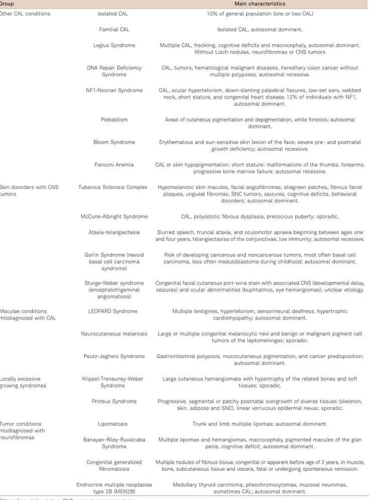
The main characteristics of other diseases, which present differential diagnosis with NF1, are summarized in Table 5.
WHEN ORDERING GENETIC TESTS
Approximately 50% of individuals with NF1 or NF2 have not inherited the disease from a parent and are result of ade novomutation. Individuals who meet the clinical diagnosis criteria for NF1, NF2 and SCH are diagnosed with confid-ence in most cases, and genetic tests are not required for diagnostic confirmation. Therefore, genetic testing may be helpful in specific cases. The decision to order or not a genetic test remains a physician/patient ( family) discussion. Genetic testing for NF is clinically available in just a few laboratories around the world. For an updated list of laboratories that offers genetic tests for NF, please refer to www.genetests.org.
NF1 VERSUS LEGIUS SYNDROME
The NF1 gene is large (280 kb and 60 exons) and most mutations are unique to a particular family, what turns the molecular diagnostic testing laborious and expensive. A definite diagnosis of NF1 can be made in most children by age four using only the NIH criteria24. Confirmatory
dia-gnostic testing can detect the mutation in 95% of cases and is indicated for suspected NF1 individuals, which do
not fulfill the NIH diagnostic criteria, most often in young children, if it can affect clinical management.
The presence of only pigmented lesions at early age is a diagnostic dilemma between NF1 and LEGIUS (caused by
SPRED1 gene mutations). The distinction between NF1
and LEGIUS in young children, in the absence of NF1 family history and neoplastic or osseous manifestations can only be achieved by genetic testing. It is cost saving and easier to start by investigation ofSPRED1gene than by NF1, because
SPREAD1contains only 8 exons7.
It is important to emphasize that currently there are only two important genotypes that correlate to phenotype in NF1. One of these is the microdeletion syndrome, in which a large part of or the wholeNF1 gene is absent and is associated with a more severe phenotype25. The other
genotype is the mutation involving the deletion of three base pairs in exon 17, which correlates with a mild phenotype26.
Therefore, discarding these two specific genotypes, it is not possible to predict the severity of the disease based on NF1 genetic testing.
NF2
It is expected that 25% to 33% of those who have NF2 and are a single occurrence in the family (simplex cases) are mosaic for NF2 mutation. The mutation detection rate by sequencing and deletion/duplication analysis is around 92% for familial cases and 72% in simplex cases. NF2 genetic testing may be helpful in specific cases and, although some genotype-phenotype correlations in NF2 are known, it can-not yet predict the severity of the disease27.
a. Pre-symptomatic testing of at-risk patients Because detection of tumors at an early stage is effective in improving the clinical management of NF2 patients, the screening for NF2 in children of affected patients can start
Table 4.Neurofibromatosis (NF1) variations. The four identified NF1 subtypes with its main clinical characteristics4.
Subtype Main characteristics
Watson
syndrome CAL, Lisch, freckling, pulmonary stenosis, short stature, macrocephaly, cognitive deficits, neurofibromas. Gene deletion
(5%) noses,NF1 gene large deletions“coarse" face becoming more marked with age, overgrowth with tall stature and large hands and feet, Pectus–dysmorphic facial features: hypertelorism, downslanting palpebral fissures, broad fleshy excavatum, broad neck, excess of tissue in hands and feet, joint hyperflexibility, muscular hypotonia, bone cysts, pes
cavus, dermal (cutaneous) neurofibromas occurring at early age and in increased numbers, including spinal neurofibromas, double lifetime risk of MPNST than NF1 population, significant delay in cognitive development, learning
difficulties, congenital heart disease and scoliosis (Huxon, The Neurofibromatoses: Differential Diagnosis and Rare Subtypes, 2011).
Mosaicism
(1%) Two or more NF1 criteria present only in specific body part (see below).
Spinal form Few CAL, normal or higher stature, few or absent cutaneous neurofibromas, but multiple spinal neurofibromas, usually bilateral and involving all 38 spinal nerve roots. MPNST higher risk.
Table 5.Differential diagnosis in neurofibromatosis (NF).
Group Main characteristics
Other CAL conditions Isolated CAL 10% of general population (one or two CAL)
Familial CAL Isolated CAL, autosomal dominant.
Legius Syndrome Multiple CAL, freckling, cognitive deficits and macrocephaly, autosomal dominant. Without Lisch nodules, neurofibromas or CNS tumors
DNA Repair Deficiency Syndrome
CAL, tumors, hematological malignant diseases, hereditary colon cancer without multiple polyposes, autosomal recessive.
NF1-Noonan Syndrome CAL, ocular hypertelorism, down-slanting palpebral fissures, low-set ears, webbed neck, short stature, and congenital heart disease. 12% of individuals with NF1,
autosomal dominant.
Piebaldism Areas of cutaneous pigmentation and depigmentation, white forelock; autosomal dominant.
Bloom Syndrome Erythematous and sun-sensitive skin lesion of the face; severe pre- and postnatal growth deficiency; autosomal recessive.
Fanconi Anemia CAL or skin hypopigmentation; short stature; malformations of the thumbs, forearms; progressive bone marrow failure; autosomal recessive.
Skin disorders with CNS tumors
Tuberous Sclerosis Complex Hypomelanotic skin macules, facial angiofibromas, shagreen patches, fibrous facial plaques, ungueal fibromas, SNC tumors, seizures, cognitive deficits, behavioral
disorders; autosomal dominant.
McCune-Albright Syndrome CAL, polyostotic fibrous dysplasia, precocious puberty; sporadic.
Ataxia-telangiectasia Slurred speech, truncal ataxia, and oculomotor apraxia beginning between ages one and four years, telangiectasias of the conjunctivae, low immunity; autosomal recessive.
Gorlin Syndrome (nevoid basal cell carcinoma
syndrome)
Risk of developing cancerous and noncancerous tumors, most often basal cell carcinoma, less often meduloblastoma during childhood; autosomal dominant.
Sturge-Weber syndrome (encephalotrigeminal
angiomatosis)
Congenital facial cutaneous port-wine stain with associated CNS (developmental delay, seizures) and ocular abnormalities (buphtalmos, eye hemangiomas); unclear etiology.
Maculae conditions misdiagnosed with CAL
LEOPARD Syndrome Multiple lentigines, hypertelorism, sensorineural deafness, hypertrophic cardiomyopathy; autosomal dominant.
Neurocutaneous melanosis Large or multiple congenital melanocytic nevi and benign or malignant pigment cell tumors of the leptomeninges; sporadic.
Peutz-Jeghers Syndrome Gastrointestinal polyposis, mucocutaneous pigmentation, and cancer predisposition; autosomal dominant.
Locally excessive growing syndromes
Klippel-Trenauney-Weber Syndrome
Large cutaneous hemangiomata with hypertrophy of the related bones and soft tissues; sporadic.
Proteus Syndrome Progressive, segmental or patchy postnatal overgrowth of diverse tissues (skeleton, skin, adipose and SNC), linear verrucous epidermal nevus; sporadic.
Tumor conditions misdiagnosed with neurofibromas
Lipomatosis Trunk and limb multiple lipomas; autosomal dominant.
Banayan-Riley-Ruvalcaba Syndrome
Multiple lipomas and hemangiomas, macrocephaly, pigmented macules of the glan penis, cognitive deficit; autosomal dominant.
Congenital generalized fibromatosis
Multiple nodules of fibrous tissue, congenital or apparent before age of 2 years, in muscle, bone, subcutaneous tissue and viscera, fatal or undergoing spontaneous remission.
Endrocrine multiple neoplasias type 2B (MEN2B)
Medullary thyroid carcinoma, pheochromocytomas, mucosal neuromas, sometimes CAL; autosomal dominant.
at birth with the search of cataracts28. If no cataracts are
found, pre-symptomatic genetic testing, if possible, should be considered.
b. Determining the diagnosis of patients who fall short of the Manchester criteria, including mosaic patients
In cases of patients that fall short of the Manchester cri-teria, genetic testing may be helpful in establishing the dia-gnosis. The diagnosis of NF2 is usually a challenge when mosaicism is present. Mutations with mosaicism levels greater than 10% can be detected in blood DNA, otherwise its iden-tification requires testing of tumor material. In these cases, the diagnosis is established if two NF2 identical mutations are found in separate tumors, but is not present in blood26.
c. Estimating the transmission risk for mosaic patients
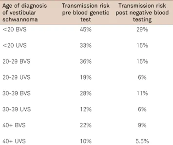
The transmission rate of NF2 is 50% in the second gen-eration, but the risk of transmission in apparently isolated patients with NF2 is less than 50% due to mosaicism. Transmission risk of NF2 decreases significantly after the proband has had negative genetic testing using a blood sam-ple (Table 6)27.
SCHWANNOMATOSIS (SCH)
Currently, it is known that germline mutations in
SMARCB1 (or INI1) gene are associated to SCH.
Nevertheless, they are found in only 40-50% of the familial cases and in 8-10% of the sporadic cases of SCH. For SCH cases unrelated to SMARCB1 germline mutations, the gen-etic alterations are not known27.
SMARCB1 gene testing may be ordered for individuals
with multiple schwannomas without evidence of vestibular tumor and no NF2 mutation detected in the blood. Nevertheless, there is a great chance of not detecting a
SMARCB1genetic alteration and the diagnosis of SCH
can-not be ruled out. SMARCB1 gene testing is also indicated for early detection of at-risk patients (because of family his-tory) for management reasons.
Tumorigenesis of schwannomas associated to SCH is believed to occur through a four-hit model, involving not only mutations in SMARCB1 alleles (hits 1 and 2), but also mutations in NF2 alleles (hits 3 and 4)30. Differently from
NF2-associated schwannomas, SCH-associated schwanno-mas of the same individual usually carry different somatic NF2 mutations. Therefore, mutational analysis of schwan-noma cells is also highly relevant for the diagnosis, even in the absence ofSMARCB1mutation in blood. The finding of differentNF2 somatic mutations in different schwannomas of the same individual, although not indicative of the pres-ence of a germlineSMARCB1, at least rules out the diagnosis of NF2 mosaicism31.
GENETIC TESTING FOR PREIMPLANTATION GENETIC DIAGNOSIS (PGD) OF NF
Another application of NF genetic test is for the PGD. It comprises a set of procedures to test a specific genetic alteration and selecting unaffected embryos for transfer to the uterus. It is a highly specialized method performed in a few centers and is available for couples when the parental mutation has already been identified. It has been shown as a promising and useful method, being the only one that prevents transmission of the mutated gene from one parent to their children32. For an updated
list of centers that perform PGD for NF, please refer to www.genetests.org
References
1. Riccardi VM. Historical background and introduction. In: Friedman JM, Gutmann DH, MacCollin M, Riccardi VM (Eds). Neurofibromatosis - Phenotype, Natural History, and Pathogenesis. 3 ed. Baltimore: The Johns Hopkins University Press, 1999:1-28.
2. Geller M, Bonalumi Filho A. Neurofibromatose: clínica, genética e terapêutica. 1 ed. Rio de Janeiro: Guanabara Koogan, 2004. 3. Huson SM. The neurofibromatoses: differential diagnosis and rare
subtypes. In: Ferner RE, Huson SM, Evans DG (Eds).
Table 6.Transmission risk of neurofibromatosis (NF2) to offspring for isolated cases of Neurofibromatosis type 2 before and after blood genetic test. Adapted from Evans and Wallace, 200829.
Age of diagnosis of vestibular schwannoma
Transmission risk pre blood genetic
test
Transmission risk post negative blood
testing
,20 BVS 45% 29%
,20 UVS 33% 15%
20-29 BVS 36% 15%
20-29 UVS 19% 6%
30-39 BVS 28% 11%
30-39 UVS 12% 6%
40+ BVS 22% 9%
40+ UVS 10% 5.5%
Neurofibromatoses in Clinical Practice. 1 ed. London: Springer-Verlag London Limited, 2011:1-46.
4. Ferner RE. Neurofibromatosis 1. In: Ferner RE, Huson SM, Evans DG (Eds.) Neurofibromatoses in clinical practice. 1 ed. London: Springer-Verlag London Limited, 2011:1-46.
5. Boyd KP, Gao L, Feng R, et al. Phenotypic variability among cafe-au-lait macules in neurofibromatosis type 1. J Am Acad Dermatol 2010;63:440-447.
6. Nunley KS, Gao F, Albers AC, Bayliss SJ, Gutmann DH. Predictive value of cafe au lait macules at initial consultation in the diagnosis of neurofibromatosis type 1. Arch Dermatol 2009;145:883-887. 7. Brems H, Pasmant E, Van Minkelen R, et al. Review and update
of SPRED1 mutations causing Legius syndrome. Hum Mutat 2012;33:1538-1546.
8. National Institutes of Health Consensus Development Conference Statement: neurofibromatosis. Bethesda, Md., USA, July 13-15, 1987. Neurofibromatosis 1988;1:172-178.
9. Trovo-Marqui AB, Goloni-Bertollo EM, et al. High frequencies of plexiform neurofibromas, mental retardation, learning difficulties, and scoliosis in Brazilian patients with neurofibromatosis type 1. Braz J Med Biol Res 2005;38:1441-1447.
10. Darrigo Jr LG, Geller M, Bonalumi Filho A, Azulay DR. Prevalence of plexiform neurofibroma in children and adolescents with type I neurofibromatosis. J Pediatr (Rio J) 2007;83:571-573.
11. Souza JFd, Toledo LLd, Ferreira MCM, Rodrigues LOC, Rezende NAd. Neurofibromatose tipo 1: mais comum e grave do que se imagina. Rev Assoc Méd Bras 2009;55:394-399.
12. Lin AL, Gutmann DH. Advances in the treatment of neurofibroma-tosis-associated tumours. Nat Rev Clin Oncol 2013;10:616-624. 13. DeBella K, Poskitt K, Szudek J, Friedman JM. Use of "unidentified
bright objects" on MRI for diagnosis of neurofibromatosis 1 in children. Neurology 2000;54:1646-1651.
14. Huson SM, Harper PS, Compston DA. Von Recklinghausen neurofi-bromatosis. A clinical and population study in south-east Wales. Brain 1988;111:1355-1381.
15. Ferner RE, Huson SM, Thomas N, et al. Guidelines for the diagnosis and management of individuals with neurofibromatosis 1. J Med Genet 2007;44:81-88.
16. Cunha KS, Geller M. Advances in neurofibromatosis research. 1 ed. New York, USA: Nova Science Publishers Inc, 2012:1-273.
17. Carranza AC, Salinas Martín MV, Polo R, Córdoba JC, González-Cámpora R. Problemas diagnósticos en tumores del nervio periférico (I and II). Rev Esp Patol 2011:97-116.
18. Park SJ, Sawitzki B, Kluwe L, Mautner VF, Holtkamp N, Kurtz A. Serum biomarkers for neurofibromatosis type 1 and early detec-tion of malignant peripheral nerve-sheath tumors. BMC Med 2013;11:109.
19. HO Classification of Tumours of the central nervous system. 4 ed: WHO; 2007:1-312.
20. Blakeley JO, Evans DG, Adler J, et al. Consensus recommenda-tions for current treatments and accelerating clinical trials for patients with neurofibromatosis type 2. Am J Med Genet A 2012;158:24-41.
21. MacCollin M. Neurofibromatosis 2: clinical aspects. In: Friedman JM, Gutmann DH, MacCollin M, Riccardi VM (Eds). Neurofibromatosis - phenotype, natural history, and pathogenesis. 3 ed. Baltimore: The Johns Hopkins University Press, 1999:299-326.
22. Baser ME, Friedman JM, Wallace AJ, Ramsden RT, Joe H, Evans DG. Evaluation of clinical diagnostic criteria for neurofibromatosis 2. Neurology 2002;59:1759-1765.
23. MacCollin M, Chiocca EA, Evans DG, et al. Diagnostic criteria for schwannomatosis. Neurology 2005;64:1838-1845.
24. Friedman JM. Neurofibromatosis 1. Seattle (WA): University of Washington, Seattle; 1993-2013; 1998 (Updated 2012) [cited 2013]. Available from: http://www.ncbi.nlm.nih.gov/books/NBK1109/. 25. Pasmant E, Sabbagh A, Spurlock G, et al. NF1 microdeletions in
neurofibromatosis type 1: from genotype to phenotype. Hum Mutat 2010;31:1506-1518.
26. Upadhyaya M, Huson SM, Davies M, et al. An absence of cutaneous neurofibromas associated with a 3-bp inframe deletion in exon 17 of the NF1 gene (c.2970-2972 delAAT): evidence of a clinically significant NF1 genotype-phenotype correlation. Am J Hum Genet 2007;80:140-151.
27. Nunes F. Research advances in mutational analysis of the NF2 gene. In: Cunha K, Geller M, editors. Advances in neurofibromatosis research. 1 ed. New York, USA: Nova Science Publishers Inc, 2012:165-80.
28. Evans DG, Raymond FL, Barwell JG, Halliday D. Genetic testing and screening of individuals at risk of NF2. Clin Genet 2012;82:416-424.
29. Evans DG, Wallace A. An update on age related mosaic and offspring risk in neurofibromatosis 2 (NF2). J Med Genet 2009;46:792-800.
30. Plotkin SR, Blakeley JO, Evans DG, et al. Update from the 2011 International Schwannomatosis Workshop: From genetics to dia-gnostic criteria. Am J Med Genet A 2013;161:405-416.
31. Papi L. Schwannomatosis: a recently recognized form of neurofi-bromatosis. In: Cunha K, Geller M (Eds). Advances in Neurofibromatosis Research. 1 ed. New York, USA: Nova Science Publishers Inc, 2012:258-267.