167
Type 1 neurofibromatosis: radiological findings of the chest
Radiol Bras. 2010 Mai/Jun;43(3):167–170
Original Article • Artigo Original
Type 1 neurofibromatosis: radiological findings
of the chest*
Neurofibromatose tipo 1: aspectos radiológicos do tórax
Marcos Pontes Muniz1, Antonio Soares Souza2, Divanei Aparecida Bottaro Criado3, José Roberto Lopes Ferraz Filho4, Rafael Marinelli Brandão5, Luciana Vargas Cardoso6, Eny Maria Goloni Bertollo7
OBJECTIVE: To identify chest radiography findings suggestive of type 1 neurofibromatosis, establishing their frequency and evaluating the possibility of including the presence of posterior mediastinal masses as a criterion for the diagnosis of type 1 neurofibromatosis. MATERIALS AND METHODS: The present study included 141 patients with type 1 neurofibromatosis assisted at the Service of Radiology of Hospital de Base and Faculdade de Medicina de São José do Rio Preto, SP, Brazil, and submitted to standard chest radiography in postero-anterior and lateral views. The results were analyzed by non-parametric methods and the level of statistical significance was set at 0.05 (p = 0.05). RESULTS: The most frequent findings were the following:
ribs erosion (19.8%), pectus excavatum (12.0%), kyphoscoliosis (3.5%) and posterior mediastinal masses (7.1%). Such results suggest that posterior mediastinal masses (neurofibroma and meningocele) should be included as a diagnostic criterion of type 1 neurofibromatosis, in conjunction with dysplasia of the sphenoid wing, pseudoarthrosis and thinning of long bone cortex, as defined by the National Institutes of Health. CONCLUSION: The presence of posterior mediastinal masses in association with the typical bone changes defined by the National Institutes of Health is a consistent finding to be considered as a diagnostic criterion of the disease.
Keywords: Type 1 neurofibromatosis; Radiology; Chest radiography; Bone abnormalities; Mediastinal neo-plasms.
OBJETIVO: Identificar alterações e frequências nas radiografias simples do tórax sugestivas de neurofibro-matose tipo 1 e avaliar a possibilidade de inclusão de massa no mediastino posterior como critério de diag-nóstico de neurofibromatose tipo 1. MATERIAIS E MÉTODOS: Foram realizadas radiografias com técnica padrão de tórax em póstero-anterior e em perfil de 141 pacientes com neurofibromatose tipo 1, atendidos no Serviço de Radiologia do Hospital de Base e Faculdade de Medicina de São José do Rio Preto, SP. Os
resultados obtidos foram avaliados por métodos não paramétricos ao nível de 0,05 de significância (p =
0,05). RESULTADOS: No presente estudo, 141 pacientes com neurofibromatose tipo 1 realizaram radiogra-fia de tórax, sendo as alterações mais frequentes: erosão óssea das costelas (19,8%), peito escavado (12,0%), cifoescoliose (3,5%) e massas no mediastino posterior (7,1%). Esses resultados sugerem que as massas (neurofibroma e meningocele) devem ser incluídas como critério diagnóstico para neurofibromatose tipo 1, juntamente com displasia do osso esfenoide, pseudoartrose e afinamento do córtex de ossos longos, con-forme definido pelo National Institutes of Health. CONCLUSÃO: A presença das massas no mediastino pos-terior associada às alterações ósseas características definidas pelo National Institutes of Health indicam ser um achado consistente para se considerar como critério diagnóstico da doença.
Unitermos: Neurofibromatose tipo 1; Radiologia; Radiografia torácica; Anormalidades ósseas; Neoplasias mediastinais.
Abstract
Resumo
* Study developed in the Diagnostic Imaging Department – College of Medicine and Hospital de Base in São José do Rio Preto, SP, Brazil.
1. PhD, Professor in Diagnostic Imaging Department, Head of Radiology Service – Hospital de Base in São José do Rio Preto, SP, Brazil.
2. PhD, Professor in Diagnostic Imaging Department, Head of Radiology Division – Hospital de Base in São José do Rio Preto, SP, Brazil.
3. Master degree, MD, Specialist in Radiodiagnosis, Diagnostic Imaging Department – College of Medicine in São José do Rio Preto, SP, Brazil.
4. Master, Specialist in Radiodiagnosis, Head of Neuroradiology Unit – Hospital de Base in São José do Rio Preto, SP, Brazil.
INTRODUCTION
Neurofibromatosis (NF) is an autoso-mal dominant disease that was first de-scribed by Friedrich von Recklinghausen in 1882. Several types of NF have been de-scribed, and type 1 (NF1), also denomi-nated peripheral or classic NF, is the most common, occuring in 1:3,000 individuals(1).
One of the main characteristics of this dis-Muniz MP, Souza AS, Criado DAB, Ferraz Filho JRL, Brandão RM, Cardoso LV, Bertollo EMG. Type 1 neurofibromatosis: radiological findings of the chest. Radiol Bras. 2010;43(3):167–170.
0100-3984 © Colégio Brasileiro de Radiologia e Diagnóstico por Imagem
5. Graduate Medicine Student – College of Medicine in São José do Rio Preto, SP, Brazil.
6. MD, Specialist in Radiodiagnosis, Responsible for the Com-puted Tomography Unit – Hospital de Base in São José do Rio Preto, SP, Brazil.
7. Professor, Molecular Biology Department – College of Medi-cine in São José do Rio Preto, SP, Brazil.
Mailing address: Dr. Marcos Pontes Muniz. Faculdade de Medicina e Hospital de Base, Departamento de Imagem. Ave-nida Brigadeiro Faria Lima, 5416, Vila São Pedro. São José do Rio Preto, SP, Brazil, 15090-000. E-mail: depimagem@famerp. br
168
Muniz MP et al.
Radiol Bras. 2010 Mai/Jun;43(3):167–170 ease is systemic and progressive
involve-ment, with impaired neurological func-tions(2) and physical deformity(3).
The diagnostic criteria for NF1 were de-fined by the National Institutes of Health (NIH)(4) in 1987 and updated three years
later(5). Main clinical presentations of NF1
include café-au-lait spots, cutaneous neu-rofibromas, ephelides or freckling in axil-lary and/or inguinal regions, plexiform neurofibromas and Lisch nodules(1,6,7).
Bone abnormalities observed in NF1 have been receiving increasing attention and must be recognized by the radiologist as some are typical of the disease and oth-ers are strongly suggestive of NF1(8,9). In
NF patients, the most frequent skeletal ab-normalities are observed in the spine and may directly affect the skeletal system due to mesodermal dysplasia (dural ectasia and bone dysplasia), or indirectly by secondary complications such as tumor compression, in particular of neurofibromas and menin-goceles(10). The commonest alterations
in-clude scoliosis, kyphoscoliosis, erosion of the posterior wall of vertebral bodies, ero-sion of the anterior wall of vertebral bod-ies, widening of intervertebral foramina, growth disorders, pseudoarthrosis, thin-ning of long bone cortices, bone cystic le-sions, costal arch erosion, thinning of pedicles, sphenoid wing dysplasia, cranial osteolytic lesions, facial/mandibular defor-mities, subperiosteal bone proliferation, bone compression due to soft-tissue tumors (neurofibromas, dura mater dysplasias, in-trathoracic meningocele)(11).
Erosion of the lower borders and irregu-larities of costal arches, rib cage deformi-ties (pectus excavatum and kyphoscoliosis) and mediastinal masses (meningocele and neurofibroma)(10) may be detected on the
chest radiographs of NF1 patients. Costal arch erosion is sometimes caused by extrinsic compression by neurofibromas which produces cortical erosion of the lower borders of the ribs, or as a conse-quence of a primary dysplastic defects in bone formation(12).
Thoracic deformities, such as pectus excavatum, observed at physical examina-tion in 31% to 50% of NF1 patients, rep-resents a depression of the lower portion of the sternum, causing excavation of the an-terior thoracic region(11). Another thoracic
deformity, caused by kyphoscoliosis, may result in evaluation distortions(11).
Thoracic meningocele is an abnormal-ity of the vertebral spine corresponding to herniation of the meninges through the in-tervertebral foramen with a benign course which remains asymptomatic in most cases(13). Approximately 70% to 80% of all
cases of thoracic meningocele occur in NF patients(13). Thoracic neurofibromas are
benign tumors involving the nerve roots of the spinal cord unilaterally or bilaterally and may affect several segments of the spine. The presence of such posterior me-diastinal masses in both postero-anterior and lateral plain chest radiographs is not enough for diagnostic differentiation, thus further investigations using computed to-mography (CT) or magnetic resonance im-aging (MRI) are required(14).
Several studies have described different imaging methods for the diagnosis of NF1(15–18). The observation of thoracic
findings and the respective sensitivity of the technique used are valid in the appli-cation of such concepts, particularly with regards to early diagnosis of the disease. The justification of the current study took into account that chest radiography is a low-cost, easy-to-perform method.
Thus, this study aimed at identifying al-terations suggestive of NF1 on postero-anterior and lateral views of plain chest ra-diographs, establishing their frequency and evaluating the possibility of including the presence of a posterior mediastinal mass as a criterion for the diagnosis of NF1. With these elements, the present study may serve as a warning to health professionals on the importance of this finding with consequent referral of their patients for more specific investigations.
MATERIALS AND METHODS
This study included 141 NF1 patients assisted in the Centro de Pesquisa e Aten-dimento em Neurofibromatose – Cepan (Research and Assistance Center for Neu-rofibromatosis), a multidisciplinary center of Hospital de Base and the College of Medicine in São José do Rio Preto (Famerp), Brazil, who were evaluated and later re-ferred to the Diagnostic Imaging Depart-ment of the same institution.
The patients underwent both postero-anterior and lateral chest radiography, per-formed with the standard technique, i.e., with the bucky focused at 1.80 m in a Philips equipment, with 35 × 35 (postero-anterior view) and 30 × 40 (lateral view) green X-ray films, developed in an auto-matic Kodak-90® processing unit. The ages of the patients ranged from 2 to 72 years (mean age, 33.1 years; standard deviation, 18.2 years).
The study was double-blinded and was approved by the Committee for Ethics in Research of Famerp. The variables ana-lyzed were the presence or absence of typi-cal thoracic bone alterations found in pa-tients diagnosed with NF1.
The obtained data were qualitatively and quantitatively evaluated by means of non-parametric statistical methods, with the significance level being set at 0.05.
RESULTS
Among the patients studied, 39% (n =
55) presented alterations at radiography, with 21 of these patients (14.9%) present-ing with more than one alteration. Pectus excavatum was observed in 17 patients (12%), costal arch alterations (irregularities and deformities) in 28 patients (19.8%) and kyphoscoliosis in five patients (3.5%). Kyphoscoliosis determines deformity of the rib cage, with reduction in the volume of the lungs and false enlargement of the cardiac area.
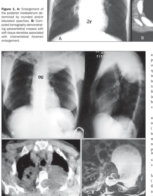
Posterior mediastinal enlargement de-termined by rounded and/or lobulated opacities with soft tissue densities next to the spine, was found in ten of the patients (7.1%) who were later submitted to CT (Figure 1), demonstrating the presence of a solid homogeneous masses in nine pa-tients and a cystic mass in one patient. The patient with the cystic mass was submitted to MRI, which demonstrated it to be a meningocele (Figure 2).
DISCUSSION
169
Type 1 neurofibromatosis: radiological findings of the chest
Radiol Bras. 2010 Mai/Jun;43(3):167–170 typical. Thus radiology may contribute to demonstrate typical findings of the disease such as sphenoid wing dysplasia, pseudoar-throsis and thinning of the long bones
cor-tex(11). Chest radiography is a complemen-tary study that, in association with the clini-cal history and physiclini-cal examination, is the main tool for evaluating thoracic alterations
and is highly sensitive. In patients with a posterior mediastinal mass associated with other clinical signs, such as café-au-lait spots, a plain chest radiography may help in the diagnosis of NF1. Whenever a pos-terior mediastinal mass is present at chest radiography, the patient must be referred for an investigation using more complex imaging methods (CT and MRI), which, besides confirming the diagnosis, may pro-vide data on possible complications.
Among the alterations found in the analysis of chest radiographs, costal arch ir-regularities were the most common ones (19.1%). This abnormality may occur due to a primary dysplastic defect in bone for-mation or by erosion of an intercostal neu-rofibroma(8,10,14). A “twisted-ribbon” ap-pearance of the ribs is frequently reported(8–
11)
. Data on the incidence of costal arch le-sions were not found in the literature re-view.
Pectus excavatum was observed on the lateral radiographic views of 17 patients (12.0%), while at physical examination this finding was observed in 31 patients. This alteration may cause clinical symptoms such as paradoxical breathing or angina-like pain(11,21,22).
Posterior mediastinal masses found in ten of the 141 patients (7.1%) were rounded or lobulated masses with soft-tissue density that emerged from the vertebral canal. Fol-lowing the division of the mediastinum in compartments(23) on plain chest radio-graphs of the 141 NF1 patients, masses were only identified in the posterior medi-astinal compartment, i.e., no masses were Figure 2.A: Postero-anterior and lateral chest radiographs showing rounded opacity in the left upper
posterior mediastinum and infiltration by tuberculosis into the upper lobe of the right lung. B: Computed tomography showing a mass with soft-tissue opacity emerging from the upper region of the intervertebral foramen of the left lung and infiltration by tuberculosis into the upper lobe of the right lung. C: Magnetic Resonance imaging demonstrating hyperintense cystic formation on T2 relaxation adjacent to the dural sac through the left neuroforamen of the upper part of the thoracic vertebral body.
170
Muniz MP et al.
Radiol Bras. 2010 Mai/Jun;43(3):167–170 found in the middle and anterior
meditina. Posterior mediastinal masses are as-sociated with NF1 and may be caused by neurofibromas that originate in spinal cord nerve roots or by intrathoracic meningo-celes(10,11,24). Plain chest radiography is not
enough for a clear differentiation between neurofibromas and meningoceles, thus there is a need for an investigation using more advanced methods, such as CT or MRI. Among the 10 patients with posterior mediastinal masses confirmed by CT, 9 presented with solid masses suggesting neurofibroma as they were related to the neuroforamen, and one patient presented with a cystic mass which was subsequently identified as a left-sided intrathoracic men-ingocele by MRI; this patient presented with residual lesions of tuberculosis on the right pulmonary apex.
About 80% of patients presenting with intrathoracic meningoceles have NF1(25,26).
Two or more skin neurofibromas or one plexiform neurofibroma are diagnostic cri-teria of NF1 as defined by the NIH(4). Thus,
the presence of neurofibromas in the paravertebral regions should be considered as of equal value in the diagnosis of NF1. No information on the incidence of poste-rior mediastinal masses in the general population was found in the literature.
Among the criteria for typical bone le-sions as defined by the NIH, sphenoid wing dysplasia was found in 1% of the cases. This finding, observed by radiography or CT, is highly specific, but is not common. The frequency of pseudoarthrosis is 1:250,000 births with approximately 50% to 90% of the cases being associated with NF1. This is considered as a relatively rare finding, with an incidence of about 3%. Thinning of the long bones cortex, ob-served in 8.5% of patients, is not a very specific finding and requires radiography of the entire appendicular skeleton, i.e., upper and lower limbs(4,11). The
identifica-tion of posterior mediastinal masses by ei-ther skeletal radiography or chest
radiog-raphy may make these findings of typical bone lesions more valuable. Thus, the in-clusion of posterior mediastinal masses as an additional criterion in the diagnosis of NF1 is suggested. At plain chest radiogra-phy, it is possible to identify bone alter-ations in NF1 patients, and the presence of posterior mediastinal masses, in addition to the characteristic bone alterations defined by the NIH(4) indicate that this is a
consis-tent finding to be proposed as a criterion for the diagnosis of NF1.
CONCLUSION
Posterior mediastinal masses associ-ated with the characteristic bone lesions as defined by the NIH, is a consistent find-ing and should be considered as a criterion for the diagnosis of type 1 neurofibroma-tosis.
REFERENCES
1. Ruggieri M. The different forms of neurofibroma-tosis. Childs Nerv Syst. 1999;15:295–308. 2. Mariaud-Schmidt RP, Rosales-Quintana S, Bitar
E, et al. Hamartoma involving the pseudoarthro-sis site in patients with neurofibromatopseudoarthro-sis type 1. Pediatr Dev Pathol. 2005;8:190–6.
3. Trovó-Marqui AB, Goloni-Bertollo EM, Valério NI, et al. High frequencies of plexiform neurofi-bromas, mental retardation, learning difficulties, and scoliosis in Brazilian patients with neurofi-bromatosis type 1. Braz J Med Biol Res. 2005;38: 1441–7.
4. [No authors listed]. Neurofibromatosis. Confer-ence Statement. National Institutes of Health Con-sensus Development Conference. Arch Neurol. 1988;45:575–8.
5. Mulvihill JJ, Parry DM, Sherman JL, et al. NIH Conference. Neurofibromatosis 1 (Recklinghausen disease) and neurofibromatosis 2 (bilateral acous-tic neurofibromatosis). An update. Ann Intern Med. 1990;113:39–52.
6. Ruggieri M, Huson SM. The neurofibromatoses. An overview. Ital J Neurol Sci. 1999;20:89–108. 7. Muniz MP, Ferraz Filho JRL, Souza AS, et al. Neurofibromatose tipo 1: aspectos clínicos e ra-diológicos. Rev Imagem. 2006;28:87–96. 8. Li Y, O’Connell P, Breidenbach HH, et al.
Ge-nomic organization of the neurofibromatosis 1 gene (NF1). Genomics. 1995;25:9–18. 9. Littler M, Morton NE. Segregation analysis of
pe-ripheral neurofibromatosis (NF1). J Med Genet. 1990;27:307–10.
10. Alwan S, Tredwell SJ, Friedman JM. Is osseous dysplasia a primary feature of neurofibromatosis 1 (NF1)? Clin Genet. 2005;67:378–90. 11. Muniz MP, Almeida JRM, Araújo Neto SA, et al.
Prevalência de achados radiográficos da neuro-fibromatose tipo 1: estudo de 82 casos. Radiol Bras. 2002;35:65–70.
12. Fortman BJ, Kuszyk BS, Urban BA, et al. Neu-rofibromatosis type 1: a diagnostic mimicker at CT. Radiographics. 2001;21:601–12. 13. Andrade GC, Braga OP, Hisatugo MK, et al.
Giant intrathoracic meningoceles associated with cutaneous neurofibromatosis type I: case report. Arq Neuropsiquiatr. 2003;61:677–81. 14. Rossi SE, Erasmus JJ, McAdams HP, et al.
Tho-racic manifestations of neurofibromatosis-I. AJR Am J Roentgenol. 1999;173:1631–8. 15. Hassell DS, Bancroft LW, Kransdorf MJ, et al.
Imaging appearance of diffuse neurofibroma. AJR Am J Roentgenol. 2008;190:582–8.
16. Lim R, Jaramillo D, Poussaint TY, et al. Superfi-cial neurofibroma: a lesion with unique MRI char-acteristics in patients with neurofibromatosis type 1. AJR Am J Roentgenol. 2005;184:962–8. 17. Shu HH, Mirowitz SA, Wippold FJ 2nd.
Neurofi-bromatosis: MR imaging findings involving the head and spine. AJR Am J Roentgenol. 1993;160: 159–64.
18. Bredella MA, Torriani M, Hornicek F, et al. Value of PET in the assessment of patients with neurofi-bromatosis type 1. AJR Am J Roentgenol. 2007; 189:928–35.
19. Khong PL, Goh WHS, Wong VCN, et al. MR imaging of spinal tumors in children with neurofi-bromatosis 1. AJR Am J Roentgenol. 2003;180: 413–7.
20. Mena E, Bookstein JJ, Holt JF, et al. Neurofibro-matosis and renovascular hypertension in chil-dren. Am J Roentgenol Radium Ther Nucl Med. 1973;118:39–45.
21. Vandenbroucke J, van Ooy A, Geukers C, et al. Dystrophic kyphoscoliosis in neurofibromatosis type I: a report of two cases and review of the lit-erature. Eur Spine J. 1997;6:272–7.
22. Zeller RD, Dubousset J. Progressive rotational dislocation in kyphoscoliotic deformities: presen-tation and treatment. Spine (Phila Pa 1976). 2000; 25:1092–7.
23. Felson B. Radiología torácica. 2ª ed. corrigida. Barcelona: Editorial Científico Médica, 1994. 24. Tsirikos AI, Ramachandran M, Lee J, et al.
As-sessment of vertebral scalloping in neurofibroma-tosis type 1 with plain radiography and MRI. Clin Radiol. 2004;59:1009–17.
25. Hunt JC, Pugh DG. Skeletal lesions in neurofibro-matosis. Radiology. 1961;76:1–20.