ARTICLE DOI: 10.1590/0004-282X20130057
White matter hyperintensities, executive
function and global cognitive performance
in vascular mild cognitive impairment
Hiper-intensidades de substância branca, função executiva e desempenho cognitivo
global no comprometimento cognitivo leve vascular
Felipe Kenji Sudo1, Carlos Eduardo Oliveira Alves1, Gilberto Sousa Alves1, Letice Ericeira-Valente1,
Chan Tiel2, Denise Madeira Moreira4, Jerson Laks1,3, Eliasz Engelhardt2
Vascular mild cognitive impairment (VaMCI) is a term that may be used to refer to a clinical state that eventually con-verts to mild dementia in the continuum of vascular cognitive impairment (VCI)1. It represents a construct derived from
mild cognitive impairment (MCI), operationally deined by Petersen2, evolving from one of its subtypes, generally the
non-amnesic single or multiple domain ones (executive func-tion, visuospatial ability, and language), that was related to incipient forms of non-Alzheimer dementias, including vas-cular dementia (VaD). his subject was also addressed with-in the VCI concept, as Vascular Cognitive Impairment No-Dementia (VaCIND)3.
1Instituto de Psiquiatria, Universidade Federal do Rio de Janeiro (UFRJ), Rio de Janeiro RJ, Brazil;
2Instituto de Neurologia Deolindo Couto, Setor de Neurologia Cognitiva e do Comportamento, UFRJ, Rio de Janeiro RJ, Brazil;
3Universidade do Estado do Rio de Janeiro, Rio de Janeiro RJ, Brazil; 4Hospital Pró-Cardíaco, Rio de Janeiro RJ, Brazil.
Correspondence: Felipe Kenji Sudo; Rua Barata Ribeiro 383 / 1.004; 22040-001 Rio de Janeiro RJ - Brasil; E-mail: [email protected] Support: Conselho Nacional de Pesquisa (CNPq) for the support to Jerson Laks, who is a Researcher 2 of this council.
Conflict of interest: There is no conflict of interest to declare.
Received 29 September 2012; Received in final form 27 February 2013; Accepted 06 March 2013. ABSTRACT
Vascular mild cognitive impairment (VaMCI) represents an early symptomatic stage of vascular cognitive impairment and might be asso-ciated to fronto-executive dysfunction. Methods: Twenty-six individuals (age: 73.11±7.90 years; 65.4% female; schooling: 9.84±3.61 years) were selected through neuropsychological assessment and neuroimaging. Clinical and neuroimaging data of VaMCI individuals (n=15) were compared to normal controls (NC, n=11) and correlated with Fazekas scale. Results: VaMCI performed significantly worse than NC in Trail-Making Test (TMT) B, errors in TMT B, difference TMT B–A and Cambridge Cognitive Examination (CAMCOG) final scores. Correlations were found among scores in modified Fazekas scale and performances in TMT B (time to complete and errors), difference TMT B–A and CAMCOG total score. Conclusion: Extension of white matter hyperintensities might be correlated to poorer global cognition and impairments in a set of fronto-executive functions, such as cognitive speed, set shifting and inhibitory control in VaMCI.
Key words: mild cognitive impairment, dementia, vascular, executive function, neuropsychology, neuroimaging, cerebrovascular disorders.
RESUMO
Comprometimento cognitivo leve vascular (CCLV) representa um estágio sintomático precoce do comprometimento cognitivo vascu-lar e associa-se à disfunção fronto-executiva. Métodos: Vinte e seis indivíduos (idade: 73,11±7,90 anos; 65,4% mulheres; escolaridade: 9,84±3,61 anos) foram selecionados por meio de avaliação cognitiva e neuroimagem. Os dados clínicos e de neuroimagem do grupo CCLV (n=15) foram comparados com controles normais (CN; n=11) e correlacionados com a escala de Fazekas. Resultados: CCLV apresentaram piores desempenhos que CN no Trail-Making Test (TMT) B, erros no TMT B, diferença TMT B–A e pontuação final do Cambridge Cognitive Examination (CAMCOG). Verificaram-se correlações entre escala de Fazekas e desempenhos no TMT B (tempo total e erros), diferença TMT B–A e a pontuação final do CAMCOG. Conclusão: A extensão das hiper-intensidades de substância branca, no grupo CCLV, correlacionou-se com pior desempenho cognitivo global e com comprometimento em um grupo de funções fronto-executivas, como velocidade e alternância cognitiva e controle inibitório.
Palavras-Chave: comprometimento cognitivo leve, demência vascular, função executiva, neuropsicologia, neuroimagem, transtornos
he American Heart Association and the American Stroke Association (AHA/ASA) workgroup approved VaMCI diagnostic criteria just as the clinical characteristics proposed in Petersen’s criteria for MCI, based on cognitive testing of a minimum of 4 cognitive domains (executive/ attention, memory, language, and visuospatial functions). he diagnostic criteria included probable, possible and unstable VaMCI. he diagnosis of probable VaMCI should be supported on (1) an assumption of decline in cognitive function from a prior baseline; (2) demonstration based on cognitive testing, of the presence of impairment in at least one cognitive domain, where the impairment is deined as a performance 1.5 standard deviation (SD) below the mean of normative values; (3) instrumental activities of daily living could be normal or mildly impaired, independently of the presence of motor/sensory symptoms; (4) history of clinical stroke or presence of subcortical cerebrovascular disease (CVD) by neuroimaging with a link between cognitive dis-order and vascular lesions4.
The importance of the necessary related vascular dis-ease must be emphasized. The heterogeneity of vascular lesions underlying VCI is already known. One of the most prevalent subtypes (36–67%, according to different au-thors), the subcortical ischemic vascular disease (SIVD), is due to gradually progressive microvascular changes that might cause insidious and subtle cognitive impairments before diagnostic of VaD becomes established. Most (83.3%) subcortical small-vessel dementias might exhibit prodromal MCI, similar to what occurs in Alzheimer’s dis-ease (AD)5. On magnetic resonance imaging (MRI), SIVD
usually appears as periventricular and deep white-matter signal hyperintensities (WMHs), visible on T2 and FLAIR images, and lesions in the prefrontal subcortical circuits are known to be involved in executive function, includ-ing control of workinclud-ing memory, organization, language, mood, regulation of attention, and constructional skills3.
These lesions have been associated to aging, systemic ar-terial hypertension, dyslipidemia, smoking and diabetes mellitus6. The assumption that SIVD may induce cognitive
changes by disrupting subcortical and cortical-cortical pathways is consensual, although correlations be-tween extensions of WMH and cognitive measures could not be well established in some studies6.
he deinition of vascular-related cognitive disorders still needs to be reined. To date, diagnostic criteria developed to characterize cognitive syndromes associated with vas-cular disease require evidence of CVD or stroke previous to cognitive impairment, focal signs on physical examination, luctuating stepwise course or dementia onset within three months of a stroke4. Although the need for a temporal
asso-ciation between stroke and cognitive changes may be rele-vant in cases of post-stroke dementia, it does not apply to cases of SIVD, in which cognitive decline may be slowly pro-gressive rather than stepwise5. herefore, diagnostic criteria
for VCI due to SIVD should embrace cognitive impairments associated to deep white matter changes. Erkinjuntti et al. proposed a diagnostic approach of subcortical VaD, incor-porating Binswanger disease and lacunar state. he neuro-imaging criteria should cover both cases having predomi-nantly white matter lesions (WML) (the “Binswanger type”), and those with predominantly lacunar infarcts (the “lacunar state type”). he “Binswanger type” cases corresponded to those with extending periventricular and deep WML, and the “lacunar state type” characterized by multiple lacunes and WML on brain images7.
One important issue that awaits clariication is the threshold of vascular load required to produce cognitive im-pairment. Large longitudinal population-based studies found evidence that mild WMH might be highly prevalent in cog-nitively normal elderly individuals6. he diiculty in deining
a non-demented pathological group with underlying small-vessel disease is to discriminate initial VCI cases, namely VaMCI individuals, from those with cognitive changes of nor-mal aging, in neuropsychological evaluations. he need for harmonized cognitive testing for VCI is recognized by some investigators. Identiication of clinical patterns of VCI, which could discern those patients from normal controls (NC) us-ing non-standardized neuropsychological instruments, has proven challenging3.
Such diiculties have been addressed in some studies. A co-hort of post-stroke patients, classiied as no-cognitive impair-ment (NCI), VaMCI, and VaD showed that VaMCI presented an intermediate load of WML compared to the other two classes (NCI=6.4±3.0 mm3; VaMCI=8.2±4.1 mm3; VaD=9.5±4.0 mm3)8.
A study that classiied individuals with the modiied Fazekas (mF) scale showed those as presenting mild (mF=1, correspond-ing to 6.49±4.7 mL of mean WMH volume), moderate (mF=2, equivalent to 18.83±7.7 mL of mean WMH volume) or severe (mF=3 or 51.35±26.1 mL of mean WMH volume) WMH9.
O’Brien6 pointed out that patients with moderate to
se-vere white matter changes might present high risk of le-sion progresle-sion and consequently of clinical decline in VCI. Moreover, Schmidt et al.10 showed that individuals
present-ing moderate and severe WMH did not signiicantly difer in performances on mini-mental state examination (MMSE) and executive function (EF) tasks.
Summing up the above indings, it seems to be possible to assume that a moderate to severe WML might be an ad-equate load to characterize VaMCI.
Although fronto-executive dysfunction has been relat-ed to VCI in studies11, data suggested that some patients
displayed dissociations in their performances on selected frontal tasks. For instance, some studies identiied subtle declines in VCI subjects relative to NC on California Card Sorting test, whereas no signiicant diferences were found in other executive tests11. hose aspects have led to the
abstract thinking, inhibition of overlearned patterns of be-havior, inhibitory control, cognitive lexibility, set shift-ing, organizational ability, plannshift-ing, regulation of working memory, and luency of thought11.
his cross-sectional study aimed to evaluate the over-all cognitive performance and the EF of a sample of VaMCI patients. he contribution of the severity of WMH on cogni-tive deicits was also assessed. Our hypothesis is that some components of EF might show impairments in early VCI, which might be correlated to the degree of vascular lesions on MRI scans.
METHODS
Subjects
Twenty-six individuals (age: 73.11±7.90 years; 65.4% fe-male; schooling: 9.84±3.61 years) were selected through neu-ropsychological assessment and neuroimaging. Clinical and neuroimaging data of VaMCI individuals (n=15) were com-pared to normal controls (NC, n=11) and correlated with Fazekas scale. hey were consecutively evaluated at the Centre for Alzheimer Disease and Related Disorders (CDA), Federal University of Rio de Janeiro (UFRJ), Brazil, between October 2008 and August 2011. he detailed sample selection criteria for this study have previously been published12.
Clinical, neuropsychological and neuroimaging assessment
Cognitive assessment, functional status and evaluation of depressive symptoms followed the procedure described in a previous paper from this group12. Working memory
was assessed through Cambridge Cognitive Examination (CAMCOG), items 159–160, corresponding to ability to count backwards from 20 to 1 and ability to subtract serial sevens backwards from 100. Performance in clock-drawing task was assessed using CLOX scoring method13.
In addition to the direct score, three variants of Trail-Making Test (TMT) scoring were calculated: diference score (TMT B–A), ratio score (TMT B:A) and logarithmic transfor-mation (Log B:A). Derived TMT scores have been proposed by some authors to better describe cognitive skills required to complete the test. For instance, TMT B–A is meant to re-move the speed component from the test evaluation. TMT B:A ratio might provide a sensitive index for task-switching ability14. he logarithmic transformation aimed to reduce
dis-persion in scores and may be useful for generalization of re-sults across diagnostic groups14.
All subjects underwent MRI scans of the brain on a 1.5T GE Signa Horizon machine. he modiied version of the Fazekas scale was applied to visually measure periventricular and deep subcortical WMH on FLAIR images9 and
individu-als were classiied as presenting absent (mF=0), mild (mF=1),
moderate (mF=2) or severe (mF=3) WMH. Hippocampal at-rophy (HA) was estimated using de Leon’s visual assessment score. For each hemisphere, the extent of HA is rated on a 4-point scale: (0-none, 1-questionable, 2-mild/moderate, and 3-severe). A cut-of score ≥2 on either hemisphere is consid-ered evidence for qualitative HA15. Both Fazekas and de Leon
were scored by a trained radiologist and a neurologist blind to the clinical and cognitive data.
Diagnosis
Subjects were diagnosed as VaMCI according the AHA/ASA criteria, in which cognitive impairment is de-ined as a performance 1.5 SD below the mean of normative values in neuropsychological assessment in at least one do-main4. Normative data available in literature for each test for
comparison to scores obtained by participants (patients and NC) in this study were used16-19.
In order to characterize the lesion load to impair cogni-tion at a VaMCI level, a cut-of score ≥2 on mF scale (moder-ate to severe degree of WMH) and HA ≤1 on de Leon score (none or questionable HA) were adopted, to ensure the inclu-sion of subjects with relevant white matter leinclu-sions, and with hippocampal size less compatible with neurodegenerative changes. Diagnostic criteria for probable VaMCI used in this study are summarized in Table 1.
he NC (n=11) did not present evidence of cognitive and functional impairment, and showed both Fazekas and de Leon scores ≤1. Individuals with signiicant depressive symp-toms (Cornell Depression Scale ≥9), and functional deicien-cy (Pfefer’s Funcional Activities Questionnaire – FAQ≥5) were excluded from this research.
Statistical analysis
he Statistical Package for the Social Sciences (SPSS) – version 20 was used for data analysis. he Mann-Whitney test was used to assess statistically signiicant diferences on neuropsychological tests, functional status and behavioral symptoms between VaMCI and NC. Pearson’s χ2 was applied
to assess statistically diferences when comparing categori-cal variables (gender, scores in de Leon and Fazekas scategori-cales)
Table 1. Diagnostic criteria for probable vascular mild cognitive impairment adopted in this study.
• Evidence, based on cognitive testing, of impairment of 1.5 SD
below the mean on one or more cognitive tests in relation to normative values for age and schooling;
• Preserved or mildly impaired functional activities,
as established with FAQ <5;
• Evidence of small-vessel disease, indicated by modiied
Fazekas scale ≥2;
• Absence of HA suggestive of neurodegenerative disease,
as defined by de Leon score ≤1.
between groups. Partial correlation was performed to verify the relationship between extension of WMH and cognitive tests elected after Mann-Whitney test, controlling for con-founding efects of schooling and age. he level of signii-cance was set at 0.05.
Ethics
his study is a branch of a project on vascular-related cog-nitive disorders, approved by the Ethics Committee of the Institute of Psychiatry, Federal University of Rio de Janeiro (IPUB-UFRJ). Informed consent was obtained from partici-pants or from a family member responsible prior to enrolment.
RESULTS
Fifteen patients fulilled previously described criteria for probable VaMCI. Table 2 illustrates socio-demographic vari-ables, Fazekas and de Leon scores of the two groups of par-ticipants. here were no diferences between NC and VaMCI in socio-demographic variables. As expected, groups difered signiicantly in Fazekas scale and Hachinski Ischemic Score (HIS), but not in de Leon scale.
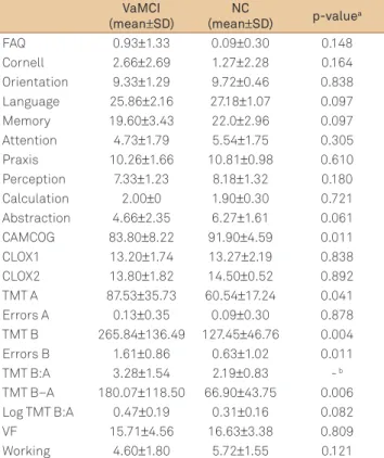
Neuropsychological assessment showed signiicant mean diferences between groups in TMT A and B, errors in TMT B, diference TMT B−A and CAMCOG total scores, for a signiicance level of 0.05. A trend for signiicance was identi-ied in abstraction. hese data are depicted in Table 3.
Based on significant results of the Mann-Whitney test, CAMCOG, TMT A and B, difference TMT B–A and errors in TMT B were selected to input to partial corre-lation test. Correcorre-lations were performed to determine whether extensions of WMH were associated with neu-ropsychological functions, with years of education and age included as control variables (Table 4). Albeit no sig-nificant differences were identified in years of school-ing or age between groups, within-group variance in
Table 2. Demographic data, Hachinski Ischemic Score, Fazekas and de Leon scores.
VaMCI
(mean±SD)
NC
(mean±SD) p-value
Gender (M/F) 6/9 3/8 0.683a
Age (years) 74.13±8.06 69.36±7.11 0.148b Schooling
(years) 8.86±4.03 11.18±2.56 0.121b MMSE 27.33±2.41 28.81±1.16 0.069b HIS 5.50±4.03 1.50±1.50 0.011b Fazekas scale
(0/1/2/3) 0/0/7/8 3/8/0/0 <0.001 b
de Leon scale
(0/1/2/3) 7/8/0/0 6/5/0/0 0.691a
aPearson’s χ2; bMann-Whitney test; VaMCI: vascular mild cognitive impairment; NC: normal controls; M: male; F: female; MMSE: Mini-Mental State Examination; HIS: Hachinski Ischemic Score; SD: standard deviation.
Table 3. Cognitive and behavioral data from vascular mild cognitive impairment and normal control groups.
VaMCI
(mean±SD)
NC
(mean±SD) p-valuea
FAQ 0.93±1.33 0.09±0.30 0.148 Cornell 2.66±2.69 1.27±2.28 0.164 Orientation 9.33±1.29 9.72±0.46 0.838 Language 25.86±2.16 27.18±1.07 0.097 Memory 19.60±3.43 22.0±2.96 0.097 Attention 4.73±1.79 5.54±1.75 0.305 Praxis 10.26±1.66 10.81±0.98 0.610 Perception 7.33±1.23 8.18±1.32 0.180 Calculation 2.00±0 1.90±0.30 0.721 Abstraction 4.66±2.35 6.27±1.61 0.061 CAMCOG 83.80±8.22 91.90±4.59 0.011 CLOX1 13.20±1.74 13.27±2.19 0.838 CLOX2 13.80±1.82 14.50±0.52 0.892 TMT A 87.53±35.73 60.54±17.24 0.041 Errors A 0.13±0.35 0.09±0.30 0.878 TMT B 265.84±136.49 127.45±46.76 0.004 Errors B 1.61±0.86 0.63±1.02 0.011 TMT B:A 3.28±1.54 2.19±0.83 - b TMT B–A 180.07±118.50 66.90±43.75 0.006 Log TMT B:A 0.47±0.19 0.31±0.16 0.082 VF 15.71±4.56 16.63±3.38 0.809 Working 4.60±1.80 5.72±1.55 0.121
aMann-Whitney test;bTMT B:A: quotient of mean values of TMT A and TMT B (>3: impairment of cognitive flexibility); VaMCI: vascular mild cognitive impairment; NC: normal controls; FAQ: Pfeffer’s Funcional Activities Questionnaire; Cornell: Cornell Depression Scale; CAMCOG: Cambridge Cognitive Examination; CLOX 1: Clock Drawing Task part 1; CLOX 2: Clock Drawing Task part 2; TMT A: Making Test part A; Errors A: errors in Trail-Making Test part A; TMT B: Trail-Trail-Making Test part B; Errors B: errors in Trail-Mak- ing Test part B; TMT B:A: ratio TMT B/TMT A; TMT B–A: difference TMT B–TMT A; Log TMT B:A: logarithmic transformation of B:A; VF: verbal fluency; Working: working memory.
Table 4. Correlations among scores on Fazekas scale and neuropsychological tests.
ra p-value rb p-value
CAMCOG -0.533 0.006 -0.609 <0.001
TMT A 0.265 0.200 0.497 0.011
TMT B 0.530 0.009 0.687 <0.001 Errors B 0.468 0.024 0.530 0.009 TMT B–A 0.502 0.015 0.669 <0.001
aControlling for years of schooling; bcontrolling for age; CAMCOG: Cambridge Cognitive Examination; TMT A: Trail-Making Test part A; TMT B: Trail-Making Test part B; Errors B: errors in Trail-Making Test part B; TMT B–A: difference TMT B–TMT A.
age, difference TMT B−A (r=0.669, p<0.001), CAMCOG (r=-0.609, p<0.001), TMT A (r=0.497; p=0.011), TMT B (r=0.687, p<0.001) and errors in TMT B (r=0.495, p=0.019) presented moderate correlations with scores in Fazekas scale.
DISCUSSION
Our results suggest the occurrence of impairments in fronto-executive tasks associated with extension of periven-tricular and deep WMH in patients with probable VaMCI. Signiicant poorer results in TMT A and B, errors in TMT B, diference TMT B–A and CAMCOG total score were identi-ied in VaMCI in relation to NC, and performances in those tests correlated with scores in Fazekas scale. A trend for sig-niicant diference was found in relation to mean scores in abstraction, compared to NC (p=0.061).
hese data are in line with results from previous research-es in which executive dysfunction was related to cerebrovas-cular disease20. A study using quantitative fractional
anisot-ropy (DTI-FA) detected higher levels of interrupted ibers in anterior ( frontal) brain regions in comparison to posterior re-gions-of-interest in patients with Binswanger disease, which might be compatible to disconnection of prefrontal-basal ganglia-thalamic circuits associated to executive function21.
Both abstract thinking subtest in CAMCOG and TMT B require integrity of the prefrontal cortex and of its connec-tions to basal ganglia. Dorsolateral and ventrolateral cor-tices were activated during tasks involving abstraction in studies using functional MRI ( fMRI)22. Impairment in
ab-stract thinking has distinguished VaMCI from NC in one study12. In addition, the Wechsler Adult Intelligence
Scale-Revised (WAIS-R) similarities subtest, which evaluates ab-stract thinking, was selectively low in initial VCI patients compared to incident AD patients, in a large prospective study23. he present data showed a trend for signiicant
diferences in abstract reasoning between VaMCI and NC, which might provide evidence of the importance of exten-sion of WMH on this cognitive function.
TMT B demands cognitive abilities such as inhibitory control, cognitive lexibility and processing speed. TMT B was related to activation in inferior dorsolateral prefrontal cortex, anterior cingulate, premotor cortex and intraparietal sulcus in fMRI studies24. Coherently, performance errors in
TMT B have been reported as a feature with high positive pre-dictive power for the presence of frontal lobe dysfunction25.
Errors in TMT B were classiied in 3 diferent types, which were consistent to dysfunction in prefrontal cortex and its subcortical connections, as follows: (1) sequential or track-ing errors (proceedtrack-ing to incorrect number or letter), (2) per-severative errors ( failure to proceed from number to letter or vice versa) and (3) proximity errors (proceeding to an incor-rect nearby number or letter)26.
In addition, TMT B:A ratio in VaMCI patients achieved mean quotient above 3, which might be suggestive of impair-ment in task-switching. Corroborating this inding, the signif-icant mean diferences in TMT B−A might also represent im-pairment in cognitive lexibility in those patients. According to a previous study, this scoring method minimizes visuoper-ceptual and working memory demands, and might corre-spond to the most reliable index of task-switching ability among all TMT direct and derived scores14. A relatively high
score in TMT B:A ratio observed in NC group, and the lack of signiicant diferences in Log B:A between VaMCI and NC are consistent with the literature27. Previous researches
that evaluated the validity of TMT derived indices indicat-ed some issues concerning the sensitivity and speciicity of those methods. For instance, the use of a cut-of score of 3 or greater for a TMT B:A ratio has been associated to high rate of false-positive results in studies27. he fact that NC
pre-sented a mean B:A ratio of 2.19±0.83 in our study indicates that some individuals in this group have surpassed the 3 cut-of, in spite of performing within normative values (i.e., total score less than 1.5 SD from the mean) for age and educational level in both TMT A and B direct scores. Further research is needed to validate its clinical utility as an index for dysexecu-tive syndrome.
Moreover, tests evaluating global cognitive function, mainly CAMCOG, were also correlated to severity of WMH in our study. Studies that analyzed CAMCOG as a screening tool for VCI showed excellent sensitivity and speciicity28.
Another aspect of our results also deserves to be ad-dressed. Impairments in abstract thinking, TMT B, diference TMT B–A and errors in TMT B with preserved CAMCOG items for working memory and verbal luency (VF) may load onto the idea of the multiple dimensions of EF, instead of an overarching executive construct. In fact, studies which per-formed factor analyses of putative EF measurements indi-cated discernible factors, such as “set shifting”, “inhibitory control”, “working memory” and “rule discovery”29. None of
the tests usually applied to evaluate EF appears to assess all those traits comprehensively13. Oosterman et al.30 noted
sig-niicant associations between WMH and fronto-executive functions in a sample with risk factors for CVD, such as in-hibitory control, planning and working memory. In the same study, HA was also associated to EF, showing a strong contri-bution to performance in working memory and set-shifting tasks. Diminished function of prefrontal-hippocampal cir-cuits associated to HA was implied as a possible mechanism for these indings30.
1. Gauthier S, Rockwood K. Does vascular MCI progress at a different rate than does amnestic MCI? Int Psychogeriatr 2003;15:257-259.
2. Petersen RC. Mild cognitive impairment as a diagnostic entity. J Intern Med 2004;256:183-194.
3. Moorhouse P, Rockwood K. Vascular cognitive impairment: current concepts and clinical developments. Lancet Neurol 2008;7:246-255.
4. Gorelick PB, Scuteri A, Black SE, et al. Vascular contributions to cognitive impairment and dementia: a statement for healthcare professionals from the American Heart Association/American Stroke Association. Stroke 2011;42:2672-2713.
5. Meyer JS, Xu G, Thornby J, et al. Is Mild cognitive impairment prodromal of vascular dementia like Alzheimer’s disease? Stroke 2002;33:1981-1985.
6. O’Brien JT. Vascular cognitive impairment. Am J Geriatr Psychiatry 2006;14:724-733.
7. Erkinjuntti T, Inzitari D, Pantoni L, et al. Research criteria for subcortical vascular dementia in clinical trials. J Neural Transm Suppl 2000;59:S23-S30.
8. Sachdev PS, Brodaty H, Valenzuela MJ, et al. Clinical determinants of dementia and mild cognitive impairment following ischemic stroke: the Sydney Stroke Study. Dement Geriatr Cogn Disord 2006;21:275-283.
9. van Straaten EC, Fazekas F, Rostrup E, et al. Impact of white matter hyperintensities scoring method on correlations with clinical data: the LADIS study. Stroke 2006;37:836-840.
10. Schmidt R, Ropele S, Ferro J, et al. Diffusion-weighted imaging and cognition in the Leukoaraiosis and Disability in the Elderly Study. Stroke 2010;41:402-408.
11. Kramer JH, Reed BR, Mungas D, et al. Executive dysfunction in subcortical ischaemic vascular disease. J Neurol Neurosurg Psychiatry 2002;72:217-220.
12. Sudo FK, Alves GS, Alves CEO, et al. Impaired abstract thinking may discriminate between normal aging and vascular mild cognitive impairment. Arq Neuropsiquiatr 2010;68:179-184.
13. Royall DR, Lauterbach EC, Cummings JL, et al. Executive control function: a review of its promise and challenges for clinical research. A report from the Committee on Research of the American Neuropsychiatric Associations. J Neuropsychiatry Clinical Neurosci 2002;14:377-405.
14. Sánchez-Cubillo I, Periáñez JA, Adrover-Roig D, et al. Construct validity of the Trail Making Test: role of task-switching, working memory, inhibition/interference control, and visuomotor abilities. J Int Neuropsychol Soc 2009;15:438-450.
15. de Leon M, Convit A, De Santi S, et al. Contribution of structural neuroimaging to the early diagnosis of Alzheimer’s disease. Int Psychogeriatr 1997;9:S183-S190.
16. Matioli MN, Caramelli P. Limitations in differentiating vascular dementia from Alzheimer’s disease with brief cognitive tests. Arq Neuropsiquiatr 2010;68:185-188.
17. Moreira IFH, Bezerra AB, Sudo FK, et al. CAMCOG subscales values in normal elderly with different educational levels. Dement Neuropsichol 2011;5:S34.
18. Brucki SMD, Malheiros SMF, Okamoto IH, Bertolucci PHF. Dados normativos para o teste de Fluência Verbal categoria animais em nosso meio. Arq Neuropsiquiatr 1997;55:56-61.
19. Tombaugh TM. Trail Making Test A and B: Normative data stratified by age and education. Arch Clin Psychol 2004;19:203-214.
20. Boone KB, Miller BL, Lesser I, et al. Neuropsychological correlates of white-matter lesions in healthy elderly subjects: a threshold effect. Arch Neurol 1992;49:549-554.
21. Engelhardt E, Moreira DM, Alves GS, et al. Binswanger’s disease: quantitative fractional anisotropy. Arq Neuropsiquiatr 2009;67:179-184.
22. Christoff K, Keramatian K, Gordon AM, Smith R, Mädler B. Prefrontal organization of cognitive control according to levels of abstraction. Brain Res 2009;1286:94-105.
23. Ingles JL, Boulton DC, Fisk JD, Rockwood K. Preclinical vascular cognitive impairment and Alzheimer disease: neuropsychological test performance 5 years before diagnosis. Stroke 2007;38:1148-1153.
24. Moll J, Oliveira-Souza R, Moll FT, et al. The cerebral correlates of set-shifting: an fMRI study of the trail-making test. Arq Neuropsiquiatr 2002;60:900-905.
25. Stuss DT, Bisschop SM, Alexander MP, et al. The Trail Making Test: a study in focal lesion patients. Psychol Assessment 2001;13:230-239.
26. Mahurin RK, Velligan DI, Hazleton B, et al. Trail Making Test errors and executive function in schizophrenia and depression. Clin Neuropsychol 2006;20:271-288.
27. Drane DL, Yuspeh RL, Huthwaite JS, Klingler LK. Demographic characteristics and normative observations for derived-trail making test indices. Neuropsychiatry Neuropsychol Behav Neurol 2002;15:39-43.
28. De Koning I, Van Kooten F, Dippel DW, et al. The CAMCOG: a useful screening instrument for dementia in stroke patients. Stroke 1998;29:2080-2086.
29. Miyake A, Friedman NP, Emerson MJ, Witzki AH, Howerter A, Wager TD. The unity and diversity of executive functions and their contributions to complex “Frontal Lobe” tasks: a latent variable analysis. Cogn Psychol 2000;41:49-100.
30. Oosterman JM, Vogels RL, van Harten B, et al. The role of white matter hyperintensities and medial temporal lobe atrophy in age-related executive dysfunctioning. Brain Cogn 2008;68:128-133.
References
clinic, and our data cannot be generalized to other popula-tions without further studies. Secondly, this study included a small sample of patients. It should also be noted that the absence of a golden-standard test for EF might cause diver-gence with results in diferent studies, according to the cho-sen neuropsychological battery.
Our findings support the importance of assessing fronto-executive functions in patients with probable ini-tial VCI. The literature highlights the difficulties of eval-uating those cognitive aspects and a set of neuropsy-chological tests recommended for extensively assessing
executive dysfunction remains to be determined. It is possible to argue that TMT B (direct score and difference B–A), CAMCOG abstract thinking subtest and CAMCOG total score might specifically distinguish VaMCI from healthy controls.