Dement Neuropsychol 2015 December;9(4):394-404
394
394 Neuroimaging criteria in Vascular MCI Sudo et al.
Original Article
Neuroimaging criteria and
cognitive performance in
vascular mild cognitive impairment
A systematic review
Felipe Kenji Sudo1, Gilberto Sousa Alves1,2, Chan Tiel3, Letice Ericeira-Valente1, Denise Madeira Moreira4, Jerson Laks1,5, Eliasz Engelhardt3
ABSTRACT. The recognition of Cerebrovascular Disease (CVD) at earlier clinical stages may favor the control of vascular risk factors and prevention of dementia. However, operational criteria for symptomatic phases at non-dementia stages are often difficult, as the current criteria normally require the evidence of extensive subcortical disease. Objective: To identify the neuroimaging profile of Vascular Mild Cognitive Impairment (VaMCI), the impact of those aspects over cognition and the neuropsychological tests that distinguished VaMCI from other groups. Methods: Searches were performed in Scopus, ISI and PsycINFO, using the following key terms: “vascular mild cognitive impairment” OR “vascular cognitive impairment no dementia” OR “vascular cognitive impairment not demented” OR “subcortical mild cognitive impairment”. Results:
Of 249 papers, 20 studies were selected. Ten of those included only patients with severe White Matter Hyperintensities (WMH), whereas 10 others admitted subjects with moderate-to-severe WMH. Both groups showed poor performances in Executive Function (EF) tasks in comparison to normal controls and other diagnostic groups. Among EF tests, those assessing “complex” EF abilities consistently distinguished VaMCI from other groups, regardless of the severity of WMH. VaMCI subjects with severe or moderate-to-severe WMH showed cognitive deficits in comparison with other groups. “Complex” EF tests were the most useful in differentiating those patients from the other groups. Conclusion: The occurrence of VaMCI may be associated with the presence of CVD at moderate levels; the detection of vascular damage at earlier stages may allow the adoption of therapeutic actions with significant effect-sizes.
Key words: cerebrovascular disorders, vascular dementia, cerebral infarction, neurological diagnostic techniques.
CRITÉRIOS DE NEUROIMAGEM E DESEMPENHO COGNITIVO NO COMPROMETIMENTO COGNITIVO LEVE VASCULAR: UMA REVISÃO SISTEMÁTICA
RESUMO. O reconhecimento precoce da Doença Cerebrovascular (DCV) pode permitir o controle de fatores de risco e a prevenção de demência. Contudo, critérios operacionais em seus estágios sintomáticos não-demenciais apresentam problemas, já que critérios atuais requerem a presença de extensa doença isquêmica subcortical. Objetivo: Identificar o perfil de neuroimagem do Comprometimento Cognitivo Leve Vascular (CCLV), o impacto destes aspectos sobre a cognição e os testes neuropsicológicos que distinguem CCLV de outros grupos. Métodos: Foram realizadas buscas no Scopus, ISI e PsycINFO, usando a estratégia: “vascular mild cognitive impairment” OR “vascular cognitive impairment no dementia” OR “vascular cognitive impairment not demented” OR “subcortical mild cognitive impairment”. Resultados: De 249 artigos, 20 foram selecionados. 10 destes incluíram apenas pacientes com hiperintensidades de substância branca (HSB) graves, enquanto 10 outros admitiram pacientes com HSB moderadas-a-graves. Ambos os grupos apresentaram desempenho pobre em tarefas de Função Executiva (FE) em comparação com controles normais e outras categorias diagnósticas. Dentre os testes de FE, aqueles que avaliam FE “complexas” diferiram consistentemente CCLV de outros grupos,
This study was conducted at the Instituto de Psiquiatria, Universidade Federal do Rio de Janeiro
1Instituto de Psiquiatria, Universidade Federal do Rio de Janeiro (UFRJ), Rio de Janeiro RJ, Brazil; 2Departamento de Medicina Clínica, Universidade Federal do
Ceará, Fortaleza CE, Brazil; 3Instituto de Neurologia Deolindo Couto, Setor de Neurologia Cognitiva e do Comportamento-INDC-CDA/IPUB, UFRJ, Rio de Janeiro
RJ, Brazil; 4Serviço de Radiologia, Instituto de Neurologia Deolindo Couto (UFRJ); Hospital Pró-Cardíaco, Rio de Janeiro RJ, Brazil. 5Universidade do Estado do Rio
de Janeiro, Rio de Janeiro RJ, Brazil.
Felipe Kenji Sudo. Av. Nossa Senhora de Copacabana, 895/302 – 22060-001 Rio de Janeiro RJ – Brazil. E-mail: [email protected]
Disclosure: The authors report no conflits of interest
Received September 09, 2015. Accepted in final form November 15, 2015.
Dement Neuropsychol 2015 December;9(4):394-404
395
Sudo et al. Neuroimaging criteria in Vascular MCI
independentemente da gravidade de HSB. Sujeitos com CCLV e HSB graves ou moderadas-a-graves apresentaram dificuldades cognitivas quando comparados aos demais grupos. Testes que avaliam FE “complexa” foram os mais úteis na diferenciação destes pacientes dos outros grupos. Conclusão: A ocorrência de VaMCI pode estar associada à presença de HSB moderadas; a detecção precoce do dano vascular permitiria a adoção de medidas terapêuticas com tamanhos de efeito significativos.
Palavras-chave: transtornos cerebrovasculares, demência vascular, infarto cerebral, técnicas de diagnóstico neurológico.
INTRODUCTION
V
ascular Cognitive Impairment (VCI) is an umbrella concept which comprises a continuum of vascular-related cognitive impairment, from high-risk preclini-cal conditions (“brain-at-risk”) to Vascular Dementia (VaD). Intermediate stages are commonly referred as Vascular Mild Cognitive Impairment (VaMCI) or Vas-cular Cognitive Impairment No-Dementia (Va-CIND).1Recent operational criteria, such as the 2011 American Heart Association (AHS)/American Stroke Association (ASA) scientiic statement on vascular contributions to cognitive impairment, suggested that the relationship between CVD and cognitive changes could be character-ized whether through the evidence of cognitive deicits succeeding a clinical stroke or through identifying vas-cular lesions on neuroimaging deemed severe enough to explain the cognitive impairment.2
More detailed neuroimaging criteria have been described in the 2014 International Society for Vas-cular Behavioral and Cognitive Disorders (VASCOG) statement for diagnosis of Vascular Cognitive Disorders (VCD). In this document, CVD was evidenced by the presence of one of the following changes: [1] extensive and conluent subcortical White Matter Hyperintensi-ties (WMH); [2] large-vessel infarcts: 1 (for Mild VCD) or ≥2 (for Major VCD); [3] 1 strategically placed infarct (in the thalamus or basal ganglia); [4] >2 lacunar infarcts outside the brainstem or at least 1 lacune combined with extensive WMH; and (5) intracerebral hemorrhages: ≥2 or 1 strategically placed.3
he VASCOG statement represented a more com-prehensive neuroimaging criterion in comparison to the AHA/ASA recommendations and a substantial change in relation to the Erkinjuntti’s neuroimaging criteria for Subcortical Ischemic VaD (2000), in which extensive and conluent WMH or moderate WMH combined with at least 5 lacunes was required to characterize CVD.4
None-theless, the persistence in the new criteria of the need for extensive and conluent WMH contrasted with some studies, which have suggested that moderate WMH with less than 5 lacunes could account for cognitive impair-ments.5 As indicated by several studies, mild WMH is
highly prevalent among normal elderly individuals and
has not been signiicantly associated with cognitive changes.6
One possible advantage in identifying CVD in its mildest clinical (VaMCI) and neuroimaging (moderate subcortical WMH and less than 5 lacunes) stages is the fact that progression of vascular damage might be pre-ventable. Early detection might allow the adoption of disease-modifying therapies that could prevent the pro-gression of vascular lesions; therefore, it might interrupt the advance of cognitive impairment that could result in VaD. Finally, recent diagnostic criteria for Va-CIND overlap with the ASA/AHA criteria for VaMCI,7 thus the
term VaMCI has been used in this review to refer to both constructs.
According to the above pondering, a systematic review was undertaken aiming: [1] to assess the neu-roimaging proile of individuals classiied as VaMCI in clinical studies; [2] to determine whether diferent neu-roimaging criteria impact over cognitive indings, and [3] to identify neuropsychological tests that could distin-guish VaMCI from normal controls or other diagnostic groups across studies using diferent criteria for CVD. he authors hypothesized that the choice of establishing the threshold of brain vascular lesions into moderate or severe stages of WMH may account for divergent cogni-tive indings among studies.
METHODS
Data search and selection. Studies were found through
searches in Scopus, ISI Web Of Knowledge and PsycINFO, using the following key terms, in all ields and published in any date: “vascular mild cognitive impairment” OR “vascular cognitive impairment no dementia” OR “vascular cognitive impairment not demented” OR “subcortical mild cognitive impairment”. his search strategy was augmented with hand searches of reference lists of included studies. More articles were obtained from directly contacting authors for relevant papers.
Dement Neuropsychol 2015 December;9(4):394-404
396 Neuroimaging criteria in Vascular MCI Sudo et al.
other diagnostic groups [VaD, AD, non-vascular MCI (non-VaMCI)] or normal controls; and written in English, French, Spanish or Portuguese.
he authors have excluded studies that: classiied individuals as VaMCI based solely on clinical/ neuropsy-chological aspects (e.g., studies in which the cognitive deicits were judged to have vascular cause through clini-cal features, such as stepwise progression, sudden onset, gait disturbances, focal neurological signs or those that applied only an ischemic score to identify the presence of cerebrovascular disease); did not assess subjects with MCI, deined as those presenting cognitive impairments that do not fulill criteria for dementia; did not acknowl-edge a detailed neuroimaging criterion for the diagnosis of VaMCI (e.g., cognitive impairment considered associ-ated with vascular lesions through subjective evaluation from an expert); did not compare cognitive performances between VaMCI and controls or other diagnostic groups; or included subjects with cortical infarction or cortical atrophy suggestive of large-vessel or neurodegenerative diseases. he current study followed the standard proto-cols of PRISMA statement.8
Data extraction. Data were extracted from full-texts by
one author (FKS) and reviewed by a second author (EE). Divergences were furtherly discussed among the entire team of authors.
RESULTS
Of a total of 249 retrieved papers, 20 studies were selected for data extraction. Figure 1 summarizes the stages of data search and selection.
Clinical criteria for MCI. Participants in the studies
presented objective cognitive deicits and preserved functional status. Mild diferences included articles that identiied those with cognitive impairments based on performances in screening tests for cognitive deicits (e.g., MMSE ≥ 24, CDR= 0.5, Clock Drawing Test scores lower than 2/6).9-14 Cognitive impairment was deined
as performances 1 to 2 standards deviations (between the 16th and the 2nd percentile) below mean normative
values, in some studies.15-21 Few studies, all of them
prior to 2009, required impairment in memory for diag-nosis of MCI;17,22,23 however, most papers did not include
any speciic cognitive domain or proposed dysexecutive symptoms as typically associated with VaMCI.
Neuroimaging criteria for subcortical vascular disease. Ten
of the studies classiied subcortical CVD as the pres-ence of white-matter changes compatible with severe WMH and/or at least 5 subcortical lacunes. Five of those followed the criteria proposed by Erkinjuntti et al. (2000) for Binswanger’s Disease, which requires the presence of severe WMH, periventricular lesions larger Figure 1. Flow-chart describing the process of study selection. Reviews, case reports, posters, conferences
and comments removed (N=29)
Studies that did not use or acknowledge a detailed neuroimaging criteria for VaMCI (N=32)
Studies that did not assess cognitive aspects of VaMCI (N=12)
Studies that assessed VaD, AD or that included VCI associated with acute stroke or large-vessel
disease (N=34)
Duplicates removed (N=13)
Excluded by title (N=90)
Excluded by language (N=27)
Articles selected for data extraction
(N=20)
Database search
Scopus (N=108); ISI (N=93); PsycInfo (N=48); Reference search (N=6);
Dement Neuropsychol 2015 December;9(4):394-404
397
Sudo et al. Neuroimaging criteria in Vascular MCI than 10 mm and deep WMH equal or over 25 mm of
diameter.11,15,16,24,25 A modiied version of the
Computer-ized Tomography (CT) criterion for Subcortical Vascular Dementia proposed by Erkinjuntti et al. (2000) was applied in two of the studies. CVD, in those cases, was represented by patchy or difuse leukoaraiosis and at least one lacunar infarct on neuroimaging.9,22 Evidence
of extensive WMH, deined as lesions larger than 3 mm of diameter in the semioval center and larger than 5 mm in the deep gray nuclei, was the criterion used in one study.26 Other methods for identiication of
individ-uals with severe WMH included semiautomatic white-matter volumetry techniques. Nordahl et al. (2005) clas-siied individuals with WMH extending for more than 19.375% of total white-matter volume as presenting severe WMH.23 Moretti et al. (2008) computed the
pres-ence of CVD by counting voxels corresponding to WMH and identifying those individuals whose lesions corre-sponded to values over the fourth quartile of volume damage.10 Table 1 illustrates those indings.
Moderate WMH and/or less than 5 lacunes were deemed suicient to characterize CVD in ten of the studies. Overall, individuals that scored 2 or more in the modiied-Fazekas Scale, corresponding to the presence of moderate periventricular WMH (“smooth halo”) with beginning conluent deep WMH, were selected for those studies. Identiication of at least 2 lacunar infarcts was an alternative criterion for diagnosis of moderately severe cerebrovascular disease. Table 2 depicts those results.
Cognitive performances and neuroimaging criteria.
Alth-ough the choice of neuropsychological tests varied across studies, cognitive assessment in most cases included tasks that measured executive function (EF), memory, language and visuospatial/ visuoconstructive abilities. Table 3 summarizes the main afected cogni-tive abilities in the selected studies. EF has been divided into 3 components, following studies that performed a latent variable approach of multiple EF measures: “shifting” (switching between tasks), “inhibition” (delib-erate overriding of prepotent responses) and “working memory/updating” (monitoring and rapidly changing new contents).27 Tests categorized as “less speciic EF
tests” included tasks that assessed multiple EF dimen-sions (e.g., Clock Drawing Test, Verbal Fluency etc.), instead of measuring one single aspect of it.28 Matching
between neuropsychological tests and cognitive domains was made in accordance with evidences in the literature.21,28-40 Table 4 summarizes the correspondence
between cognitive domains and neuropsychological tests used in the studies.
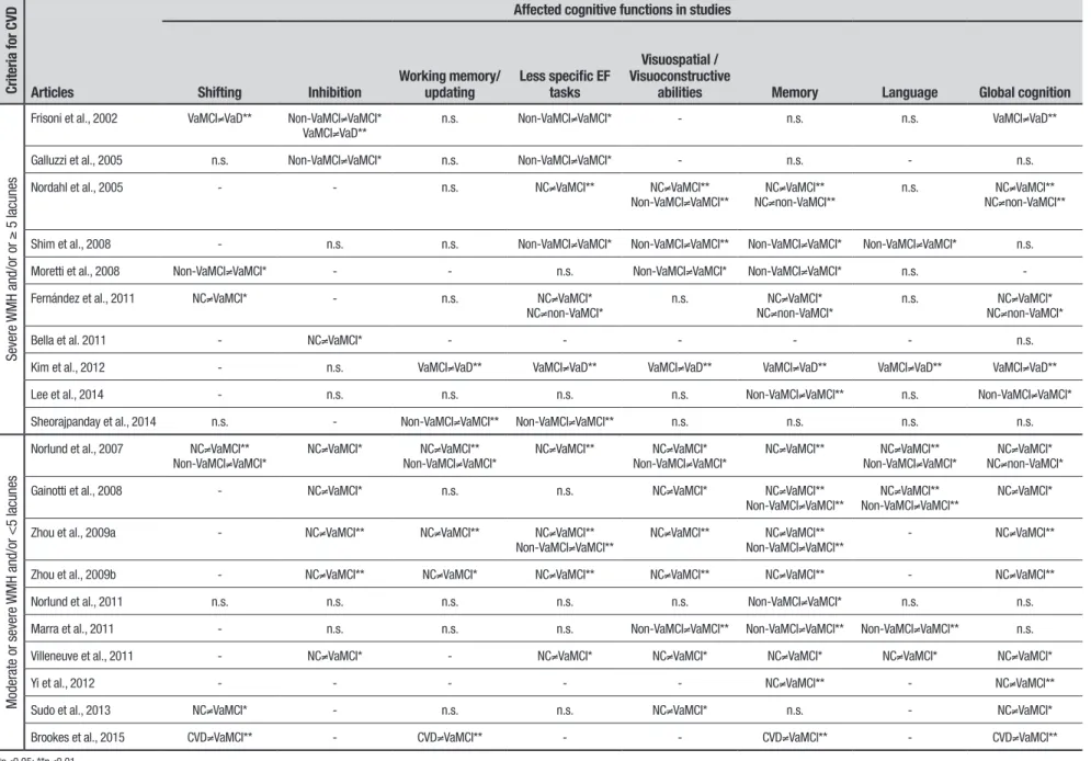
Studies using the severe WMH and/or more than 5 lacunes criteria evidenced signiicant diferences among VaMCI, VaD and controls in EF, Memory and Visuo-spatial/ Visuoconstructive tasks. Tests that measured “impure” and unspeciic EF dimensions, labeled herein as “less speciic EF tasks”, consistently distinguished VaMCI from the other groups, while Working Memory Tasks appear to be less sensitive for detection of VaMCI. As expected, performances in Memory tests identiied non-VaMCI from VaMCI, but also diferentiated VaMCI from controls in some studies. Global cognitive measures were more accurate in distinguishing VaMCI from con-trols and VaD than from non-VaMCI.
When moderate-to-severe WMH and/or less than 5 lacunes were used as criteria for CVD, EF, Memory, Visuospatial abilities tests, as well as Global Cognitive assessment, diferentiated VaMCI from controls in most studies. Memory and Language tests were accurate mea-sures in distinguishing VaMCI from non-VaMCI. Among EF dimensions, Inhibition and unspeciic EF tests con-sistently detected VaMCI from controls in the selected studies.
DISCUSSION
he idea that VCI comprises a spectrum of diferent stages of vascular-related cognitive impairment may suggest that dementia can be preceded by subtle cogni-tive changes associated with CVD.41 However, the
boundaries of vascular burden that mark the earliest clinical stages of CVD still need to be deined. he importance of establishing the milder pathological clin-ical phase of VCI resides in the fact that early identii-cation of cognitive decline associated with CVD might allow adequate control of vascular risk factors, so as to prevent progression to dementia. In this perspective, the adoption of the neuroimaging criteria proposed by Erkinjuntti et al. for Binswanger Disease (2000) iden-tiied cases in which white-matter injury is already extensive, that may limit the efect-sizes of prophylactic actions. he present article reviewed data suggestive of expressive cognitive changes associated with moderate-to-severe WMH and less than 5 lacunes. Identiication of those subjects might allow more efective actions in preventing progression of cognitive decline.
discrimi-Dement Neuropsyc
hol 2015 December;9(4):394-404
398
Neuroimaging criteria in
Vascular MCI
Sudo et al.
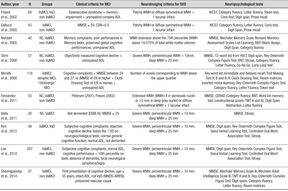
Table 1. Studies that included severe (largely confluent) WMH and/or at least 5 lacunes for diagnosis of SVD.
Author, year N Groups Clinical criteria for MCI Neuroimaging criteria for SVD Neuropsychological tests
Frisoni et al., 2002
64 VaMCI, VaD,
non-VaMCI
Dysexecutive syndrome + memory impairment + unimpaired complex ADL
Patchy WMH or diffuse symmetrical WMH + 1 lacunar infact
WCST, Category fluency, Letter fluency, Token test, Corsi test, Digit span, Prose recall
Galluzzi et al., 2005
43 VaMCI,
non-VaMCI
MMSE ≥ 24, CDR=0.5 Patchy WMH or diffuse symmetrical WMH +
1 lacunar infact
WCST, Category fluency, Letter fluency, Corsi test, Digit Span, Prose recall
Nordahl et al., 2005
42 NC, VaMCI,
non-VaMCI
Memory complaints, poor perfomances in Memory tasks, preserved global cognitive
performances, unimpaired ADL
WMH extension above the 75th percentile (WMH above 19.375% of total white matter volume)
MMSE, Wechsler Memory Scale Revised, Memory Assessment Scales List Learning, BNT, Block design,
Digit Span, Category fluency
Shim et al., 2008
57 NC, VaMCI,
non-VaMCI
Objectively measured cognitive decline + unimpaired ADL
Severe WMH, periventricular WMH > 10mm,
deep WMH ≥ 25 mm
MMSE, 12-word list from HVLT, Digit span, Rey-Osterrieth Complex Figure Test, BNT, Stroop, Category fluency,
Letter Fluency, Go-No Go, Luria Loop test
Moretti et al., 2008
116 VaMCI,
atrophic MCI, “cholinergic”
MCI
Cognitive complaints + MMSE between 24 and 27, or (MMSE of 28 or higher + Clock
Drawing Test of 2/6 or worse) + unimpaired ADL
Number of voxels corresponding to WMH above the upper quartile
Rey word list immediate and delayed recall, Trail Making Test A, B and B-A, Clock Drawing Test, Raven matrices, Inverted motor learning, Rey-Osterrieth Complex Figure Test,
Category fluency, Letter Fluency, Token test
Fernández et al., 2011
53 NC, VaMCI,
non-VaMCI
Petersen (2001), Frisoni (2002) Extensive WMH (WMH>3 in semiovale nuclei
or >5 mm in deep grey nuclei) or diffuse symmetrical WMH + 1 lacunar infact
MMSE, CERAD (Category fluency, BNT, Word list memory test, constructional praxis, TMT A and B), Digit Span,
Abstraction, Letter fluency
Bella et al., 2011
20 NC, VaMCI Not demented (DSM-IV), MMSE ≥ 24 Severe WMH, periventricular WMH > 10 mm,
deep WMH ≥ 25 mm
MMSE, Stroop
Kim et al., 2012
48 VaMCI, VaD Subjective cognitive complaints; objective
cognitive decline below the 1 SD on neuropsychological tests; normal general cognitive function; normal ADL; not demented
Severe WMH, periventricular WMH > 10 mm,
deep WMH ≥ 25 mm
MMSE, Digit span, Rey-Osterrieth Complex Figure Test, Seoul Verbal Learning Test, Controlled Oral Word
Association Test, Stroop
Lee et al., 2014
207 VaMCI,
non-VaMCI
Subjective cognitive complaints, normal ADL, cognitive performance < 16th percentile on tests, absence of dementia, focal neurological
symptoms/signs
Severe WMH, periventricular WMH > 10 mm,
deep WMH ≥ 25 mm
MMSE, Digit span, Rey-Osterrieth Complex Figure Test, Seoul Verbal Learning Test, Controlled Oral Word
Association Test, Stroop
Sheorajpanday et al., 2014
57 VaMCI,
non-VaMCI
First presentation of cognitive decline, age ≥
55 years, intact ADL, not VaD (NINDS-AIREN), presumed vascular cause
Severe WMH, periventricular WMH > 10 mm,
deep WMH ≥ 25 mm
MMSE, Wechsler Memory Scale III, Wechsler Adult Intelligence Scale III, TMT A and B, Rey-Osterrieth Complex
Dement Neuropsyc
hol 2015 December;9(4):394-404
399
Sudo
et
al.
Neuroimaging criteria in
Vascular MCI
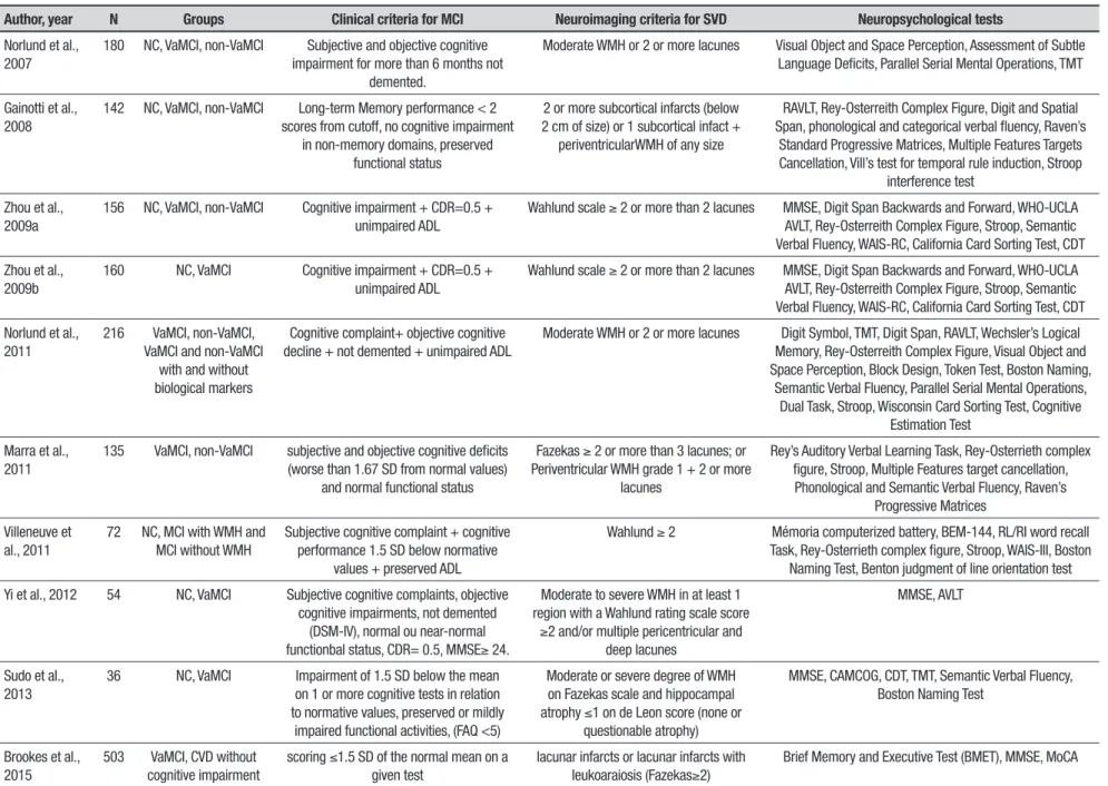
Table 2. Studies that included moderate (beginning confluent; smooth halo) WMH and/or less than 5 lacunes required for diagnosis of SVD.
Author, year N Groups Clinical criteria for MCI Neuroimaging criteria for SVD Neuropsychological tests
Norlund et al., 2007
180 NC, VaMCI, non-VaMCI Subjective and objective cognitive
impairment for more than 6 months not demented.
Moderate WMH or 2 or more lacunes Visual Object and Space Perception, Assessment of Subtle
Language Deficits, Parallel Serial Mental Operations, TMT
Gainotti et al., 2008
142 NC, VaMCI, non-VaMCI Long-term Memory performance < 2
scores from cutoff, no cognitive impairment in non-memory domains, preserved
functional status
2 or more subcortical infarcts (below 2 cm of size) or 1 subcortical infact +
periventricularWMH of any size
RAVLT, Rey-Osterreith Complex Figure, Digit and Spatial Span, phonological and categorical verbal fluency, Raven’s
Standard Progressive Matrices, Multiple Features Targets Cancellation, Vill’s test for temporal rule induction, Stroop
interference test
Zhou et al., 2009a
156 NC, VaMCI, non-VaMCI Cognitive impairment + CDR=0.5 +
unimpaired ADL
Wahlund scale ≥ 2 or more than 2 lacunes MMSE, Digit Span Backwards and Forward, WHO-UCLA
AVLT, Rey-Osterreith Complex Figure, Stroop, Semantic Verbal Fluency, WAIS-RC, California Card Sorting Test, CDT
Zhou et al., 2009b
160 NC, VaMCI Cognitive impairment + CDR=0.5 +
unimpaired ADL
Wahlund scale ≥ 2 or more than 2 lacunes MMSE, Digit Span Backwards and Forward, WHO-UCLA
AVLT, Rey-Osterreith Complex Figure, Stroop, Semantic Verbal Fluency, WAIS-RC, California Card Sorting Test, CDT
Norlund et al., 2011
216 VaMCI, non-VaMCI,
VaMCI and non-VaMCI with and without biological markers
Cognitive complaint+ objective cognitive decline + not demented + unimpaired ADL
Moderate WMH or 2 or more lacunes Digit Symbol, TMT, Digit Span, RAVLT, Wechsler’s Logical
Memory, Rey-Osterreith Complex Figure, Visual Object and Space Perception, Block Design, Token Test, Boston Naming,
Semantic Verbal Fluency, Parallel Serial Mental Operations, Dual Task, Stroop, Wisconsin Card Sorting Test, Cognitive
Estimation Test
Marra et al., 2011
135 VaMCI, non-VaMCI subjective and objective cognitive deficits
(worse than 1.67 SD from normal values) and normal functional status
Fazekas ≥ 2 or more than 3 lacunes; or
Periventricular WMH grade 1 + 2 or more lacunes
Rey’s Auditory Verbal Learning Task, Rey-Osterrieth complex figure, Stroop, Multiple Features target cancellation, Phonological and Semantic Verbal Fluency, Raven’s
Progressive Matrices
Villeneuve et al., 2011
72 NC, MCI with WMH and
MCI without WMH
Subjective cognitive complaint + cognitive performance 1.5 SD below normative
values + preserved ADL
Wahlund ≥ 2 Mémoria computerized battery, BEM-144, RL/RI word recall
Task, Rey-Osterrieth complex figure, Stroop, WAIS-III, Boston Naming Test, Benton judgment of line orientation test
Yi et al., 2012 54 NC, VaMCI Subjective cognitive complaints, objective
cognitive impairments, not demented (DSM-IV), normal ou near-normal
functionbal status, CDR= 0.5, MMSE≥ 24.
Moderate to severe WMH in at least 1 region with a Wahlund rating scale score
≥2 and/or multiple pericentricular and
deep lacunes
MMSE, AVLT
Sudo et al., 2013
36 NC, VaMCI Impairment of 1.5 SD below the mean
on 1 or more cognitive tests in relation to normative values, preserved or mildly
impaired functional activities, (FAQ <5)
Moderate or severe degree of WMH on Fazekas scale and hippocampal
atrophy ≤1 on de Leon score (none or
questionable atrophy)
MMSE, CAMCOG, CDT, TMT, Semantic Verbal Fluency, Boston Naming Test
Brookes et al., 2015
503 VaMCI, CVD without
cognitive impairment
scoring ≤1.5 SD of the normal mean on a
given test
lacunar infarcts or lacunar infarcts with
leukoaraiosis (Fazekas≥2)
Dement Neuropsyc
hol 2015 December;9(4):394-404
400
Neuroimaging criteria in
Vascular MCI
Sudo et al.
Table 3. Summary of cognitive findings in the selected studies according with the neuroimaging criteria for CVD.
Criteria for CVD Articles
Affected cognitive functions in studies
Shifting Inhibition Working memory/ updating Less specific EF tasks
Visuospatial / Visuoconstructive
abilities Memory Language Global cognition
Severe
WMH and/or or
≥
5 lacunes
Frisoni et al., 2002 VaMCI≠VaD** Non-VaMCI≠VaMCI* VaMCI≠VaD**
n.s. Non-VaMCI≠VaMCI* - n.s. n.s. VaMCI≠VaD**
Galluzzi et al., 2005 n.s. Non-VaMCI≠VaMCI* n.s. Non-VaMCI≠VaMCI* - n.s. - n.s. Nordahl et al., 2005 - - n.s. NC≠VaMCI** NC≠VaMCI**
Non-VaMCI≠VaMCI**
NC≠VaMCI** NC≠non-VaMCI**
n.s. NC≠VaMCI** NC≠non-VaMCI**
Shim et al., 2008 - n.s. n.s. Non-VaMCI≠VaMCI* Non-VaMCI≠VaMCI** Non-VaMCI≠VaMCI* Non-VaMCI≠VaMCI* n.s. Moretti et al., 2008 Non-VaMCI≠VaMCI* - - n.s. Non-VaMCI≠VaMCI* Non-VaMCI≠VaMCI* n.s. -Fernández et al., 2011 NC≠VaMCI* - n.s. NC≠VaMCI*
NC≠non-VaMCI*
n.s. NC≠VaMCI* NC≠non-VaMCI*
n.s. NC≠VaMCI* NC≠non-VaMCI*
Bella et al. 2011 - NC≠VaMCI* - - - n.s.
Kim et al., 2012 - n.s. VaMCI≠VaD** VaMCI≠VaD** VaMCI≠VaD** VaMCI≠VaD** VaMCI≠VaD** VaMCI≠VaD** Lee et al., 2014 - n.s. n.s. n.s. n.s. Non-VaMCI≠VaMCI** n.s. Non-VaMCI≠VaMCI* Sheorajpanday et al., 2014 n.s. - Non-VaMCI≠VaMCI** Non-VaMCI≠VaMCI** n.s. n.s. n.s. n.s.
Modera
te or severe
WMH and/or <5 lacunes
Norlund et al., 2007 NC≠VaMCI** Non-VaMCI≠VaMCI*
NC≠VaMCI* NC≠VaMCI** Non-VaMCI≠VaMCI*
NC≠VaMCI** NC≠VaMCI* Non-VaMCI≠VaMCI*
NC≠VaMCI** NC≠VaMCI** Non-VaMCI≠VaMCI*
NC≠VaMCI* NC≠non-VaMCI* Gainotti et al., 2008 - NC≠VaMCI* n.s. n.s. NC≠VaMCI* NC≠VaMCI**
Non-VaMCI≠VaMCI**
NC≠VaMCI** Non-VaMCI≠VaMCI**
NC≠VaMCI*
Zhou et al., 2009a - NC≠VaMCI** NC≠VaMCI** NC≠VaMCI** Non-VaMCI≠VaMCI**
NC≠VaMCI** NC≠VaMCI** Non-VaMCI≠VaMCI**
- NC≠VaMCI**
Zhou et al., 2009b - NC≠VaMCI** NC≠VaMCI* NC≠VaMCI** NC≠VaMCI** NC≠VaMCI** - NC≠VaMCI**
Norlund et al., 2011 n.s. n.s. n.s. n.s. n.s. Non-VaMCI≠VaMCI* n.s. n.s.
Marra et al., 2011 - n.s. n.s. n.s. Non-VaMCI≠VaMCI** Non-VaMCI≠VaMCI** Non-VaMCI≠VaMCI** n.s. Villeneuve et al., 2011 - NC≠VaMCI* - NC≠VaMCI* NC≠VaMCI* NC≠VaMCI* NC≠VaMCI* NC≠VaMCI*
Yi et al., 2012 - - - NC≠VaMCI** - NC≠VaMCI**
Sudo et al., 2013 NC≠VaMCI* - n.s. n.s. NC≠VaMCI* n.s. - NC≠VaMCI*
Brookes et al., 2015 CVD≠VaMCI** - CVD≠VaMCI** - - CVD≠VaMCI** - CVD≠VaMCI**
Dement Neuropsychol 2015 December;9(4):394-404
401
Sudo et al. Neuroimaging criteria in Vascular MCI
Table 4. Cognitive domains and corresponding neuropsychological tasks.
Cognitive functions Tests
Executive Function (EF) Shifting Wisconsin Card Sorting Test (WMST): perseveration, Trail-Making Test (TMT) B, Dual task, Num-ber-Letter sequencing
Inhibition WCST: non-perseverative errors and categories, Go/No go, Fist/Edge/Palm sequence, Stroop test
Working Memory/Update
Digit Span forward and backwards, Corsi test, Parallel Serial Mental Operations, CAMCOG: Work-ing Memory Subtest, Number and Letter sequencWork-ing.
Less specific EF tests Category and Letter verbal fluency, Luria loop, Raven matrices, Barcelona test (Abstraction sub-test), CAMCOG: Abstraction subtest, COWAT, Digit-Symbol substitution test, Cognitive estimation test, WAIS-III(picture interpretation and arrangement, Clock Drawing Test/CLOX 1 (spontaneous drawing), California Card Sorting Test
Visuospatial/visuoconstructive abilities Block design, Rey figure: copy, TMT A, Visual Object and Space Perception, Lines cancellation test, Clock Drawing Test/CLOX 2. (copy), Multiple Features Target Cancellation
Memory Prose recall, Babcock Story Recall test, Wechsler Memory Scale-Revised, Memory Assessment Scales, Hopkins Verbal Learning Test, Rey figure: recall and recognition, CAMCOG: Memory sub-test, Five-item memory test
Language Token test, Boston Naming test, Assessment of Subtle Language Deficits
Global Cognition MMSE, CAMCOG, BMET
MMSE: Mini-Mental State Examination; CAMCOG: Cambridge Cognitive Examination part of the Cambridge Examination for. Mental Disorders of the Elderly (CAMDEX).; BMET: Brief Memory and Executive Test; WAIS-III: Wechsler Adult Intelligence Scale, 3rd Edition.
nating VaMCI from controls than speciic and “pure” EF measures, even in the group with moderate WMH. Data from functional neuroimaging studies suggested that those “higher level” EF may recruit diverse areas in the prefrontal, parietal, medial and superior temporal corti-ces, and subcortical structures (amygdala, thalamus and cerebellum).42,43 hese indings indicate that complex EF
may result from the ine integration of many diferent cortical areas and subcortical regions, which depends on an extensive and delicate network of neural projections.44
Moderate white-matter changes, represented by periven-tricular smooth halo and beginning conluent deep WMH on neuroimaging, may be suicient to interrupt segments of inter-cortical and/or cortical-subcortical loops, leading to disconnection of areas associated with complex EF.45
On the other hand, data on the accuracy of more speciic EF measures in distinguishing controls, VaMCI and non-VaMCI appeared to be inconsistent, as observed in relation to shifting tasks. Performances in inhibition tasks were signiicantly worse in VaMCI subjects than in controls in most of the studies with moderate-to-severe CVD. his inding might suggest an early impairment of inhibitory control in VCI patients, which is in line with a previous prospective study.46 Interconnections among
prefrontal cortex, subcortical regions and posterior areas might be interrupted in those patients, leading to loss of prefrontal inhibitory inputs over cortical-subcortical networks associated with task-irrelevant distracters.47,48
Among the severe CVD group, only two studies per-formed a similar analysis, showing conlicting results. Furthermore, working memory tasks were consistently inaccurate in diferentiating VaMCI from non-VaMCI in most studies. Reports of impairments in working mem-ory in amnestic MCI are abundant in the literature; thus, both Vascular and amnestic MCI might share, through diferent pathological mechanisms, similar prefron-tal and cingulate dysfunction associated with working memory abilities.49
Non-executive cognitive domains were also tested in the studies. As expected, episodic memory tasks were more impaired in “atrophic” MCI than in VaMCI, in most of the studies. Yet, the inding that episodic memory per-formances were signiicantly poorer in VaMCI than in controls may highlight the role of the prefrontal cortex for the retrieval of information. Recent evidence suggested that left prefrontal cortex may participate in the recall process through the use of environmental cues and the ability to inhibit irrelevant memories during a task.50 Also,
not surprisingly, impairments in visuospatial and visuo-constructive abilities were more prominent in VaMCI than in non-VaMCI and controls. hose alterations have been associated with CVD in diferent studies.51,52 Finally,
Dement Neuropsychol 2015 December;9(4):394-404
402 Neuroimaging criteria in Vascular MCI Sudo et al.
neuropsychological batteries, many studies reported ceiling-efects for MMSE in samples comprising single-domain MCI subjects. However, evidence suggested that it may present similar accuracy in detecting multidomain impairments as compared with the Montreal Cognitive Assessment (MoCA) and the Addenbrooke’s Cognitive Examination–Revised (ACE-R).53
Some other issues should be addressed. Despite slight variations, specially related to the instruments used to detect cognitive impairment and to the degree of devia-tion from normal cognidevia-tion necessary to characterize the disorder, the clinical criteria proposed by Petersen et al. for MCI (2001) were adopted almost unchanged by most of the authors.54 his fact might indicate that,
albeit past criticisms were directed to the disorder’s con-struct validity, the use of the clinical entity described by Petersen et al. has largely prevailed among clinical stud-ies.55 Conversely, other operational criteria have shown
to be not optimal to identify MCI associated with CVD. Salvadori et al. (2015) reported that the criteria proposed by Winblad et al. (2004) might overlook non-amnestic MCI presentations.52
here are limitations in this review that need to be commented. Diferent terminologies used to describe periventricular and deep WMH and imprecise expres-sions (e.g., “patchy WMH”, “difuse WMH”, “smooth halo” and “caps”), present in diferent criteria make it diicult to compare lesion loads across studies. Further-more, the characterization of periventricular/deep WMH itself has been object of divergence by some authors, who adopted diferent distances between the ventricle’s margin and the lesion to deine it as “periventricular” or “deep”.56,57 Moreover, tasks classiied as assessing a
spe-ciic aspect of EF may not be pure measures of that pro-cess, since they commonly require other EF and non-EF features. Models of EF as a unique or multiple constructs have been proposed and there is no agreement regarding
neuropsychological tests that may thoroughly assess all of its aspects. Further studies using conirmatory factor-analysis of EF measures may allow the establishment of cognitive batteries comprising tests that evaluate com-plementary processes of EF.
he present review evidenced that the choice of neu-roimaging criteria to characterize CVD in MCI subjects did not result in groups with diferent cognitive pro-iles. One possible hypothesis is the complex nature of subcortical disease, in which vascular and non-vascular (e.g., Alzheimer’s disease, multiple sclerosis) events often interact, ultimately resulting in WM disconnection and cognitive impairment.58,59 In addition, as suggested by
Pasi et al. (2015), that may also be due to the fact that cognitive tests may lose their accuracy in distinguishing groups of patients once certain degree of vascular lesions is reached.60
In conclusion, evidence in the literature suggested that the use of moderate-to-severe WMH and less than 5 lacunar infarcts as the earliest pathological neuroimag-ing presentation of CVD appear to be appropriate. Future operational criteria for VCI, especially for VaMCI, should place more emphasis in the clinical relevance of the early diagnosis. As mentioned, this measure may allow early intervention over risk-factors, with opportune efect in preventing progression to VaD.
Support. Conselho Nacional de Pesquisa (CNPq) for the
support to Jerson Laks, who is a Researcher 2 of this council. Fundação de Apoio à Pesquisa do Estado do Rio de Janeiro (FAPERJ): APQ1 (Proc. 111.327/2014).
Authors contributions. Design of the study: FKS, EE, JL.
Analysis of the data: FKS, EE. Intellectual contribution to the writing of the manuscript: FKS, GSA, LEV, CT, DMM, EE. Manuscript written by: FKS. Interim and inal revision: EE, GSA, JL.
REFERENCES
1. Garrett KD, Browndyke JN, Whelihan W, et al. The neuropsychological profile of vascular cognitive impairment-no dementia: comparison to patients at risk for cerebrovascular disease and vascular dementia. Arch Clin Neuropsych 2004;19:745-757.
2. Gorelick PB, Scuteri A, Black SE, et al. Vascular contributions to cogni-tive impairment and dementia: a statement for healthcare professionals from the American Heart Association/American Stroke Association. Stroke 2011;42:2672-2713.
3. Sachdev P, Kalaria R, O’Brien J, et al. Diagnostic criteria for vascular cognitive disorders: a VASCOG statement. Alzheimer Dis Assoc Disord 2014;28:206-218.
4. Erkinjuntti T, Inzitari D, Pantoni L, et al. Research criteria for subcortical vascular dementia in clinical trials. J Neural Transmission 2000; 59(Suppl 1):23-30.
5. Schmidt R, Ropele S, Ferro J, et al. Diffusion-weighted imaging and cognition in the Leukoaraiosis and Disability in the Elderly Study. Stroke 2010;41:e402-8.
6. O’Brien JT. Vascular cognitive impairment. Am J Geriatr Psychiatry 2006;14:724-733.
7. Zhao Q, Zhou Y, Wang Y, Dong K, Wang Y. A new diagnostic algorithm for vascular cognitive impairment: the proposed criteria and evaluation of its reliability and validity. Chin Med J 2010;123:311-319.
Dement Neuropsychol 2015 December;9(4):394-404
403
Sudo et al. Neuroimaging criteria in Vascular MCI
10. Moretti DV, Pievani M, Fracassi C, et al. Brain vascular damage of cholinergic pathways and EEG markers in mild cognitive impairment. J Alzheimers Dis 2008;15:357-372.
11. Bella R, Ferri R, Pennisi M, et al. Enhanced motor cortex facilitation in patients with vascular cognitive impairment-no dementia. Neurosci Lett 2011;503:171-15.
12. Zhou A, Jia J. A screen for cognitive assessments for patients with vascular cognitive impairment no dementia. Int J Geriatr Psychiatry 2009;24:1352-1357.
13. Zhou A, Jia J. Different cognitive profiles between mild cognitive impair-ment due to cerebral small vessel disease and mild cognitive impairimpair-ment of Alzheimer’s disease origin. J Int Neuropsychol Soc 2009;15:898-905. 14. Yi L, Wang J, Jia L, et al. Structural and functional changes in subcortical vascular mild cognitive impairment: a combined voxel-based morphom-etry and resting-state fMRI study. PLoS One 2012;7:e44758. 15. Kim SH, Kang HS, Kim HJ, et al. The effect of ischemic cholinergic
damage on cognition in patients with subcortical vascular cognitive impairment. J Geriatr Psychiatry Neurol 2012;25:122-127.
16. Lee MJ, Seo SW, Na DL, et al. Synergistic effects of ischemia and
β-amyloid burden on cognitive decline in patients with subcortical vascular mild cognitive impairment. JAMA Psychiatry 2014;71:412-422. 17. Gainotti G, Ferraccioli M, Vita MG, Marra C. Patterns of
neuropsycho-logical impairment in MCI patients with small subcortical infarcts or hippocampal atrophy. J Int Neuropsychol Soc 2008;14:611-619. 18. Marra C, Ferraccioli M, Vita MG, Quaranta D, Gainotti G. Patterns of
cognitive decline and rates of conversion to dementia in patients with degenerative and vascular forms of MCI. Curr Alzheimer Res 2011; 8:24-31.
19. Villeneuve S, Massoud F, Bocti C, Gauthier S, Belleville S. The nature of episodic memory deficits in MCI with and without vascular burden. Neuropsychologia 2011;49:3027-3035.
20. Sudo FK, Alves CE, Alves GS, et al. White matter hyperintensities, exec-utive function and global cognitive performance in vascular mild cognitive impairment. Arq Neuropsiquiatr 2013;71:431-436.
21. Brookes RL, Hollocks MJ, Khan U, Morris RG, Markus HS. The Brief Memory and Executive Test (BMET) for detecting vascular cogni-tive impairment in small vessel disease: a validation study. BMC Med 2015;13:51.
22. Frisoni GB, Galluzzi S, Bresciani L, Zanetti O, Geroldi C. Mild cognitive impairment with subcortical vascular features: clinical characteristics and outcome. J Neurol 2002;249:1423-1432.
23. Nordahl CW, Ranganath C, Yonelinas AP, DeCarli C, Reed BR, Jagust WJ. Different mechanisms of episodic memory failure in mild cognitive impairment. Neuropsychologia. 2005;43:1688-1697.
24. Shim YS, Yoon B, Shon YM, Ahn KJ, Yang DW. Difference of the hippo-campal and white matter microalterations in MCI patients according to the severity of subcortical vascular changes: neuropsychological correlates of diffusion tensor imaging. Clin Neurol Neurosurg 2008; 110:552-561.
25. Sheorajpanday RV, Mariën P, Nagels G, Weeren AJ, Saerens J, van Putten MJ, De Deyn PP. Subcortical vascular cognitive impairment, no dementia: EEG global power independently predicts vascular impairment and brain symmetry index reflects severity of cognitive decline. J Clin Neurophysiol 2014;31:422-428.
26. Fernández PJ, Campoy G, García Santos JM, et al. Is there a specific pattern of attention deficit in mild cognitive impairment with subcortical vascular features? Evidence from the Attention Network Test. Dement Geriatr Cogn Disord 2011;31:268-275.
27. Miyake A, Friedman NP. The Nature and Organization of Individual Differences in Executive Functions: Four General Conclusions. Curr Dir Psychol Sci 2012;21:8-14.
28. Snyder HR, Miyake A, Hankin BL. Advancing understanding of executive function impairments and psychopathology: bridging the gap between clinical and cognitive approaches. Front Psychol 2015;6:328. 29. Vakil E, Blachstein H. Rey Auditory-Verbal Learning Test: structure
anal-ysis. J Clin Psychol 1993;49:883-890.
30. Greve KW, Farrell JF, Besson PS, Crouch JA. A psychometric anal-ysis of the California Card Sorting Test. Arch Clin Neuropsychol 1995; 10:265-278.
31. Chapman LL, White DA, Storandt M. Prose recall in dementia. A comparison of delay intervals. Arch Neurol 1997;54:1501-1504. 32. Cappa SF, Binetti G, Pezzini A, Padovani A, Rozzini L, Trabucchi M.
Object and action naming in Alzheimer’s disease and frontotemporal dementia. Neurology 1998;50:351-355.
33. Saxton J, Ratcliff G, Munro CA, et al. Normative data on the Boston Naming Test and two equivalent 30-item short forms. Clin Neuropsychol 2000;14:526-534.
34. Ferber S, Karnath HO. How to assess spatial neglect-line bisection or cancellation tasks? J Clin Exp Neuropsychol 2001;23:599-607. 35. Peña-Casanova J, Monllau A, Böhm P, et al.; Grupo NORMACODEM.
Correlations between cognition and function in Alzheimer’s disease: based on the abbreviated Barcelona Test (a-BT). Neurologia 2005; 20:4-8.
36. Shin MS, Park SY, Park SR, Seol SH, Kwon JS. Clinical and empirical applications of the Rey-Osterrieth Complex Figure Test. Nat Protoc 2006;1:892-899.
37. Piccardi L, Iaria G, Ricci M, Bianchini F, Zompanti L, Guariglia C. Walking in the Corsi test: which type of memory do you need? Neurosci Lett 2008;432:127-131.
38. Sánchez-Cubillo I, Periáñez JA, Adrover-Roig D, et al. Construct validity of the Trail Making Test: role of task-switching, working memory, inhibi-tion/interference control, and visuomotor abilities. J Int Neuropsychol Soc 2009;15:438-450.
39. Paula JJ, Bertola L, Nicolato R, Moraes EN, Malloy-Diniz LF. Evaluating language comprehension in Alzheimer’s disease: the use of the Token test. Arq Neuropsiquiatr 2012;70:435-440.
40. Marra C, Gainotti G, Scaricamazza E, Piccininni C, Ferraccioli M, Quar-anta D. The Multiple Features Target Cancellation (MFTC): an attentional visual conjunction search test. Normative values for the Italian population. Neurol Sci 2013;34:173-180.
41. Meyer JS, Xu G, Thornby J, Chowdhury MH, Quach M. Is Mild cognitive impairment prodromal of vascular dementia like Alzheimer’s disease? Stroke 2002;33:1981-1985.
42. Noveck IA, Goel V, Smith KW. The neural basis of conditional reasoning with arbitrary content. Cortex 2004;40(4-5):613-622.
43. Gold JI, Shadlen MN. The Neural Basis of Decision Making. Ann Rev Neurosci 2007;30:535-574.
44. Liang P, Wang Z, Yang Y, Jia X, Li K. Functional disconnection and compensation in mild cognitive impairment: evidence from DLPFC connectivity using resting-state fMRI. PLoS One 2011;6:e22153. 45. Malloy P, Correia S, Stebbins G, Laidlaw DH. Neuroimaging of white
matter in aging and dementia. Clin Neuropsychol 2007;21:73-109. 46. Jokinen H, Kalska H, Ylikoski R, et al. Longitudinal cognitive decline in
subcortical ischemic vascular disease--the LADIS Study. Cerebrovasc Dis 2009;27:384-391.
47. Knight RT, Staines WR, Swick D, Chao LL. Prefrontal cortex regulates inhibition and excitation in distributed neural networks. Acta Psychol (Amst) 1999;101(2-3):159-178.
48. Aron AR, Robbins TW, Poldrack RA. Inhibition and the right inferior frontal cortex. Trends Cogn Sci 2004 Apr;8:170-177.
49. Migo EM, Mitterschiffthaler M, O’Daly O, et al. Alterations in working memory networks in amnestic mild cognitive impairment. Neuropsychol Dev Cogn B Aging Neuropsychol Cogn 2015;22:106-127.
50. Dobbins IG, Foley H, Schacter DL, Wagner AD. Executive control during episodic retrieval: Multiple prefrontal processes subserve source memory. Neuron 2002;35:989-996.
51. Rasquin SM, Lodder J, Visser PJ, Lousberg R, Verhey FR. Predictive accuracy of MCI subtypes for Alzheimer’s disease and vascular dementia in subjects with mild cognitive impairment: A 2-year follow-up study. Dement Geriatr Cogn Disord 2005;19:113-119.
Dement Neuropsychol 2015 December;9(4):394-404
404 Neuroimaging criteria in Vascular MCI Sudo et al.
impairment-Tuscany study. Alzheimers Dement 2015; doi: 10.1016/j. jalz.2015.02.010.
53. Pendlebury ST, Mariz J, Bull L, Mehta Z, Rothwell PM. MoCA, ACE-R, and MMSE versus the National Institute of Neurological Disorders and Stroke-Canadian Stroke Network Vascular Cognitive Impairment Harmo-nization Standards Neuropsychological Battery after TIA and stroke. Stroke 2012;43:464-469.
54. Petersen RC, Doody R, Kurz A, et al. Current concepts in mild cognitive impairment. Arch Neurol 2001;58:1985-1992.
55. Gauthier S, Touchon J. Mild cognitive impairment is not a clinical entity and should not be treated. Arch Neurol 2005;62:1164-1166. 56. Schmidt R, Fazekas F, Kleinert G, et al. Magnetic resonance imaging
signal hyperintensities in the deep and subcortical white matter. A comparative study between stroke patients and normal volunteers. Archiv Neurol 1992;49:825-827.
57. Scheltens P, Barkhof F, Leys D, et al. A semiquantative rating scale for the assessment of signal hyperintensities on magnetic resonance imaging. J Neurol Sci 1993;114:7-12.
58. Nordlund A, Göthlin M, Wallin A. Vascular disease, Alzheimer’s disease biomarkers and cognition in mild cognitive impairment: additive or syner-getic effects? Dement Geriatr Cogn Disord 2011;32:250-256. 59. Nordlund A, Rolstad S, Klang O, Lind K, Hansen S, Wallin A. Cognitive
profiles of mild cognitive impairment with and without vascular disease. Neuropsychology 2007;21:706-712.