Role of Echocardiography in the Ventricular Assessment of the
Transplanted Heart Versus Heart Rejection
Gabriel Antonio Stanisci Miguel
1-3, Salomón Soriano Ordinola Rojas
1,2, Reinaldo Wilson Vieira
2, José Pedro da
Silva
1, Henry Abensur
1Real e Benemérita Associação Portuguesa de Beneficência de São Paulo - Hospital São Joaquim1, São Paulo, SP; Faculdade de Ciências Médicas da Universidade Estadual de Campinas (UNICAMP) - Comissão de Pós-Graduação2, Campinas, SP; Cardio Life - Centro de Cardiologia e Medicina Avançada LTDA3, Anápolis, GO – Brazil
Mailing Address: Gabriel Antonio Stanisci Miguel •
Av. Senador José Lourenço Dias, 798, sala 12, Centro Médico Maria Amélia, Centro. Postal Code 75020-010, Anápolis, GO – Brazil
E-mail: [email protected], [email protected]
Artigo recebido em 20/03/12; revisado em 22/03/12; aceito em 04/07/12.
Abstract
Background:Heart transplantation is an alternative for individuals with end-stage heart disease. However, episodes of heart rejection (HR) are frequent and increase morbidity and mortality, requiring the use of an accurate non-invasive exam for their diagnosis, since endomyocardial biopsy (EMB) is not a complication-free procedure.
Objective: To compare the parameters obtained by use of Doppler echocardiography in a group of transplanted patients with HR (TX1) and another group of transplanted patients without rejection (TX0), having as reference a control group (CG) and observing the behavior of the left ventricular systo-diastolic function expressed as the myocardial performance index (MPI)
Methods: Transthoracic echocardiographies were performed from January 2006 to January 2008 to prospectively assess 47 patients divided into three groups: CG (36.2%); TX0 (38.3%); and TX1 (25.5%). The MPI was compared between the groups, and data were analyzed by use of Fisher exact test and nonparametric Kruskal-Wallis test, both with significance level of 5%.
Results:The groups did not differ regarding age, weight, height, and body surface. When compared to GC, TX0 and TX1 showed a change in the left ventricular systo-diastolic function, expressed as an increase in MPI, which was greater in TX1 [0.38 (0.29 – 0.44); 0.47 (0.43 – 0.56); 0.58 (0.52 – 0.74), respectively; p < 0.001].
Conclusion: Echocardiography was a very accurate test to detect changes in the systo-diastolic function of the transplanted heart; however, it did not prove to be reliable to replace BEM in the safe diagnosis of HR. (Arq Bras Cardiol 2012;99(5):1031-1039)
Keywords: Echocardiography; heart transplantation; graft rejection / complications; ventricular function.
The beginning of the 1980s represents the beginning of the second phase of HT. It is known as the post-cyclosporine era or the era of HR control, in which an increase in the following was observed: the number of HT4; and one-year and five-year survival rates to approximately 80% and 65%, respectively, according to the most recent registry of the International Society for Heart and Lung Transplantation (ISHLT). However, although HR is no longer a restriction for transplantation, it still accounts for 12% of the adult deaths in the period between 30 and 360 days after HT5.
In Brazil, according to the Brazilian Society of Cardiology (SBC, in Portuguese) first guideline for HT registry, the one-year and four-year survival rates are 66% and 54%, respectively. Heart rejection has accounted for 18% of the deaths in that case series of 778 patients6.
Several non-invasive techniques for the diagnosis of rejection have been investigated, but, in clinical practice, none has proved to be sufficiently reliable to replace endomyocardial biopsy (EMB). However, routine biopsy is extremely inconvenient to patients, has risks, and increases costs. The strategy of performing biopsy one year after transplantation has been questioned, and attempts to reduce its frequency have been reported. However,
Introduction
Heart failure (HF) is a clinical condition of high morbidity and mortality that affects approximately 23 million people worldwide1. In Brazil, approximately 6.4 million individuals are estimated to have HF. According to the Brazilian Ministry of Health, from January to July 2008, there were 147,348 hospitalizations due to HF, with mortality rate of 8.1% (11,978 patients) and estimated expenditure of R$ 132 million; in the Brazilian state of São Paulo, that cost exceeded R$ 25 million2.
the identification of rejection is based on performing biopsy at predefined intervals7.
Echocardiography is fundamental to manage transplanted patients. In the immediate postoperative period it enables the assessment of global systolic and diastolic functions, segmentary contractility, and hemodynamic parameters of both ventricles. In addition, echocardiography provides extremely accurate information about the severity of pulmonary hypertension and the diagnosis of valvular or pericardial changes, being ideal for follow-up and serial assessments8. Echocardiography has advantages over EMB because it is less expensive, not restricted to large centers, rapidly performed, and has no complications. However, it cannot diagnose the type of rejection and requires properly trained professionals.
Objective
The objective of this study was to compare Doppler echocardiographic parameters in transplanted patients with HR (TX1) and without HR (TX0), using a control group (CG) as reference, and assessing the behavior of the left ventricular systo-diastolic function expressed by use of myocardial performance index (MPI).
Methods
From January 2006 to January 2008, patients who had undergone orthotopic HT according to the bicaval bipulmonary technique were assessed. They were recruited during hospitalization at the Real e Benemérita Associação Portuguesa de Beneficência de São Paulo, in the city of São Paulo, and were referred to EMB, which is the reference standard for the diagnosis of HR, at the Integrated Pathology Center of that hospital. All patients provided written informed consent, and the study was approved by the Ethics Committee in Research (nº 376-08) on 5/28/2008.
The patients used the triple regimen consisting of corticosteroids, a calcineurin inhibitor and antiproliferative drugs as follows: prednisolone (1 mg/kg/day)9; mycophenolate mofetil (500 mg, every 12 h); and cyclosporine (2 mg/kg/day). Some patients used azathioprine and methotrexate.
The inclusion criteria were as follows: patients with orthotopic HT due to cardiomyopathy of any etiology, at least seven days after surgery; age greater than 18 years; echocardiography performed at least seven days after EMB; and regular heart rhythm. Patients with the following characteristics were excluded: echocardiographic window technically inadequate to the satisfactory quality of the test; irregular heart rhythm; cardiac pacemaker (PM); EMB with insufficient material or inconclusive diagnosis; and fever at the time of the test.
The recipients had no information about the donors or their families.
Study design
This was a prospective study, controlled with a group of healthy individuals. All transplanted patients underwent echocardiography and EMB, and the researchers involved in each of the two techniques knew nothing about the results
of the other. The echocardiographic findings were correlated with the results of the EMB, considering the presence or absence of HR.
The CG consisted of healthy asymptomatic volunteers, with normal clinical exam, resting electrocardiography (ECG) and Doppler echocardiography, and history of neither cardiovascular nor systemic disease. The group of transplanted patients was subdivided into transplanted patients with HR (TX1) and without HR (TX0). The demographic data analyzed were as follows: sex (men, women); and color (white, mixed, and black). The groups had the same distribution of age, weight, height, and body surface (BS).
Clinical assessment
At the time EMB was indicated, the patients underwent anamnesis and clinical examination, and were classified according to New York Heart Association (NYHA) functional class to quantify the extension of HF, by use of the functional assessment regarding physical activity10. The etiology of the cardiomyopathy was investigated for HT indication.
Right ventricular endomyocardial biopsy
The EMBs were performed one day after echocardiography, via internal jugular vein puncture with radioscopic monitoring. Fragments of ventricular myocardium were obtained, sent to anatomical-pathological study by an experienced pathologist, and immediately reviewed by another pathologist, who ignored the report of the first pathologist. If the results disagreed, the samples would be sent to analysis of concordance by a third professional, which, however, was not necessary.
Cellular rejection was graded according to the ISHLT criteria as follows11: grade 0 = no rejection; grade IA = focal (perivascular or interstitial) infiltrate with no myocyte damage; grade IB = diffuse but sparse infiltrate with no myocyte damage; grade II = one focus only with aggressive infiltrates and/or myocyte damage; grade IIIA = multifocal aggressive infiltrates and/or myocyte damage; grade IIIB = diffuse inflammatory process with myocyte necrosis; and grade IV = diffuse aggressive polymorphous infiltrate with hemorrhage and myocyte necrosis. The biopsy specimens were considered adequate when at least three myocardial fragments were obtained for optical microscopy analysis, after formalin fixation and slide staining with hematoxylin and eosin. After sheath withdrawal, hemostasis was obtained with local compression.
Echocardiographic assessment
Echocardiography was performed with the Toshiba Nemio 30 Ultrasound System (Otawara-Shi, Tochigi, Japan), equipped with a 2.5- to 5.0-MHz multifrequency transducer. The patients were positioned in a left lateral decubitus for image acquisition in the parasternal and apical views. During the exam, heart rhythm and frequency were monitored by use of an electrocardiographic lead. All heart structures were measured according to the recommendations of the American Society of Echocardiography (ASE)12-14.
to exclude intra- and interobserver variability. If the results disagreed, the exam would be performed by a third observer for concordance analysis. The echocardiographers were the same during the entire study, were highly experienced, with SBC level III training15. Three measurements for each echocardiographic variable were obtained, and their arithmetic mean was used for analysis.
The variables assessed were as follows: left ventricular diastolic diameter (LVDD); left ventricular diastolic volume (LVDV); interventricular septum thickness (IVST); left ventricular posterior wall thickness (LVPWT); relative wall thickness (RWT); ventricular mass indexed to body surface (VM/bs); left ventricular ejection fraction (LVEF); E wave deceleration time (EWDT); isovolumetric relaxation time (IVRT); ratio between the early and late ventricular filling velocities (E/A); peak early diastolic velocity on tissue Doppler echocardiography (E’); ratio between the early diastolic velocity of mitral flow and early diastolic velocity on tissue Doppler echocardiography (mitE/E’); left ventricular systole (LVS); left ventricular ejection time (LVET); and MPI.
Myocardial performance index
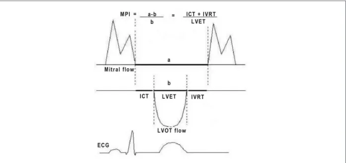
A new index for assessing the ventricular systo-diastolic function has been recently proposed, the MPI. Obtained by using Doppler echocardiography, and combining systolic and diastolic intervals of the cardiac cycle, the MPI value is lower than 0.39 ± 0.05 in normal individuals16, and increases as left ventricular function worsens (Figure 1). That index allows assessing ventricular function at rest or during pharmacological stress17.
The MPI does not require the analysis of ventricular borders, which is indispensable for calculating EF. Its measurement depends only on transmitral and transaortic flows, consisting
in adding the left ventricular isovolumetric relaxation and contraction times, divided by LVET. It is adequate for diagnosing global cardiac dysfunction, because the greater its numerical value, the greater the grade of left ventricular dysfunction and worse the patient’s clinical status, being considered an indicator of high risk for congestive HF and death due to cardiac cause18. The MPI seems to be independent of the geometry of the chamber, of changes in heart rate or blood pressure19,20, and of the severity of mitral regurgitation, although it depends partially on variations in filling20. The MPI is a reproducible method that closely correlates with invasive measures of systolic and diastolic functions.
To determine mitral transvalvular flow, the volume sample was positioned at the level of the extremities of the mitral cusps, and a biphasic pattern (above baseline) was obtained. The beginning of the flow reflects mitral valve opening, the first wave corresponding to the rapid ventricular filling phase, and the second wave corresponding to atrial contraction. To determine aortic transvalvular flow, the volume sample was positioned immediately below the valvular plane, and a monophasic pattern (below baseline), corresponding to ventricular ejection, was obtained21,22.
• left ventricular systole (a) - interval between the end of the second diastolic wave and the beginning of the first diastolic wave in mitral transvalvular flow.
• left ventricular ejection time (b) - interval between the beginning and end of the wave in aortic transvalvular flow.
Statistical analysis
Data analysis was performed by using the SPSS (Statistical Package for Social Sciences) software for Windows, version 12,
and R: A language and environment for statistical computing23.
Figure 1 – Schematic representation for calculating the myocardial performance index, showing the mitral low (top) and aortic low (bottom) of the cardiac cycle, a:
All tests were performed considering bilateral hypotheses and adopting the significance level of α = 5%.
Initially, descriptive statistics was used to assess the frequency, median and interquartile interval of the variables studied. Quantitative data were shown as median and interquartile interval. Qualitative data were shown as frequencies.
Fisher exact test was used to assess the homogeneity of the proportions of men and women and of white and black/mixed individuals in the groups CG, TX0 and TX1. The nonparametric test was used, because the requirements for the parametric test were not met.
The medians of the variables age, height, weight, and BS in the three groups were compared by use of the Kruskal-Wallis test, and, in the presence of a statistically significant difference, that test was followed by the multiple comparison test to identify which groups differed. A nonparametric test was also used, because those variables had no normal distribution, and the normality of the data was tested by use of the Kolmogorov-Smirnov test24,25.
In our sample, the proportion between women and men in groups CG, TX0 and TX1 differed. Thus, the groups were not homogeneous regarding sex. The same did not apply to the variable ethnicity, according to which the groups were homogeneous (Table 1).
In addition, the groups did not statistically differ regarding age, weight, height, and BS, being thus similar (Table 2).
At the time of EMB, of the TX0 patients, 15 were NYHA functional class II and three were NYHA functional class III; of the TX1 patients, two were NYHA functional class I, nine were NYHA functional class II, and one was NYHA functional class III (Chart 1). Of the 30 transplanted patients studied, HR was as follows: no HR, 18 patients (60%); grade IA HR, four patients (13.3%); grade IB HR, six patients (20%); and grade II HR, two patients (6.6%).
The most frequent etiologies of heart disease with HT indication were as follows: idiopathic dilated cardiomyopathy (31%); Chagas cardiomyopathy (29%); ischemic cardiomyopathy (25%); valvular heart disease (7%); restrictive cardiomyopathy (4%); and undetermined cause (4%).
Results
This study’s sample comprised 47 patients as follows: 17 (36.2%) healthy individuals (CG); and 30 transplanted individuals, 18 (38.3%) in the TX0 group and 12 (25.50%) in the TX1 group.
Of the transplanted patients, six were excluded from the study and not considered in the statistical analysis as follows: one due to technically inadequate echocardiographic window; two due irregular heart rhythm; one due to the presence of PM; and two due to insufficient EMB material.
Some echocardiographic variables were also compared (Table 3).
Aiming at assessing the left ventricular systo-diastolic function by use of MPI, the LVS and the LVET of the cardiac cycle were assessed (Table 4).
A statistically significant difference was observed in the medians of the variables IVST, LVPWT, VM/bs, RWT and LVEF in the three groups as follows: the medians of the variables
IVST, LVPWT, and VM/bs were lower in the CG than those in the TX0; the median of the variable LVEF was greater in the CG than that in the TX0.
Regarding the variable RWT, its median in the CG was lower than those in the TX1 and TX0.
A statistically significant difference was also observed in the medians of the variables E’, EWDT and E/A in the three groups as follows: the median of the variable E’ was greater in the CG than that in the TX0; the median of the variable E/A was lower in the CG than those in the TX1 and TX0.
Regarding specifically the calculation of MPI, there was a statistically significant difference in the medians of the variables LVS, LVET and MPI between the three groups as follows: the median of the variable LVS was greater in CG than that in TX0; the median of the variable LVET was greater in CG than those in TX0 and TX1; the median of the variable MPI was lower in CG than those in TX0 and TX1. However, no difference in the medians of the two groups of transplanted patients was observed (Chart 2).
Assessing only the groups of transplanted patients, 40% had rejection and 60%, no rejection. Regarding the TX1, HR according to the ISHLT criteria was as follows: grade IB, 50%; grade IA, 33.33%; and grade II, 16.66%. No other HR grade was observed on EMB.
Discussion
Since the first HT was performed in South Africa in 1967, advances have provided longer survival to transplanted patients. The major improvement was the introduction, in the 1980s, of the immunosuppressive agent cyclosporine, which has boosted the interest in restarting the large-scale HT program, today limited due especially to the scarcity of donors available26.
In the era of cyclosporine, rejection began to cause less myocardial edema, and, thus, measuring left ventricular mass has become obsolete as an isolated criterion to assess HR27. Our study has shown greater medians of IVST, LVPWT and VM/bs for transplanted patients as compared with those in the CG below the limits established for ventricular hypertrophy; however, no statistical significance was observed for the median of LVDD and for the calculation of LVDV, allowing us to infer that the increase in ventricular mass in transplanted patients occurs due to ventricular wall thickening.
Left ventricular diastolic dysfunction can be assessed by use of E/A, EWDT, IVRT and E’.
In our case series, no statistically significant difference was observed between the medians of the CG, TX0 and TX1 regarding the analysis of IVRT; however, some statistically significant change in diastolic function could be identified, by comparing the medians and the interquartile intervals of the variables E’, EWDT and E/A between the three groups of patients.
Table 2 – Comparison between the groups of the medians, interquartile intervals and p-values of the personal variables
Variables
CG TX0 TX1
p-value* Median
(IQI)
Median (IQI)
Median (IQI)
Age (years) (29.5 – 44.0)34.0 (33.5 – 51.5)46.0 (32.5 – 54.5)39.0 0.354
Weight (kg) 70.0
(57.5 – 85.0)
66.5 (57.5 – 88.0)
61.0
(57.5 – 73.5) 0.661
Height (m) 1.65
(1.59 – 1.73)
1.66 (1.64 – 1.70)
1.69
(1.64 – 1.72) 0.821
BS (m2) 1.84
(1.58 – 1.98)
1.75 (1.62 – 2.03)
1.69
(1.63 – 1.88) 0.758
* Kruskal-Wallis test; CG: control group; TX0: transplanted patients without rejection; TX1: transplanted patients with rejection; IQI: interquartile interval (1st quartile – 3rd quartile); BS: body surface.
Chart 1 – Distribution of the 30 transplanted patients according to NYHA functional class (I, II and III) and presence of heart rejection. TX0: transplanted patients without
rejection; TX1: transplanted patients with rejection.
Table 1 – Frequency and percentage of patients in the groups according to sex and ethnicity
GC TX0 TX1
p-value*
N (%) N (%) N (%)
Sex Female 8 (47.1) 1 (5.6) 2 (16.7) 0.012
Male 9 (52.9) 17 (94.4) 10 (83.3)
Ethnicity White 15 (88.2) 16 (88.9) 12 (100.0) 0.660
Black/Mixed 2 (11.8) 2 (11.1) 0 (0.0)
Assessing diastolic function is more sensitive in detecting rejection, because, according to the literature, diastolic changes precede any evidence of systolic dysfunction9. This is in accordance with data from this case series, whose medians of global left ventricular systolic function were increased (LVEF > 50%) in all groups assessed.
Episodes of rejection are frequent after HT, and, when repeated, can cause fibrosis and ventricular cavity changes. By use of dobutamine, Bellotti et al.28 have reported normal contractility in transplanted patients with no rejection; however, in the presence of rejection, contractility was reduced29, suggesting that dobutamine echocardiography could aid in correlating with episodes of rejection.
The accuracy of MPI and its comparison with the method considered gold standardfor quantifying EF have been studied by
Lax et al.30, who have developed the formula, EF = 60 - (34 x MPI), transforming MPI into a widely known variable among clinicians. In recent years, a lot of effort has been put into discovering a non-invasive technique to replace EMB in patients undergoing HT. Because systolic and diastolic dysfunctions are present during episodes of HR, the Tei index has been investigated as a potential predictor of acute rejection31.
In our case series, MPI was greater in TX0 and in TX1 than in CG, both with statistical significance. In addition, MPI was greater in TX1 than in TX0, suggesting a possible worsening of systo-diastolic function in cases of HR; that hypothesis, however, could not be confirmed, because there was no statistical significance. Bellotti et al.28 have reported that, in the presence of an episode of rejection, the patients can have a reduction in the contractility reserve.
Table 3 – Echocardiographic characteristics of the patients studied
Variables
CG TX0 TX1
p-value* Post Hoc+
Median (IQI) Median (IQI) Median (IQI)
LVDD (mm) 48.0
(44.0 – 50.0)
45.3 (42.7 – 47.3)
46.0
(40.2 – 51.8) 0.344 –
LVDV (ml)
107.5 (87.6 – 118.2)
93.9 (81.9 – 105.5)
97.4
(71.0 – 128.7) 0.344 –
IVST (mm)
8.0 (7.5 – 9.0)
10.7 (8.3 – 12.0)
9.0
(8.0 – 11.7) 0.002
CG ≠ Tx0
CG = Tx1
Tx0 = Tx1
LVPWT (mm)
8.0 (7.5 – 8.5)
10.4 (8.3 – 11.0)
9.5
(7.5 – 11.7) 0.006
CG ≠ Tx0
CG = Tx1
Tx0 = Tx1
RWT 0.32
(0.31 – 0.34)
0.46 (0.35 – 0.50)
0.42
(0.36 – 0.46) <0.001
CG ≠ Tx0 CG ≠ Tx1
Tx0 = Tx1
VM/bs (g/m²)
89.05 (70.9 – 101.2)
116.6 (90.8 – 120.8)
102.3
(84.2 – 149.8) 0.010
CG ≠ Tx0
CG = Tx1
Tx0 = Tx1
LVEF (%)
61.0 (58.0 – 64.0)
55.5 (40.5 – 60.2)
55.5
(51.0 – 62.2) 0.014
CG ≠ Tx0
CG = Tx1 Tx0 = Tx1
EWDT (ms)
206.0 (184.0 – 240.0)
180.0 (104.0 – 216.0)
167.5
(126.2 – 190.7) 0.030
CG = Tx0
CG ≠ Tx1
Tx0 = Tx1
IVRT (ms) 104.0
(88.0 – 116.0)
96.0 (80.0 – 107.2)
91.0
(80.0 – 101.0) 0.209 –
E/A (cm/s)
1.46 (1.20 – 1.79)
1.86 (1.57 – 3.14)
2.23
(2.16 – 3.00) 0.001
CG ≠ Tx0
CG ≠ Tx1
Tx0 = Tx1
E’ (cm/s)
17.0 (14.6 – 17.7)
13.0 (11.1 – 15.3)
15.6
(11.3 – 16.2) 0.038
CG ≠ Tx0
CG = Tx1 Tx0 = Tx1
mit E/E’ (3.7 – 5.3)4.3 (4.3 – 6.1)4.7 (4.0 – 6.7)5.3 0.283 –
Chart 2 – Top: Box plot of the variables used to calculate the myocardial performance index. Left: box plot of the left ventricular systole (LVS) (p = 0.008), and right: box plot of the left ventricular ejection time (LVET) (p < 0.001) of control patients, and transplanted patients without rejection (TX0) and transplanted patients with rejection (TX1). Bottom: Box plot of the already analyzed myocardial performance index. TX0: transplanted patients without rejection; TX1: transplanted patients with rejection (p < 0.001)
LV
S
LV
E
T
M
P
I
Control Control
Control
Tx0 Tx1 Tx0 Tx1
Tx0 Tx1
Variables
GC TX0 TX1
p-value* Post Hoc+
Median (IQI)
Median (IQI)
Median (IQI)
LVS 416.0
(397.5 – 426.5)
384.0 (340.7 – 400.0)
406.6
(360.0 – 480.2) 0.008
GC ≠ Tx0
GC = Tx1
Tx0 = Tx1
LVET 298.0
(285.0 – 312.0)
246.6 (230.7 – 277.7)
245.0
(218.0 – 267.6) <0.001
GC ≠ Tx0
GC ≠ Tx1 Tx0 = Tx1
MPI 0.38
(0.29 – 0.44)
0.47 (0.43 – 0.56)
0.58
(0.52 – 0.74) <0.001
GC ≠ Tx0
GC ≠ Tx1
Tx0 = Tx1 MPI calculation; * Kruskal-Wallis test; IQI: interquartile interval (1st quartile – 3rd quartile); CG: control group; TX0: transplanted patients without rejection; TX1: transplanted patients with rejection; LVS: left ventricular systole; LVET: left ventricular ejection time; MPI: myocardial performance index
A possible explanation for the increased values of MPI lies in the fact that the LVET is shorter in transplanted patients, and much shorter in the presence of HR, because of the pathophysiology of the graft, with no significant change in the IVRT, corroborating the findings of the study by Toumanidis et al. in 200832.
The good correlation between echocardiographic changes and EMB results have suggested that Doppler echocardiography should be used as a first choice technique to aid in the non-invasive diagnosis of HR33.
Conclusion
It has been shown that MPI is increased in transplanted patients as compared with a control group, and no significant difference was observed between the two groups of transplanted patients. Echocardiography showed good accuracy to detect changes in the systo-diastolic function of
the transplanted heart; however, it did not prove to be reliable to replace BEM in the safe diagnosis of HR.
Potential Conflict of Interest
No potential conflict of interest relevant to this article was reported.
Sources of Funding
There were no external funding sources for this study.
Study Association
This article is part of the doctoral thesis submitted by Gabriel Antonio Stanisci Miguel, from Faculdade de Ciências Médicas da Universidade Estadual de Campinas (UNICAMP) - Comissão de Pós-Graduação em Cirurgia.
1. Mcalister FA, Ezekowitz JH, Hooton N, Vandermeer B, Spooner C, Dryden DM, et al. Cardiac resynchronization therapy for patients with left ventricular systolic dysfunction: a systematic review. JAMA.2007;297(22):2502-14.
2. Ministério da Saúde – Sistema de Informações hospitalares do SUS. Datasus. [acesso em 2009 fev 1]. Disponível em http://www.datasus.gov.br.
3. Miniati DN, Robbins RC, Reitz BA. Heart and heart-lung transplantation In: Braunwald E, Zipes DP, Libby P. Heart disease: a textbook of cardiovascular medicine. 6th ed. Philadelphia: W B Saunders Company; 2001. p. 615-34.
4. Kostakis AJ, White DJG, Calne RY. Prolongation of the rat heart allograft survival by cyclosporine A. IRCS Med Sci. 1977;5:280.
5. Taylor DO, Edwards LB, Boucek MM, Trulock EP, Keck BM, Hertz MI. The registry of International Society for Heart and Lung Transplantation: twenty-first. Official adult heart transplant report-2004. J Heart Lung Transplant. 2004;23(7):796-803.
6. Bocchi EA, Fiorelli AI. The first guidelines group for heart transplantation of the Brazilian Society of Cardiology. The Brazilian experience with heart transplantation: a multicenter report. J Heart Lung Transplant. 2001;20(6):637-45.
7. Resende MV, Vieira ML, Bacal F, Andrade JL, Stolf NA, Bocchi EA. Tissue doppler echocardiography in the diagnosis of heart transplantation rejection. Arq Bras Cardiol. 2011;97(1):8-16.
8. Bacal F, Souza-Neto JD, Fiorelli AI, Mejia J, Marcondes-Braga FG, Mangini S, et al.; Sociedade Brasileira de Cardiologia. II Diretriz brasileira de transplante cardíaco. Arq Bras Cardiol. 2009;94(1 supl.1):e16-e73.
9. Valantine HA, Schnittger I. The role of echocardiography in the evaluation of patients after heart transplantation. In: Otto CM. (editor). The practice of clinical echocardiography. Philadelphia: W.B. Saunders Company; 2002. p. 658-78.
10. Lindenfeld J, Miller G, Shakar SF, Zolty R, Lowes BD, Wolfel EE, et al. Drug therapy in the heart transplant recipient. Part I: Cardiac rejection and immunosuppressive drugs. Circulation. 2004,110(24):3734-40.
11. Billingham ME, Cary NR, Hammond ME, Kemnitz J, Marboe C, McCallister HA, et al. A working formulation for the standardization of nomenclature in the diagnosis of heart and lung rejection: Heart Rejection Study Group. The International Society for Heart Transplantation. J Heart Transplant. 1990;9(6):587-93.
12. Sahn DJ, De Maria A, Kisslo J, Weyman A. Recommendations regarding quantitation in M-mode echocardiography: results of a survey of echocardiographic measurements. Circulation. 1978;58(6):1072-83.
13. Schiller NB, Shah PK, Crawford M, De Maria A, Devereux R, Feingenbaum H, et al. Recommendations for quantitation of the left ventricle by two-dimensional echocardiography. American Society of Echocardiography Committee on Standars, Subcommittee on quantitation of two-dimensional echocardiograms. J Am Soc Echocardiogr. 1989;2(5):358-67.
14. Lang RM, Bierig M, Devereux RB, Flachskampf FA, Foster E, Pellikkav PA, et al. Recommendations for chamber quantification: A report from the American Society of Echocardiography’s Guidelines and Standards Committee and the Chamber Quantification Writing Group, developed in conjunction with the European Association of Echocardiography, a branch of the European Society of Cardiology. J Am Soc Echocardiogr. 2005;18(12):1440-63.
15. Sociedade Brasileira de Cardiologia. Diretriz de angina estável. Arq Bras Cardiol. 2004;83(supl. 2):6-44.
16. Tei C, Ling LH, Hodge DO, Bailey KR, Oh JK, Rodeheffer RJ, et al. New index of combined systolic and diastolic myocardial performance: a simple and reproductible measure of cardiac function – a study in normal and dilated cardiomyopathy. J Cardiol. 1995;26(6):357-66.
17. Gulati VK, Katz WE, Follansbee WP, Gorcsan J 3rd. Mitral annular decent velocity by tissue Doppler echocardiography as an index of global left ventricular function. Am J Cardiol. 1996;77(11):979-84.
18. Oki T, Tabata T, Yamada H. Clinical application of pulsed Doppler tissue imaging for assessing abnormal left ventricular relaxation. Am J Cardiol. 1997;79(7):921-8.
19. Srivastava PM, Burell LM, Calafiore P. Lateral vs medial mitral annular tissue Doppler in the echocardiographic assessment of diastolic function and filling pressures: which should we use? Eur J Echocardiogr. 2005;6(2):97-106.
20. Schertel ER. Assessment of left ventricular function. Thorac Cardiovasc Surg. 1998;46 Suppl 2:248-54.
21. Azevedo J, García-Fernandez MA, Puerta P. Patterns pulsed Doppler tissue imaging of regional ventricular wall diastolic velocities in na normal population: its relation with the left ventricular Doppler inflow profile [abstract]. Eur Heart J. 1995;16(Suppl.):451.
22. Waggoner AD, Bierig SM. Tissue Doppler imaging: a useful echocardiographic method for the cardiac sonographer to assess systolic and diastolic ventricular function. J Am Soc Echocardiogr. 2001;14(12):1143-52.
23. R Development Core Team. R: A language and environment for statistical computing. R Development Core Team: a language and environment for statistical computing. Vienna (Austria): Foundation for Statistical Computing; 2008. URL http://www.R-project.org.
24. Noether GE, Dulker M. Introduction to statistics: the nonparametric way. New York: Springer-Verlag; 1990.
25. Siegel C, Castellan NJ Jr. Non parametric statistics for the behavioral sciences. New York: McGraw Hill Int; 1988. p. 213-4.
26. Abensur H. Transplante cardíaco. In: Silva CES. Ecocardiografia: princípios e aplicações clínicas. Rio de Janeiro (RJ): Revinter; 2007. p. 1017-21.
27. St Goar FG, Gibbons R, Schnittger I, Valantine HA, Popp RL. Left ventricular diastolic function. Doppler echocardiographic changes soon after cardiac transplantation: Circulation. 1990;82(3):872-8.
28. Bellotti G, Moraes AV, Bocchi EA, Graziozi P, Medeiros CC, Cerri GG, et al. Efeitos da rejeição na reserva de contratilidade do enxerto após o transplante cardíaco. Arq Bras Cardiol. 1996;67(1): 5-9.
29. Salles AF, Machado CV, Cordovil A, Leite WA, Moisés VA, de Almeida DR, et al. A elevação da pressão arterial sistólica durante o teste ergométrico após transplante cardíaco: correlação com o quadro clínico e a função ventricular avaliada pela ecocardiografia sob estresse com dobutamina. Arq Bras Cardiol. 2006;87(5):628-33.
30. Lax JA, Bermann AM, Cianciulli TF, Morita LA, Masoli O, Prezioso HA. Estimation of the ejection fraction in patients with myocardial infarction obtained from thecombined index of systolic and diastolic left ventricular function: a new method. J Am Soc Echocardiogr. 2000;13(2):116-23.
31. Mooradian S, Goldberg C, Crowley D, Ludomirsky A. Evaluation of noninvasive index of global ventricular function to predict rejection after pediatric cardiac transplantation. Am J Cardiol. 2000;86(3):358-60.
32. Toumanidis S Th, Papadopoulou ES, Saridakis NS, Agapitos E, Nanas JN, Stamatelopoulos SF. The myocardial performance index in cardiac transplantation. Hellenic J Cardiol. 2002;43:194-201.