Authors
Salustiano Araujo1
Helton P. Lemes1
Danny A. Cunha1
Vinicius S. Queiroz1
Daniela D. Nascimento1
Sebastião Rodrigues Ferreira Filho1
1Universidade Federal de
Uberlândia – UFU, Uberlândia, MG, Brazil
Submitted on: 01/10/2010. Approved on: 06/01/2011.
Corresponding author: Sebastião Rodrigues Ferreira Filho Rua Paraíba 3055, Umuarama
Uberlândia – MG – Brazil CEP: 38400-000 E-mail:
This study was carried out at Universidade Federal de Uberlândia, Minas Gerais, Brazil.
Authors declare no conflict of interest.
A
BSTRACTIn patients with chronic renal failure on hemodialysis, left ventricular hypertrophy is related to the increase in total peripheral vascular resistance and volume overload. The presence of residual diuresis enables greater control of the volemia of these. We evaluated the morpho-functional chan-ges of the left ventricle in patients with chronic kidney disease on hemodyalisis treatment with and without residual diu-resis. A total of 31 non diabetic patients were studied and they were divided into two groups: with residual diuresis (RD+) (n = 17) and without residual diuresis (RD-) (n = 14). In both groups, RD+ vs. RD-, using data from a Doppler echocar-diogram differences were found, respecti-vely, in the cardiac index (3.9 ± 0.2 vs. 3.0 ± 0.2 L/min/m2; p = 0.0056), systolic index (54 ± 2.9 vs. 45 ± 3.3 mL/b/m2; p = 0.04), end diastolic volume (141 ± 6.7 vs. 112 ± 7.6 mL; p = 0.008), end diastolic diameter (52 ± 0.7 vs. 48 ± 1.1 mm; p = 0.0072) and total peripheral resistance index (1121 ± 56 vs. 1529 ± 111 dyne.sec.cm-5; p = 0.001). RD+ had lower relative wall thickness than RD- (0.38 ± 0.01 vs. 0.45 ± 0.01; p = 0.0008). The ejection fraction and the left ventricular mass index were similar in both groups. The urinary 24-hour volume correlated with the rela-tive wall thickness (r = -0.42; p = 0.0186) and with peripheral resistance index (r = -0.48; p = 0.0059). In conclusion, there were distinct ventricular geometric patterns and different functional performances be-tween RD+ and RD- groups. The presence of residual diuresis can be responsible by these modifications in systolic function.
Keywords: chronic kidney failure, ventri-cular remodelling, stroke volume.
[J Bras Nefrol 2011;33(1): 60-67]©Elsevier Editora Ltda.
Cardiac morphology and function in patients with and
without residual diuresis on hemodialysis
I
NTRODUCTIONP
ATIENTSA
NDM
ETHODSPATIENTS/PROTOCOL
This is a cross-sectional study whose patients were re-cruited in a private hemodialysis clinic. CKD patients who received regular hemodialysis treatment three times a week and agreed to participate in the study met the inclusion criteria. The exclusion criteria were patients in hemodialysis treatment for less than three months, patients with a previous clinical history of cardiovas-cular disease, diabetic patients and uncontrolled blood pressure. Using the echocardiogram, those patients with an ejection fraction ≤ 55% and with segmental changes in LV contractility were also excluded 22,23. Thus, two groups were created: RD+ comprised pa-tients with daily residual diuresis ≥ 100 mL/24 hrs (n = 17; 11♂ and 6♀), and RD- comprised patients wi-th daily residual diuresis < 100 mL/24 hrs (n = 14; 9♂ and 5♀) (Table 1). The following clinical and labora-tory data of the groups were assessed: systolic arterial pressure (SAP), diastolic arterial pressure (DAP), length of hemodialysis treatment (LHT), urinary 24-hour vo-lume (UV24hs), hemoglobin (Hb), serum calcium (Ca), parathormone (PTH), serum albumin (Alb) and total
peripheral vascular resistance index (TPVRi). This stu-dy was previously approved by the Ethics Committee of the Universidade Federal de Uberlândia.
METHODS
The SAP and DAP were immediately obtained before the HD session using the arm opposite the AV fistu-la and represented the average of the fistu-last ten HD ses-sions. Mean arterial blood pressure (MAP) was calcu-lated using the formula DAP + (SAP-DAP/3), and the total peripheral vascular resistance index (TPVRi) was calculated using the formula (MAP-CVP/CD) x 80, where CVP is the central venous pressure, which was always considered a zero value (dyne.seg.cm-5).24;25 The pressure values were expressed in mmHg. The UV24hs (mL) was collected during the interdialytic period. Interdialytic weight gain (iWG) represents the diffe-rence between body weight immediately after the HD session, and the weight obtained immediately before the next HD session. The iWG value was considered the arithmetic average of the last ten HD sessions. The assessment of adequacy of dialysis was done using the Kt/V index. Kt/V is defined as the dialyzer clearance of urea (K, obtained from the manufacturer in mL/min),
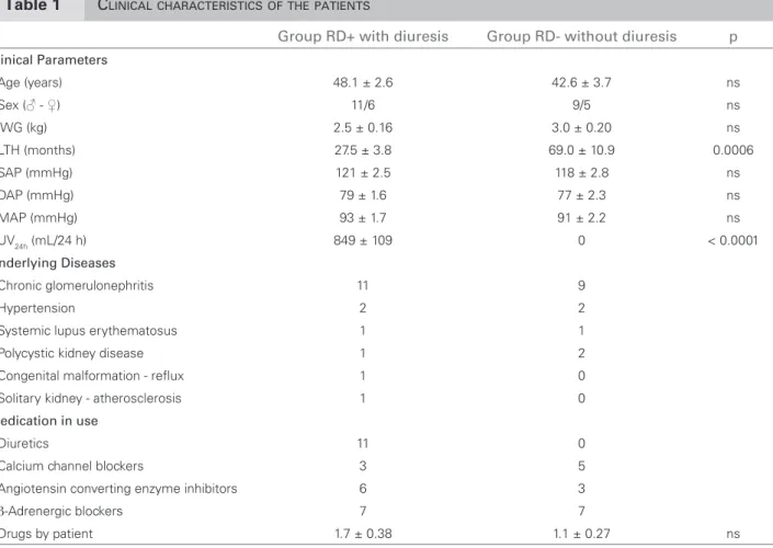
Table 1 CLINICALCHARACTERISTICSOFTHEPATIENTS
Group RD+ with diuresis Group RD- without diuresis p
Clinical Parameters
Age (years) 48.1 ± 2.6 42.6 ± 3.7 ns
Sex (♂ - ♀) 11/6 9/5 ns
iWG (kg) 2.5 ± 0.16 3.0 ± 0.20 ns
LTH (months) 27.5 ± 3.8 69.0 ± 10.9 0.0006
SAP (mmHg) 121 ± 2.5 118 ± 2.8 ns
DAP (mmHg) 79 ± 1.6 77 ± 2.3 ns
MAP (mmHg) 93 ± 1.7 91 ± 2.2 ns
UV24h (mL/24 h) 849 ± 109 0 < 0.0001
Underlying Diseases
Chronic glomerulonephritis 11 9
Hypertension 2 2
Systemic lupus erythematosus 1 1
Polycystic kidney disease 1 2
Congenital malformation - reflux 1 0
Solitary kidney - atherosclerosis 1 0
Medication in use
Diuretics 11 0
Calcium channel blockers 3 5
Angiotensin converting enzyme inhibitors 6 3
β-Adrenergic blockers 7 7
multiplied by the duration of the dialysis treatment (t, in minutes) divided by the volume of distribution of urea in the body (V, in mL). Biochemical plasma values were obtained using standardized methods that have been previously described.26-28 The Doppler echocardiogram used the Acuson–Aspen ® C2-4 Mhz transducer conducted in the interdialytic period. The exam was performed twenty hours after finishing the hemodialysis session, in the interdialytic period, by an experienced examiner unaware of the protocol. The Doppler echocardiographic variables were analyzed according to the criteria from the American Society of Echocardiography.22;29-31 For determination of pos-terior relative wall thickness (RWT) it was used the measurement of posterior wall thickness (PWT) of the left ventricle.32-35 The following parameters were also evaluated: the ejection fraction (EF), systolic index (SI), cardiac index (CI), end diastolic volume of the left ven-tricle (EDV), end diastolic diameter of the left venven-tricle (EDD), interventricular septum thickness (IVS) and left ventricular mass index (LVMi) 22,24,35. Additionally, the blood flow of the arteriovenous fistula (BFAV) was as-sessed with the same echocardiograph equipment.37;38
STATISTICALANALYSIS
For the statistical analysis, we used the GraphPad Prism program, version 5.0, for Windows (GraphPad Software, San Diego California USA). We conduct-ed t-tests and Wilcoxon tests to compare averages between the groups. Values lower than 0.05 were considered significant. The correlation coefficient used was Pearson’s correlation coefficient. The data were presented in average ± standard error (Χ ± SE). A multiple regression test was performed when H0: vascular resistance was not a dependent variable of treatment time and/or residual diuresis and/or PTH and/or mean arterial pressure; while H1: vascular re-sistance was dependent on at least one of those vari-ables cited above. For these calculations, the Bioestat 5.0 was used and alpha error = 0.05 was considered as a decision level.
R
ESULTSClinical and laboratory characteristics of both groups are presented in Tables 1 and 2.
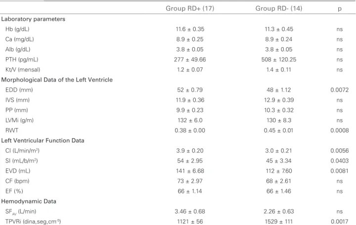
Group RD+ (17) Group RD- (14) p
Laboratory parameters
Hb (g/dL) 11.6 ± 0.35 11.3 ± 0.45 ns
Ca (mg/dL) 8.9 ± 0.25 8.9 ± 0.24 ns
Alb (g/dL) 3.8 ± 0.05 3.8 ± 0.05 ns
PTH (pg/mL) 277 ± 49.66 508 ± 120.25 ns
Kt/V (mensal) 1.2 ± 0.07 1.4 ± 0.11 ns
Morphological Data of the Left Ventricle
EDD (mm) 52 ± 0.79 48 ± 1.12 0.0072
IVS (mm) 11.9 ± 0.36 12.9 ± 0.39 ns
PP (mm) 9.9 ± 0.23 10.3 ± 0.32 ns
LVMi (g/m) 132 ± 6.0 130 ± 8.3 ns
RWT 0.38 ± 0.00 0.45 ± 0.01 0.0008
Left Ventricular Function Data
CI (L/min/m2) 3.9 ± 0.20 3.0 ± 0.21 0.0056
SI (mL/b/m2) 54 ± 2.95 45 ± 3.34 0.0403
EVD (mL) 141 ± 6.68 112 ± 7.60 0.0081
CF (bpm) 73 ± 2.97 68 ± 2.61 ns
EF (%) 66 ± 1.14 66 ± 1.46 ns
Hemodynamic Data
SFAV (L/min) 3.46 ± 0.68 2.26 ± 0.63 ns
TPVRi (dina,seg,cm-5) 1121 ± 56 1529 ± 111 0.0017
ns = p > 0.05. Hb: hemoglobin, Ca: calcium, Alb: albumin, PTH: parathyroid hormone, EDD: end diastolic diameter, IVS: intraventricle septum, PWT: posterior wall thickness; LVMi: left ventricular mass index; RWT: relative wall thickness, CI: cardiac index; SI: systolic index, EDV: end diastolic volume; HR: heart rate; EF: ejection fraction; BFAV: fistulae blood flow; TPVRi: total peripheral resistance index.
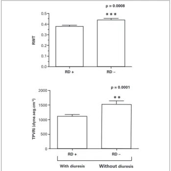
Figure 1. Relative wall thickness (RWT) and total peripheral vascular resistance index (TPVRi) in RD+ (with diuresis) and RD- (without diuresis) groups.
RD +
RWT
* * *
0.5 0.4 0.3 0.2 0.1 0.0
RD – p = 0.0008
RD +
With diuresis
TPVRi (dyne.seg.cm
-5) * *
2000
1500
1000 500
0
RD –
Without diuresis p = 0.0001
0.60
0.40
0.20
0,00 2500
2000
1500
1000
500
0
0 500 1000 1500 2000
Figure 2. Residual diuresis (RD), total peripheral vascular resistance index (TPVRi) and relative wall thickness (RWT) correlations.
r = - 0.49 p = 0.0054 r = - 0.42 p = 0.01
RWT
TPVRi (dyne.seg.cm
5)
Residual diuresis 24 hs (ml)
MORPHOLOGICALDATAOFTHELEFTVENTRICLE
The relative wall thickness was significantly lower in group RD+ than in group RD- (0.38 ± 0.01 vs. 0.45 ± 0.01; p = 0.0008) (Figure 1). Significant correla-tions were found between relative wall thickness and: hemodialysis time (r = 0.40; p = 0.024), systolic in-dex (r = - 0.61; p = 0.0003), cardiac inin-dex (r = -0.56; p = 0.001) and UV24hs (r = -0.42; p = 0.01). The end diastolic diameter was higher in RD+ (52 ± 0.7 vs. 48 ± 1.1; p = 0.0072), and we found significant posi-tive correlations between end diastolic diameter and UV24hs (r = 0.41; p = 0.022) and negative correlations between end diastolic diameter and relative wall thi-ckness (r = -0.60; p = 0.0003).
It was observed a significant positive correlation between the systolic index and left ventricular mass index (r = 0.51; p = 0.0039), between the total pe-ripheral resistance index and relative wall thickness (r = 0.51; p = 0.0036) and systolic index and relative wall thickness (r = - 0.61; p = 0.0003). The TPVRi correlated with the CI, SI, RWT and UV24h (Figures 2 and 3) (Table 2). For the multiple regression analy-sis the TPVRi was considered as a dependent variable while time of treatment, residual diuresis, PTH and MAP were considered as variables that could influ-ence the peripheral resistance index. We found that F regression = 4.26, p = 0.041, rejected H0 and the following partial coefficients: time (t: -1.47; p = 0.152); residual diuresis (t: -3.35; p = 0.0023); MAP (t: 0.490; p = 0.490) and PTH (t: = 0.498 p = 0.6225).
LEFT VENTRICULAR FUNCTIONAND HEMODYNAMIC
DATA
The cardiac index of the patients was significantly hi-gher in group RD+ than in group RD- (3.9 ± 0.2 vs. 3.0 ± 0.2 L/min/m2; p = 0.0056). A significant negative correlation between the cardiac index and relative wall thickness was observed (r = - 0.56; p = 0.001). The sys-tolic index was significantly higher in group RD+ (54 ± 2.9 vs. 45 ± 3.3 mL/b/m2; p = 0.0403), as was the end diastolic volume (141 ± 6 vs. 112 ± 7 mL; p = 0.0081). The total peripheral resistance index was significantly lower in group RD+ (1121 ± 56 vs. 1529 ± 111 di-na.seg.cm-5; p = 0.001) (Figure 1, Table 1). The BF
AV was not statistically different between groups RD+ and RD- (1.8 ± 0.6 vs. 1.4 ± 0.6 L/min/m2; p = ns).
D
ISCUSSIONFigure 3. Total peripheral vascular resistance index (TPVR), cardiac index (CI), systolic index (SI) and relative wall thickness (RWT) correlations.
0.60
0.40
0.20
0.00
RTW
r = 0.51 p = 0.0036
r = 0.66 p < 0.0001
r = - 0.86 p < 0.0001
100
80
60
20 40
0
0
0 500 1000 1500 2000 2500
2 4 6
SI (mlb/m2)
CI (l m
2)
wall thickness, suggesting that, for different values of resistance, there are different values of thickness on the posterior wall (Figure 2). The absence of residual diuresis may have contributed to higher peripheral resistance values and hypervolemic status in the anu-ric group. Although the interdialytic weight gain was not significant between the groups due to the small size of this sample, the group without diuresis were about 20 per cent heavier than the group with uri-nary volume. Our data also showed that low-volume diuresis coexisted with high values of peripheral resis-tance (Figure 2). Wang et al. found that left ventricu-lar mass increased in peritoneal dialysis patients who had lower glomerular filtration rates.9 However, these authors found similar end diastolic diameters among the assessed groups, which differs from the findings in our study. One of the possible reasons for these
different findings could be due to the type of dialysis, while another reason may be that the authors did not correlate modifications in the left ventricle with the residual 24-hour urinary volume of their patients.
With respect to the functional characteristics of the left ventricle, we observed that CI and SI were lower in the anuric patients (RD-) (Table 2). This fin-ding could be explained by the lower values found in the end ventricular diastolic diameter in RD- (Table 2). The morphological differences of left ventricles be-tween the two groups were responsible for distinct va-lues of cardiac and systolic indexes. Therefore, CKD patients on hemodialysis treatment who lose residual diuresis may have changes in the geometry and func-tion of the left ventricle.
Although Faguli et al. demonstrated LVH in the majority of the hemodialyzed patients studied, these authors did not exclude patients with pathologies that could interfere with the myocardium remodeling pro-cess, such as diabetes mellitus, ischemic cardiomyopa-thy or systemic arterial hypertension; further, they did not correlate their findings with the residual urinary volume.40
In CKD patients, RD progressively decreased with time in replacement therapy. In our study, the anuric group (RD-) spent more time in HD treatment than the group with dieresis (RD+), and the loss of urina-ry volume only happened in patients with the largest length time on treatment. The reasons for losing the residual diuresis may be explained by the changes re-lated to the underlying disease that caused the CKD or by a continuous inflammatory process, anemia and associated co-morbidities.12,17,41-43 The length of time spent on hemodialysis could increase the poten-tial risks to the left ventricle. These risks come from the treatment itself and from the progression of the illness. However, applying multiple regression and defining the load imposed on the left ventricle as a dependent variable we found a statistical significance only for residual diuresis (p = 0.05). The length of time spent on dialysis therapy, PTH and mean arterial pressure did not have significant values. These data were more relevant because patients were normoten-sive, thus the volume component of blood pressure could be greater than the resistive component in these patients.
In this study, the morpho-functional cardiac chan-ges could not be attributed to the blood pressure levels found, because the groups RD+ and RD- de-monstrated similar diastolic and systolic blood pres-sure values (Table 1). The same could be said with regard to the use of anti-hypertensive drugs, as there
was no difference in the number of hypertensive me-dications used in both groups (Table 1). Gunal et al. demonstrated that the use of anti-hypertensive drugs contributed to LVMi reductions in only 6% of the patients on hemodialysis (11). However, our results demonstrated that both groups RD+ and RD- did not show differences in their LVMi values (p > 0.05) (Table 2), indicating that the effect of the drugs used did not substantially interfere with our results.
The presence of anemia is another factor that could be related to the presence of LVH.44;45 The role of anemia as a cause of LVH in patients undergoing dialysis has been well established; correction of ane-mia with erythropoietin results in the partial regres-sion of LVH (46). In this study, the Hb level remai-ned relatively normal, and there was no difference in Hb level between the groups studied (Table 2).
It has already been shown that hypoalbumi-nemia is involved in ventricular hypertrophy and dilatation, and in heart failure, and, ultimately de-creases patients’ survival.47-51 The CANUSA study demonstrated that serum Alb concentration strongly correlated with mortality.3,52 In our study, the se-rum Alb values were normal; we did not find sig-nificant differences between the two groups (Table 2). Therefore, we believe that the cardiac changes found in our study could not have been caused by this variable. Persistent hypocalcemia is considered a pathogenic factor in the reduction of left ventricular systolic function.53 However, serum calcium in our study remained at normal values; there was no diffe-rence between the two groups (Table 2). Secondary hyperparathyroidism is common in uremic patients, and this complication has contributed to the develo-pment of left ventricular systolic dysfunction54 and LVH.55 PTH serum levels between the groups we-re statistically non significant (Table 2). The BFAV is another important factor that contributes to cardiac morphology and function.38;56 In our study, all fistu-las were examined by using their blood flows; there was no statistically significant difference between the groups (Table 2). In addition, morphological and functional changes may be attributed to the different removal rates of solutes in the hemodialysis process; however, they were not different in RD+ and RD-. It is worth noting that the Kt/V measurement in our study did not take into account solute removals pro-moted by residual diuresis (Table 2).
Based on our study findings, we conclude that the different CI and SI values found in these groups can be attributed to distinct ventricular geometric patterns that are determined by total peripheral
resistance and by the presence of residual diuresis. Therefore, the preservation of diuresis appears to in-fluence left ventricular morphology and function in patients with chronic kidney failure.
As limitations of this study it is necessary to ob-serve that it is a cross sectional study and the data of the patients represent the moment when the va-riables were observed, and they could not express the exactly progression of the patient disease. Beside this, the small size of samples could induce an alpha error in its statistics analyses.
We believe that more prospective studies should be conducted in order to confirm such a hypothesis.
R
EFERENCES1. Wang AY. The “heart” of peritoneal dialysis. Perit Dial Int 2007;2:S228-S232.
2. Maiorca R, Brunori G, Zubani R et al. Predictive value of dialysis adequacy and nutritional indices for mortality and morbidity in CAPD and HD patients. A longitudinal study. Nephrol Dial Transplant 1995; 10:2295-305. 3. Adequacy of dialysis and nutrition in continuous
peri-toneal dialysis: association with clinical outcomes. Canada-USA (CANUSA) Peritoneal Dialysis Study Group. J Am Soc Nephrol 1996; 7:198-207.
4. Termorshuizen F, Korevaar JC, Dekker FW, van Manen JG, Boeschoten EW, Krediet RT. The relative impor-tance of residual renal function compared with perito-neal clearance for patient survival and quality of life: an analysis of the Netherlands Cooperative Study on the Adequacy of Dialysis (NECOSAD)-2. Am J Kidney Dis 2003; 41:1293-302.
5. Nolph KD, Prowant BF, Moore HL, Reyad SE. Hematocrit and residual renal creatinine clearance in patients undergoing continuous ambulatory peritoneal dialysis (CAPD). Perit Dial Int 1990; 10:279-82. 6. K/DOQI clinical practice guidelines for chronic kidney
disease: evaluation, classification, and stratification. Am J Kidney Dis 2002; 39:S1-266.
7. Foley RN, Parfrey PS, Harnett JD, Kent GM, Murray DC, Barre PE. The prognostic importance of left ven-tricular geometry in uremic cardiomyopathy. J Am Soc Nephrol 1995; 5:2024-31.
8. Levy D, Garrison RJ, Savage DD, Kannel WB, Castelli WP. Prognostic implications of echocardiographically determined left ventricular mass in the Framingham Heart Study. N Engl J Med 1990; 322:1561-6.
9. Wang AY, Wang M, Woo J et al. A novel association between residual renal function and left ventricular hypertrophy in peritoneal dialysis patients. Kidney Int 2002; 62:639-47.
10. Parfrey PS, Harnett JD, Griffiths SM et al. The clini-cal course of left ventricular hypertrophy in dialysis pa-tients. Nephron 1990; 55:114-20.
12. Wang AY, Wang M, Woo J et al. Inflammation, residu-al kidney function, and cardiac hypertrophy are interre-lated and combine adversely to enhance mortality and cardiovascular death risk of peritoneal dialysis patients. J Am Soc Nephrol 2004; 15:2186-94.
13. Verdecchia P, Carini G, Circo A et al. Left ventricu-Left ventricu-lar mass and cardiovascuventricu-lar morbidity in essential hy-pertension: the MAVI study. J Am Coll Cardiol 2001; 38:1829-35.
14. Lang SM, Bergner A, Topfer M, Schiffl H. Preservation of residual renal function in dialysis patients: effects of dialy-sis-technique-related factors. Perit Dial Int 2001; 21:52-7. 15. Horinek A, Misra M. Does residual renal function de-cline more rapidly in hemodialysis than in peritoneal di-alysis? How good is the evidence? Adv Perit Dial 2004; 20:137-40.
16. Lysaght MJ, Vonesh EF, Gotch F et al. The influence of dialysis treatment modality on the decline of remaining renal function. ASAIO Trans 1991; 37:598-604. 17. Marron B, Remon C, Perez-Fontan M, Quiros P, Ortiz
A. Benefits of preserving residual renal function in peri-toneal dialysis. Kidney Int Suppl 2008; 108:S42-S51. 18. Levin A. Clinical epidemiology of cardiovascular
dis-ease in chronic kidney disdis-ease prior to dialysis. Semin Dial 2003; 16:101-5.
19. McMahon LP, Roger SD, Levin A. Development, pre-vention, and potential reversal of left ventricular hy-pertrophy in chronic kidney disease. J Am Soc Nephrol 2004; 15:1640-7.
20. Harnett JD, Parfrey PS, Griffiths SM, Gault MH, Barre P, Guttmann RD. Left ventricular hypertrophy in end-stage renal disease. Nephron 1988; 48:107-15.
21. London GM, Parfrey PS. Cardiac disease in chronic uremia: pathogenesis. Adv Ren Replace Ther 1997; 4:194-211.
22. Lang RM, Bierig M, Devereux RB et al.
Recommendations for chamber quantification: a re-port from the American Society of Echocardiographys Guidelines and Standards Committee and the Chamber Quantification Writing Group, developed in conjunction with the European Association of Echocardiography, a branch of the European Society of Cardiology. J Am Soc Echocardiogr 2005; 18:1440-63.
23. Nishimura RA, Tajik AJ, Shub C, Miller FA, Jr., Ilstrup DM, Harrison CE. Role of two-dimensional echocar-diography in the prediction of in-hospital complications after acute myocardial infarction. J Am Coll Cardiol 1984; 4:1080-7.
24. Marchais SJ, Guerin AP, Pannier BM, Levy BI, Safar ME, London GM. Wave reflections and cardiac hy-pertrophy in chronic uremia. Influence of body size. Hypertension 1993; 22:876-83.
25. Ferreira Filho SR, de Castro Rodrigues FF, Oliveira PC, Nery MA. Systemic hemodynamic changes in older hypertensive patients after drinking water or eating a meal. Hypertension 2007;49:e31.
26. Burnett RW, Covington AK, Fogh-Andersen N et al. Use of ion-selective electrodes for blood-electrolyte analy-sis. Recommendations for nomenclature, definitions and conventions. International Federation of Clinical Chemistry and Laboratory Medicine (IFCC). Scientific Division Working Group on Selective Electrodes. Clin Chem Lab Med 2000; 38:363-70.
27. Chromy V, Rozkosna K, Sedlak P. Determination of serum creatinine by Jaffe method and how to calibrate to eliminate matrix interference problems. Clin Chem Lab Med 2008; 46:1127-33.
28. Javed MU, Michelangeli F. Enzymatic method for as-saying calcium in serum with Ca++ -ATPase. Exp Mol Med 2003; 35:17-22.
29. Harnett JD, Murphy B, Collingwood P, Purchase L, Kent G, Parfrey PS. The reliability and validity of echo-cardiographic measurement of left ventricular mass in-dex in hemodialysis patients. Nephron 1993; 65:212-4. 30. Schiller NB, Shah PM, Crawford M et al. Recommendations
for quantitation of the left ventricle by two-dimensional echocardiography. American Society of Echocardiography Committee on Standards, Subcommittee on Quantitation of Two-Dimensional Echocardiograms. J Am Soc Echocardiogr 1989; 2:358-67.
31. Sahn DJ, DeMaria A, Kisslo J, Weyman A. Recommendations regarding quantitation in M-mode echocardiography: results of a survey of echocardio-graphic measurements. Circulation 1978; 58:1072-83. 32. Ganau A, Devereux RB, Roman MJ et al. Patterns of
left ventricular hypertrophy and geometric remodel-ing in essential hypertension. J Am Coll Cardiol 1992; 19:1550-8.
33. Ford LE. Heart size. Circulation Res 1976; 39:297-303. 34. Gaash WH. LV radius to wall thickness ratio. Am J
Cardiol 1979;43:1189-94.
35. Fast JH. Limits of reproducibility of left ventricular wall thickness and mass by M-mode echocardiography. Neth J Med 1989; 34:297-301.
36. Zoccali C, Benedetto FA, Mallamaci F et al. Prognostic impact of the indexation of left ventricular mass in patients undergoing dialysis. J Am Soc Nephrol 2001; 12:2768-74.
37. Miller PE, Tolwani A, Luscy CP et al. Predictors of adequacy of arteriovenous fistulas in hemodialysis pa-tients. Kidney Int 1999; 56:275-80.
38. Robbin ML, Chamberlain NE, Lockhart ME et al. Hemodialysis arteriovenous fistula maturity: US evalu-ation. Radiology 2002; 225:59-64.
39. de SG. Left ventricular geometry and hypotension in end-stage renal disease: a mechanical perspective. J Am Soc Nephrol 2003; 14:2421-7.
40. Fagugli RM, Pasini P, Quintaliani G et al. Association between extracellular water, left ventricular mass and hypertension in haemodialysis patients. Nephrol Dial Transplant 2003; 18:2332-8.
41. Jacobs LH, van de Kerkhof JJ, Mingels AM et al. Inflammation, overhydration and cardiac biomarkers in haemodialysis patients: a longitudinal study. Nephrol Dial Transplant 2010; 25:243-8.
42. Krediet RT. How to preserve residual renal function in patients with chronic kidney disease and on dialysis? Nephrol Dial Transplant 2006; 21:42-ii46.
43. Yang PY, Lin JL, Lin-Tan DT et al. Residual daily uri-ne volume association with inflammation and nutrition status in maintenance hemodialysis patients. Ren Fail 2009; 31:423-30.
45. Stojimirovic B, Petrovic D, Obrenovic R. Left ventricu-lar hypertrophy in patients on hemodialysis: importan-ce of anemia. Med Pregl 2007; 60:155-9.
46. Silberberg J, Racine N, Barre P, Sniderman AD. Regression of left ventricular hypertrophy in dialysis patients following correction of anemia with recombi-nant human erythropoietin. Can J Cardiol 1990; 6:1-4. 47. Foley RN, Parfrey PS, Harnett JD, Kent GM, Murray
DC, Barre PE. Hypoalbuminemia, cardiac morbidi-ty, and mortality in end-stage renal disease. J Am Soc Nephrol 1996; 7:728-36.
48. Moon KH, Song IS, Yang WS et al. Hypoalbuminemia as a risk factor for progressive left-ventricular hyper-trophy in hemodialysis patients. Am J Nephrol 2000; 20:396-401.
49. Suda T, Hiroshige K, Ohta T et al. The contribution of residual renal function to overall nutritional sta-tus in chronic haemodialysis patients. Nephrol Dial Transplant 2000; 15:396-401.
50. Fine A, Cox D. Modest reduction of serum albumin in continuous ambulatory peritoneal dialysis patients is common and of no apparent clinical consequence. Am J Kidney Dis 1992; 20:50-4.
51. Struijk DG, Krediet RT, Koomen GC, Boeschoten EW, Arisz L. The effect of serum albumin at the start of con-tinuous ambulatory peritoneal dialysis treatment on pa-tient survival. Perit Dial Int 1994; 14:121-6.
52. Bargman JM, Thorpe KE, Churchill DN. Relative contribution of residual renal function and peritoneal clearance to adequacy of dialysis: a reanalysis of the CANUSA study. J Am Soc Nephrol 2001; 12:2158-62. 53. Feldman AM, Fivush B, Zahka KG, Ouyang P,
Baughman KL. Congestive cardiomyopathy in patients on continuous ambulatory peritoneal dialysis. Am J Kidney Dis 1988; 11:76-9.
54. Drueke T, Fauchet M, Fleury J et al. Effect of para-thyroidectomy on left-ventricular function in haemo-dialysis patients. Lancet 1980; 1:112-4.
55. London GM, De Vernejoul MC, Fabiani F et al. Secondary hyperparathyroidism and cardiac hyper-trophy in hemodialysis patients. Kidney Int 1987; 32:900-7.