Left Atrial Volume as an Index of Diastolic Function
Antônio Carlos Sobral Sousa 8QLYHUVLGDGH)HGHUDOGH6HUJLSHH+RVSLWDO6mR/XFDVGH$UDFDM~$UDFDM~6(%UD]LO
M a i l i n g A d d r e s s : C a r l o s S o b r a l S o u s a Ć $ Y ' H S 6 L O Y L R 7 H L [ H L U D ă $ U D F D M ~ 6 ( % U D ] L O
Email: [email protected] 5HFHLYHGLQ$FFHSWHGLQ Congestive heart failure (CHF) is one of the main
causes of death and hospital admissions in our country,
according to data from Datasus1. This clinical syndrome
is progressive and characterized by complex cardiac and systemic adaptations, which vary during disease evolution2.
It has been observed, however, that in approximately 30% to 50% of the individuals who develop CHF, the systolic function estimated through the left ventricular
ejection fraction (LVEF) is normal or relatively normal3.
Thus, the cause of cardiac decompensation in these patients is the left ventricular diastolic dysfunction, justifying the term “diastolic cardiac failure”4.
Little is known on the natural history of this disease, particularly regarding the mechanisms causing affected
patients’ death5, although its prevalence is known
among certain groups, such as the elderly and women, as well as the fact that it usually precedes the systolic dysfunction in most cardiac affections, including Chagas’
myocardiopathy.
There have been few studies published in literature regarding the incidence of CHF in patients with diastolic dysfunction evidenced by Doppler echocardiography. It has been demonstrated, in a population of individuals older than 65 years with no clinical evidence of cardiac disease, that the detection of this type of dysfunction through Doppler echocardiography has a predictive value for the development of CHF in 11 a 15% of the cases within a five-year period10.
D
IASTOLE AND TYPES OF DIASTOLIC DYSFUNCTIONAccording to the most generally employed clinical concept11, the diastolic phase of the cardiac cycle, starting with the closing of the semilunar valves, comprises the largest part of the active ventricular relaxation, with periods of isovolumetric relaxation, and rapid ventricular filling, as well as diastasis or passive filling and, finally, the period that involves atrial contraction. The illustration of this concept is depicted in Figure 1.
Even though several independent factors affect the diastolic properties of the left ventricle (LV) its actions converge to the transmitral pressure gradient, which, in fact, is the physical determinant of the left ventricular filling12.
During the isovolumetric relaxation period, the LV behaves as an isolated chamber, as the aortic and mitral valves are closed; thus, its volume is not altered when there is a progressive decrease of intracavitary pressure. The heart relaxation is also the main determinant of the rapid ventricular filling, which is caused by the opening of the mitral valve as a result of the pressure decrease inside the LV, which is lower than that observed in the left
atrium (LA)13. It is an energy-dependent process, which
corresponds to the active sequestration, contragradient, of calcium ions released from troponin during the contractile activation14.
The rapid ventricular filling, which under normal circumstances is responsible for 80% of the ventricular filling, is also due to the pressure in the left atrium at the moment of the mitral valve opening (preload) and elastic recoil (suction) of the left ventricle. This phenomenon occurs because the shortened muscle fibers at the end of the systole, together with the collagen matrix, act as a compressed coil to generate recoil forces at the initial
phase of the diastole14, resulting in a decrease of LV
pressure, despite the progressive increment of its volume. The LA emptying provides the manometric equalization between the left chambers, comprising the diastasis phase.
In this phase, which is influenced by the LV complacency (pressure/volume ratio), ventricular filling is basically originated from the pulmonary venous flow, as the LA behaves as a “passive conduit”, enabling the direct passage of blood from the pulmonary valves to the
LV15. The atrial contraction, which happens at the end
of the diastolic period, contributes for 15% to 20% of
ventricular filling in normal individuals16, and depends
on interactions of the LV with the pericardium and the right ventricle (RV), on the atrioventricular synchronism (PR interval of the electrocardiogram), on the cardiac rhythm (loss of the atrial contraction in the presence of arrhythmias such as atrial fibrillation) and on the LA and LV pressures14,17,18.
The normal LV must be capable of accommodating a significant volume of blood without causing the diastolic pressure elevation. Hence, the proportion of ventricular filling during the initial and final phases of diastole depends on the myocardial relaxation, the elastic retraction, the LV complacency and the LA pressure, which originate from
the interaction between heart disease and volemia14.
by mitral stenosis, LA myxoma, constrictive pericarditis
and cardiac tamponade.
E
VALUATION OF THE DIASTOLIC FUNCTION Part of the difficulty in studying the diastolic function of the LV is due to the fact that its main determinants (relaxation and complacency) act in different phases of the diastole, with an overlap of the time of action. In addition to undergoing mutual interaction, they are also influenced, as mentioned above, by the LV systolic function, by the heart frequency (HF) and the heart conduction system.The analysis of the ventricular filling pattern provides important information on the diastolic performance of the LV. Historically, the hemodynamic study through cardiac catheterism has been the reference standard
for the acquisition of such information21. However, its
invasive nature makes this methodology unfeasible in routine clinical practice. Diastolic function assessments can also be obtained by non-invasive techniques, such as the determination of the LV volume of dimensions during
the cardiac cycle, by radioisotope angiography22, or by
Doppler echocardiography12,23.
Many patients with CHF and preserved systolic function predominantly present the diastolic mechanisms that determine the symptoms of dyspnea and fatigue. In these individuals, the LV is not dilated and contracts normally;
however, the diastolic function is compromised. In
diastolic heart failure, the LV has decreased complacency and it is unable to fill adequately with normal pressures. This condition results in the reduction of the final diastolic volume, which causes the decrease in the systolic volume and symptoms of low cardiac output and/or final diastolic pressure elevation, which, in turn, determines the onset of symptoms of pulmonary congestion. Thus, the characteristics of CHF (incapacity of the heart in pumping blood to maintain the tissue metabolic needs, preserving the filling pressures) can be mainly due to lusitropic abnormalities.
There are three types of diastolic dysfunction: a) increase of ventricular rigidity, which is common in ischemic heart disease in the elderly, in cardiac amyloidosis and endomyocardiofibrosis; b) ventricular hypertrophy, of which the main cause is systemic arterial hypertension (SAH); c) mechanical obstructions, caused
Fig. 1 – Cardiac cycle divided in periods corresponding to the clinical definition and the conception of the heart as a muscular propulsion system, of the systolic (S) and diastolic (D) events; P=ventricular pressure curve; V=ventricular volume curve; IC=isovolumetric contraction; IR=isovolumetric relaxation; RVF= rapid ventricular filling period. Observe that, in the mechanical conception of the heart, the systole comprehends not only the contraction and ejection phases, but also the whole of the active relaxation period, including, as well, the rapid ventricular filling. Adapted from Marin-Neto and Sousa (1988).
CONTRACTION RELAXATION COMPLACENCY
IC EJECTION RI PER DIASTASIS
ATRIAL CONTRACTION CLINICAL DEFINITION
MUSCULAR PROPULSION
SYSTEM
The use of Doppler echocardiography in diastolic function assessment started in the seventies with
Gibson & Brown24, who developed an analysis method
of the continuous variation of the LV dimension, using a computerized system of reutilization of echocardiographic tracings. In our country, this technique was standardized by
Marin-Neto and Sousa25 and utilized in the demonstration
of early diastolic dysfunction in Chagas disease6, showing it to be reproducible, but also time-consuming, which, in a way, limits its routine utilization.
L
EFT ATRIUM VOLUMEAs mentioned before, alterations in the relaxation and complacency of the LV secondary to a defect in the actin-myosin interaction and increase in collagen deposition or the viscoelastic properties of the heart cause an elevation of the left ventricular end-diastolic pressure (LVDP) and consequently, elevation of pressure in the LA to maintain the ventricular filling26. The parietal tension increase leads to the dilation of the atrial chamber, which consequently
reflects the diastolic dysfunction of the LV27,28. The LA
behaves as a reservoir during the ventricular systole, as a conduit that allows the passage of blood from the pulmonary veins to the LV at the beginning of the diastole, and as an active contractile chamber at the end of the diastole.
During the diastolic period, this atrial chamber is directly exposed to the pressures of the LV through the
open mitral valve28, as shown in Fig. 2. Hence, the
size of the LA is greatly influenced by same factors that
determine ventricular filling26, so constituting a stable
parameter that reflects the duration and severity of
the lusitropic dysfunction. Consequently, it has been
considered that the LA dimension is a potent predictor
of adverse events in several clinical situations such as: a) ischemic stroke30; b) chronic atrial fibrillation (AF)31; c) left ventricular failure32; d) mitral regurgitation33 and
e) diastolic dysfunction.
The routinely available methods for the determination of the LA size through Doppler echocardiography are the measurement of the anteroposterior dimension obtained at the parasternal projection of its long axis and volume calculation, also utilizing the apical two-chamber and the
apical four-chamber views34. The uniplane measurement
of the anteroposterior dimension has decreased accuracy and low reproducibility in the quantification of the left atrial dimension, which are caused by technical limitations such as the ultrasound beam angulation, the irregular geometry of the LA and the fact that the growth of this chamber is not a uniform one, due to the physical limitation imposed
by the sternum and vertebral column35.
This can, in part, explain the conflicting results described in the specialized literature regarding the size of the LA when assessed by its anteroposterior dimension, as a variable, in order to establish the prognosis in certain clinical situations; in patients with chronic AF, who took
part in the AFASAK36 study, the anteroposterior dimension
did not have a predictive value for thromboembolism
(TE), whereas another study (SPAF)37 showed that this
variable was the best predictor for stroke, when assessing a similar population.
On the other hand, the LA volume (LAV) can generally
be obtained by several means: a) the cube38 method,
according to which the atrial chamber has a spherical
shape, through the following formula: LA=4/3 S x
(anteroposterior dimension/2)3; b) ellipsoid method,
assuming an elliptical shape of the LA, according to the
formula: 4/3 S x anteroposterior dimension/2 x lenght/2
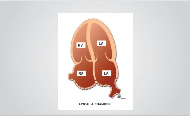
x mediolateral dimension/2, in which the length is the Fig. 2 – Schematic representation of the bidimensional echocardiogram obtained at the apical four-chamber position. Observe that in the diastolic period, with the mitral valve open, the left atrium is directly exposed to the filling pressures of the left ventricular chamber.
RV LV
RA
APICAL 4 CHAMBER
LA
distance between the point of coaptation of the mitral valve cuspids and the upper wall of the LA and the mediolateral dimension is the transverse dimension, obtained at the apical 4-chamber position. All calculations must be performed at the end of the systole; c) the biplane
area-length measurement40, using the formula 0.85 x
4-chamber area x 2-chamber area/perpendicular axis, in which the areas are obtained at the apical positions, by excluding the left atrial appendix and the confluence of the pulmonary veins; the perpendicular length is measured between the plane of the mitral valve (MV) ring and the upper portion of the LA; d) uniplane or biplane Simpson’s
disks method41, in which the technician performs the
tracing of the left atrial endocardium at the apical 2- and 4-chamber positions, as shown in Fig. 3.
The methodologies for measurement of the left atrium volume (LAV) that best apply to clinical practice are
those that utilize Simpson’s technique, either uniplane34
or biplane42.
Lester and cols.34 observed that the uniplane
technique, derived from the apical 4-chamber position, represents an accurate measurement of the factual size of the LA. According to Schiller & Foster43, the LA volume, normalized for the body surface, is an index of the left atrial size that seems to be a better indicator of the factual size of this cavity, and concluded, based on personal experience, that the normal value for the LA volume
index in both sexes was 21 ml/m2, with 32 ml/m2 being
the upper limit of normality (with a confidence interval
RI RI WKH th percentile). Simpson’s method for obtaining LAV has been validated in clinical trials utilizing
the biplane angiography technique44 and cine computed
tomography (CT)45. More recently, Khankirawatana e
cols.42, comparing several methods of measuring LAV, also
demonstrated the great accuracy of Simpson’s method.
Some studies46,47 reported that the LA size would
naturally increase with age. These observations are supported by the evidence that 70% of the patients
with AF are 65 years or older48. Therefore, senescence
can cause alterations that would eventually culminate with LA dilation and dysfunction, thus increasing the
predisposition to atrial arrhythmias. However, these
studies utilized M-mode-derived parameters to evaluate atrial dimension, which is a technique that has been
shown to be geometrically less exact. Thomas and cols.50
did not find significant variations in LAV produced by aging when utilizing Simpson’s method, thus suggesting that the confirmation of LA increase indicates a pathological manifestation, and not a physiological aging process. The same authors verified that, in order to compensate for the decrease of ventricular filling that occurs in the initial phases of diastole (which depends on the relaxation), the elderly individual increases the volume of active emptying volume (atrial contraction), so that the total draining volume remains unaltered. The increased velocity of the A wave of the mitral flow chart observed in elderly individuals (see below) reflects such phenomenon.
The LAV has been considered to be an index independent from the acute variations of pre-load, and therefore, it provides a more accurate evaluation of
ventricular dysfunction18,34. However, Barberato and
cols.51, in a recent publication, demonstrated for the first time that the index of LAV is affected by acute pre-load modifications, by utilizing a clinical model of volume variation offered by hemodialysis. Nevertheless, it is noteworthy that this observed pre-load dependence was less than that observed in Doppler-derived indexes.
Fig. 3 – Left atrium volume (LAV) measurement utilizing the uniplane Simpson’s method at the end of the systole, in the apical four-chamber view. A 25-year old normal individual is depicted on the left, presenting LAV=40 mL and LAV index =21 mL/m2; the schematic representation of the procedure can be seen on the right.
RV LV
RA LA
APICAL 4 CHAMBER
Several clinical studies have demonstrated the usefulness of LAV in the prognosis of several pathologies.
Tsang and cols.52, evaluating a group of patients with
no history of valvulopathies, showed that this variable translates a sensitive morphophysiological expression of the degree of LV diastolic dysfunction, and also constitutes
a useful marker of cardiovascular risk. Cioffi e cols.53,
studying the connection between left ventricular geometry and LA dimension in patients with systemic arterial hypertension, verified that the concentric LV hypertrophy is associated to a higher LAV, thus indicating a higher degree of diastolic function involvement, also evaluated by the mitral flow chart, than the eccentric hypertrophy. In this study, the degree of the atrial chamber increment was similarly correlated to the LV mass at the two patterns of ventricular geometry. However, the same group, in a previous study, had demonstrated that in individuals with chronic aortic valve disease, the LA volumetric increase is directly associated with LV mass, within the context of concentric geometry. Its value as an AF predictor has also
been disclosed. Tsang and cols.54, by studying an elderly
population of both gender who were, in the beginning of the investigation, in sinusal rhythm and did not present significant cardiac diseases, observed that an increase of 30% of the LAV was followed by an increment of 43% in the risk of presenting AF. The authors also verified that the predictive value of this variable for AF occurrence in apparently healthy elderly individuals is higher than that obtained by a combination of clinical factors and M-mode
LA measurement. Moller and cols.55 concluded that the
LAV index is a strong predictor of early mortality after acute myocardial infarction (AMI) and that it also added prognostic information to the clinical parameters and the conventional measurements of systolic and diastolic LV function. This investigation also showed that, if this index was normal, the patient’s prognosis was favorable, even when his or her systolic function was compromised. Beinart and cols.56 observed, in a similar population, that
when this index was obtained within the first 48 hours of hospital admission, it was also an independent predictor of late mortality, incorporating additional information to the clinical and echocardiographic ones. Recently,
Sabharwal57 reported that, in ischemic cardiomyopathy
() WKH /$9 ZDV DQ LQGHSHQGHQW IDFWRU
in predicting mortality among several clinical and echocardiographic parameters evaluated, which is in accordance to the results previously described by Rossi
and cols.58 in patients with dilated cardiomyopathy.
Barnes and cols., studying a population of elderly
individuals, demonstrated the importance of LAV as an independent predictor of the first ischemic stroke and death, in individuals with no previous AF. Although the conception that the increase of LA dimension gives rise to a higher incidence of AF, and consequently, a higher incidence of stroke is appealing, the investigators postulate the existence of another mechanism to explain such association, as only 15% of all ischemic brain strokes are attributed to AF60. In a recently published population-based study, Prichett e cols.61 observed that the diastolic dysfunction is associated to the increase of LAV, regardless of the presence of cardiovascular disease, left ventricular systolic dysfunction and ventricular hypertrophy, and also that this index is a reliable indicator of the presence of lusitropic dysfunction of a significant degree.
C
ONCLUSIONSThe left atrial volume (LAV) evaluation by Doppler echocardiography is a sensitive index that expresses the severity of the left ventricular diastolic function, as well as providing prognostic information on several cardiopathies. This methodology is increasingly being incorporated into the routine practice, due to its relative technical simplicity and its potential for providing relevant information, making the management of the patient undergoing the technique easier for the physician.
R
EFERENCES'$7$686'HSDUWDPHQWRGHLQIRUPiWLFDGR6860LQLVWpULRGD6D~GH 'LVSRQtYHOHPKWWSZZZGDWDVXVJRYEU!
2. Jessup M, Brozena S. Heart failure. N Engl J Med 2003; 348: 2007-18.
3. Aurigemma GP, Gaasch WH. Diastolic heart failure. N Engl J Med 2004;
4. Zile MR, Baicu CR, Gaasch WH. Diastolic heart failure. Abnormalities in active relaxation and passive stiffness of the left ventricle. N Engl J Med
5. Redfield MM. Understanding “diastolic” heart failure. N Engl J Med
6. Sousa ACS, Marin-Neto JA, Maciel, BC, Gallo Jr L, Amorim DS, Barreto-0DUWLQV/('LVIXQomRVLVWyOLFDHGLDVWyOLFDQDVIRUPDVLQGHWHUPLQDGD GLJHVWLYDHFDUGtDFDFU{QLFDGDPROpVWLDGH&KDJDV$UT%UDV&DUGLRO
7. Vasan RS, Larson MG, Benjamin EJ, et al. Congestive heart failure in subjects with normal versus reduced left ventricular ejection fraction. Prevalence and mortality in a population-based cohort. J Am Coll Cardiol
8. Gottdiener JS, McClelland RL, Marshall R. outcome of congestive heart failure in elderly persons: influence of left ventricular systolic function: WKHFDUGLRYDVFXODU+HDOWK6WXG\$QQ,QWHUQ0HG *DDVFK:+=LOH05/HIWYHQWULFXODUGLDVWROLFG\VIXQFWLRQDQGGLDVWROLF
KHDUWIDLOXUH$QQX5HY0HG
10. Aurigemma GP, Gottdiener JS, Shermanski L, Gardin J, Kitzman D. Predictive value of systolic and diastolic function for incident congestive heart failure in the elderly: The Cardiovascular Health Study. J Am Coll Cardiol 2001; 37: 1042-8.
11. Brutsaert DL, Sys SU, Gillebert TC. Diastolic failure: Pathophysiology DQGWKHUDSHXWLFLPSOLFDWLRQV-$P&ROO&DUGLRO 12. Appleton CP, Firstenberg MS, Garcia MJ, Thomas JD. The echo-Doppler
evaluation of left ventricular diastolic function. A current perspective. Cardiol Clin 2000; 18: 513-46.
13. Grossman W, McLaurin LP. Diastolic properties of the left ventricle. Ann ,QW0HG
14. Opie LH. Mechanisms of cardiac contraction and relaxation. In: Zipes DP, Libby P, Bonow RO & Braunwald E. (ed.) Braunwald’s Heart Disease. A Textbook of Cardiovascular Medicine. 7 th Edition. s. l.: Elsevier 6DXQGHUV
15. Gilbert JC, Glantz SA. Determinants of left ventricular filling and of the GLDVWROLFSUHVVXUHYROXPHUHODWLRQV&LUF5HV 16. Mirsky I, Pasipoularides A. Clinical assessment of diastolic function.
3URJ&DUG'LV
17. Manning WJ, Silverman DI, Katz SE, Douglas OS. Atrial ejection force: a noninvasive assessment of atrial systolic function. J Am Coll Cardiol
18. Hurrell DG, Nishimura RA, Ilstrup DM, Appleton CP. Utility of preload alteration in assessment of left ventricular filling pressure by Doppler
HFRFDUGLRJUDSKLFVWXG\-$P&ROO&DUGLRO &ROXFFL:6%UDXQZDOG(3DWKRSK\VLRORJ\RIKHDUWIDLOXUH,Q=LSHV'3
Libby P, Bonow RO & Braunwald E, ed. Braunwald’s Heart Disease. A Textbook of Cardiovascular Medicine. 7 th Edition. Elsevier Saunders;
20. Grossman W. Diastolic dysfunction in congestive heart failure. N Engl J 0HG
21. Vasan RS, Levy D. Defining diastolic heart failure: a call for standardized diagnostic criteria. Circulation 2000; 101: 2118-21.
22. Bonow RO. Radionuclide angiographic evaluation of left ventricular GLDVWROLFIXQFWLRQ&LUFXODWLRQ
6RXVD$&61RYRVPpWRGRVGHDYDOLDomRGDIXQomRGLDVWyOLFD5HYLVWD Brasileira de Ecocardiografia 2001; 3: 13-28.
24. Gibson DG, Brown, D. Measurement of instantaneous left ventricular dimension and filling rate in man, using echocardiography. Br Heart J
0DULQ1HWR-$6RXVD$&6$YDOLDomRGRGHVHPSHQKRGLDVWyOLFRGR YHQWUtFXORHVTXHUGRPHGLDQWHWpFQLFDHFRFDUGLRJUiILFDFRPSXWDGRUL]DGD $UT%UDV&DUGLRO
26. Basnight MA, Gonzalez MS, Kershenovich SC, Appleton CP. Pulmonary venous flow velocity: relation to hemodinamics, mitral flow velocity and OHIWDWULDOYROXPHDQGHMHFWLRQIUDFWLRQ-$P6RF(FKRFDUGLRJU 4: 547-58.
27. Greenberg BH, Chatterjee K, Parmley W, Werner J, Holly A. The influence of left ventricular filling pressure on atrial contribution to cardiac output.
$P+HDUW-28. Appleton CP, Galloway JM, Gonzalez MS, Gaballa M, Basnight MA. Estimation of left ventricular filling pressures using two-dimensional and Doppler echocardiography in adult patients with cardiac disease. Additional value of analyzing left atrial size, left atrial ejection fraction and difference in duration of pulmonary venous and mitral flow velocity DWDWULDOFRQWUDFWLRQ-$P&ROO&DUGLRO 6LPHN&/)HOGPDQ0'+DEHU+/5HODWLRQVKLSEHWZHHQOHIWYHQWULFXODU
wall thickness and left atrial size: comparison with other measures of GLDVWROLFIXQFWLRQ-$06RF(FKRFDUGLRJU 30. Benjamin EJ, D’Agostinho RB, Belanger AJ, Wolf PA, Levy D. Left atrial
size and the risk of stroke and death. The Framingham Heart Study. &LUFXODWLRQ
31. Vaziri SM, Larson MG, Benjamin EJ. Echocardiographic predictors of nonrheumatic atrial fibrillation: the Framingham Heart Study. Circulation
32. Modena MG, Muia N, Sgura FA. Left atrial size is the major predictor of cardiac death and overall clinical outcome in patients with dilated FDUGLRP\RSDWK\DORQJWHUPIROORZXSVWXG\&OLQ&DUGLRO 553-60.
33. Pizzarello RA, Turnier J, Padhmanabhan VT. Left atrial size, pressure, and V wave height in patients with isolated, severe, pure mitral regurgitation. &DWKHW&DUGLRYDVF'LDJQ
34. Lester SJ, Ryan EW, Schiller NB, Foster E. Best method in clinical practice and in research studies to determine left atrial size. Am J Cardiol
35. Wade M, Chandraratana P, Ried C. Accuracy of nondirected and directed M-mode echocardiography as an estimate of left atrial size. Am J Cardiol
36. Petersen P, Kastrup J, Larsen SH. Risk factors for thromboembolic complications in chronic atrial fibrillation: the Copenhagen AFASAK VWXG\$UFK,QWHUQ0HG
37. Stroke Prevention in Atrial Fibrillation investigators. Predictors of thromboembolism in atrial fibrillation, II: echocardiographic features of SDWLHQWVDWULVN$QQ,QWHUQ0HG
38. Schabelman S, Schiller NB, Silverman NH. Left atrial volume estimation E\WZRGLPHQVLRQDOHFKRFDUGLRJUDSK\&DWKHW&DUGLRYDVF'LDJQ 7: 165-78.
:H\PDQ$(/HIWYHQWULFXODULQIORZWUDFW,,WKHOHIWDWULXPSXOPRQDU\ veins, and coronary sinus. In: Weyman AE, ed. Principles and Pratice RI(FKRFDUGLRJUDSK\QG(G3KLODGHOSKLD3D/HD )HELJHU
40. Ren J-F, Kotler MN, DePace NL, Mintz GS, Kimbiris D, Kalman P, Ross J. Two-dimensional echocardiographic determination of left atrial emptying volume: a noninvasive index in quantifying the degree of nonrheumatic PLWUDOUHJXUJLWDWLRQ-$P&ROO&DUGLRO
41. Gutman J, Wang Y, Wahr D, Schiller N. Normal left atrial function GHWHUPLQHGE\GLPHQVLRQDOHFKRFDUGLRJUDSK\$P-&DUGLRO 51: 336-40.
42. Khankirawatana B, Khankirawatana RN, Porter T. How should left atrial size be reported? Comparative assessment with use of multiple HFKRFDUGLRJUDSKLFPHWKRGV$P+HDUW-43. Schiller N, Foster E. Analysis of left ventricular systolic function. Heart
VXSSO
44. Bartunek J, Vantrimpont PJ, De-Bruyne B. Left atrial-volume determination by echocardiography: validation by biplane angiography LQWKHVHWWLQJRIEDOORRQYDOYRSODVW\,QW-&DUG,PDJLQJ 8.
45. Kircher B, Abbott JA, Pau S, et al. Left atrial volume determination by biplane two-dimensional echocardiography: validation by cine
FRPSXWHGWRPRJUDSK\$P+HDUW-46. Gardin JM, Henry WL, Savage DD, Ware JH, Burn C, Borer JS. Echocardiographic measurements in normal subjects: evaluation of an adult population without clinically apparent heart disease. J Clin 8OWUDVRXQG
47. Triposkiadis F, Tentolouris K, Androulakis A. Left atrial mechanical function in the health elderly: new insights from a combined assessment of changes in atrial volumes and transmitral flow velocity. J Am Soc (FKRFDUGLRJU
48. Benjamin EJ, Levy D, Vaziri SM D’, Agostinho RB, Belanger AJ, Wolf PA. Independent risk factors for atrial fibrillation in a population-based FRKRUW-$0$
*RVVHOLQN$7&ULMLQV+-+DPHU+3+LOOHJH+/LH.,&KDQJHVLQOHIW and right atrial size after cardioverson of atrial fibrillation: role of mitral YDOYHGLVHDVH-$P&ROO&DUGLRO
50. Thomas L, Levett K, Boyd A, Leung DYC, Schiller NB, Ross DL. Compensatory changes in atrial volumes with normal aging; is atrial enlargement inevitable? J Am Coll Cardiol 2002; 40: 1630-5. 51. Barberato SH, Mantilla DEV, Misocami M, Gonçalves SM, Bignelli AT,
Riella MC, Pecoits-Filho R. Effect of preload reduction by hemodialysis on left atrial volume and echocardiographic Doppler parameters in SDWLHQWVZLWKHQGVWDJHUHQDOGLVHDVH$P-&DUGLRO 10.
52. Tsang TSM, Gersh BJ, Appleton CP, Tajik AJ, Barnes ME, Bailey KR, Seward JB. Left atrial volume as a morphophysiologic expression of left ventricular diastolic dysfunction and relation to cardiovascular risk EXUGHQ$P-&DUGLRO
53. Cioffi G, Mureddu GF, Stefenelli C, Simine G. Relationship between left ventricular geometry and left atrial size anf function in patients with V\VWHPLFK\SHUWHQVLRQ-K\SHUWHQV
54. Tsang TSM, Barnes ME, Gersh BJ, Bailey KR, Oh JK, Leibson C, Montgomery SC, Seward JB. Left ventricular diastolic dysfunction as a predictor of the first diagnosed nonvalvular atrial fibrillation in 840 elderly men and women. J Am Coll Cardiol 2002; 40: 1636-44. 55. Moller JE, Hillis GS, Oh JK, Seward JB, Reeder GS, Wright S, Park SW,
Bailey KR, Pellika PA. Left atrial volume. A powerful predictor of survival after acute myocardial infarction. Circulation 2003; 107: 2207-12. 56. Beinart R, Boyko V, Schwammenthal E, Kuperstein R, Sagie A, Hod
H, Matetzky S, Behar S, Eldar M, Feinberg MS. Long-term prognostic significance of left atrial volume in acute myocardial infarction. J Am Coll Cardiol 2004; 44: 327-34.
57. Sabharwal N, Cemin R, Rajan K, Hickman M, Lahiri A, Senior R. Usefulness of left atrial volumes as a predictor of mortality in patients ZLWKLVFKHPLFFDUGLRP\RSDWK\$P-&DUGLRO 58. Rossi A, Cicoira M, Zanolla L, Sandrini R, Golia G, Zardini P,
Enriquez-Sarano M. Determinants and prognostic value of left atrial volume in patients with dilated cardiomyopathy. J Am Coll Cardiol 2002; 40: 1425-30.
%DUQHV0(0L\DVDND<6HZDUG-$*HUVK%-5RVDOHV$*%DLOH\.5 Petty GW, Wiebers DO, Tsang TSM. Left atrial volume in the prediction of first ischemic stroke in an elderly cohort without atrial fibrillation. Mayo &OLQ3URF
60. American Heart Association. Heart Disease and Stroke Statistics – 2004 Uptate. Dallas, Tex.
61. Pritchett AM, Mahoney DW, Jacobsen SJ, Rodeheffer RJ, Karon BL, Redfield MM. Diastolic dysfunction and left atrial volume. J Am Coll &DUGLRO