Spontaneous Respiratory Modulation Improves Cardiovascular
Control in Essential Hypertension
Carlos Hermano da Justa Pinheiro, Renato Antônio Ribeiro Medeiros, Denise Gonçalves Moura Pinheiro, Maria de Jesus
Ferreira Marinho
Laboratório de Cardiopneumologia e Fisiologia do Exercício da Universidade de Fortaleza, Escola de Saúde Pública do Estado do Ceará, Imperial College of Science Technology and Medicine - IC - Fortaleza, CE - Brazil - London - England
Summary
Background: Recent studies show that controlled breathing improves barorelex and heart rate variability and lowers blood pressure in hypertensive patients.
Objective: To evaluate the efects of slow breathing training on cardiorespiratory system modulation of patients (n=10, men and women, ages ranging from 45 to 60) with essential hypertension seen in an outpatient setting.
Methods: According to the study design, each patient was used as his/her own control, and data were collected before and after the intervention. The following parameters were assessed: heart rate variability (HRV), systolic blood pressure (SBP), diastolic blood pressure (DBP), mean arterial blood pressure (MAP), respirometry, chest expansion measurement, and statistical data analysis. Respiratory training was performed in 30-minute sessions held twice a week over one month using slow breathing exercises.
Results: Our results were as follows: a reduction in SBP, DPB, and MAP (p < 0.05 vs control); an increase in heart rate variability, as evidenced by greater RR interval variation and SDNN index; a decline in respiratory rate (p < 0.01 vs control); and an increase in tidal volume (p < 0.01 vs control) and thoracic expansibility (p < 0.01 vs control).
Conclusion: Respiratory retraining using the slow breathing technique appears to be a useful adjunctive for cardiorespiratory control in hypertensive patients. (Arq Bras Cardiol 2007;88(6):576-583)
Key words: Respiration; blood pressure; hypertension; breathing exercises.
Mailing address: Carlos Hermano da Justa Pinheiro • Avenida Jaguaré, 249/124 - 05346-000 - São Paulo, SP - Brazil E-mail: [email protected]
Manuscript received September 3, 2006; revised received November 8, 2006; accepted December 14, 2006.
Introduction
Systemic hypertension is one of the major risk factors for cardiovascular disease (CVD), and represents a public health problem in Brazil, where it afects 22% of the population of both genders1. Worldwide, according to the Seven Countries
Study2, each 10 mm Hg increase in blood pressure doubles
the risk of death in hypertensive patients. As hypertension is an independent risk factor for the two leading causes of death in Brazil, namely myocardial infarction and stroke3, its primary
prevention by pharmacological and non-pharmacological interventions reduces the morbidity and mortality associated with the disease4,5. Among non-pharmacological therapies,
changes in lifestyle, such as weight loss, alcohol and dietary salt restriction, and moderate-intensity exercise are recommended for the clinical control of the disease6. In this respect, recent
evidence has emerged that a decrease in respiratory rate thanks to new electronic devices that interact with the patients, guiding them towards slow breathing, lowers blood pressure
in patients with mild and moderate hypertension and those with resistant hypertension, without changes in medication7,8.
In a recent study carried out by our group, slow breathing stimulation through greater chest expansion successfully lowered blood pressure and heart rate of a hypertensive patient in the postoperative period after myocardial resvascularization surgery9. Scientiic evidence suggests that
slow breathing improves barorelex sensitivity in healthy10 and
hypertensive subjects11. Therefore, respiratory modulation may
be of therapeutic value in controlling hypertension. According to Schein et al7, such improvement is associated with a change
in patient’s breathing pattern, which would breathe at lower frequency and higher amplitude. However, the few studies described in the international literature report only acute changes, or those that take place in a single session, in the respiratory system, with repercussions on the cardiovascular system. In view of this problem, our study sought to evaluate the efects of slow breathing in respiratory retraining and modulation of cardiovascular and respiratory control of patients with essential hypertension.
Methods
Cardiopulmonary and Exercise Physiology laboratory of the University of Fortaleza (Laboratório de Cardiopneumologia e Fisiologia do Exercício da Universidade de Fortaleza). Informed consent was obtained from each subject, and all the principles postulated in the Declaration of Helsinki in 197412 and revised
in 198313 were followed.
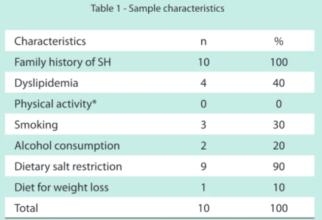
Ten patients (ive men and ive women, ages ranging from 45 to 60) diagnosed with essential hypertension and under clinical and pharmacological treatment (Table 1 and Figure 1) were included in the study. All of them were seen at the Integrated Medical Assistance Nucleus (Núcleo de Assistência Médica Integrada - NAMI) of the University of Fortaleza.
No patient was obese (mean BMI was 26 ± 1.4) or had other comorbidities associated with hypertension, except for dyslipidemia (Table 1). Subjects with essential hypertension and under clinical treatment were considered eligible. Study patients were selected based on their clinical follow-up forms. After the inclusion and exclusion criteria described below were applied, interviews were conducted to collect data on subjects’ clinical and pharmacological treatment and their epidemiological proile.
Exclusion criteria were the following: patients with secondary hypertension; renal, heart or liver failure; recent cardiovascular event; pulmonary disease, diabetes mellitus, neuropathies, autoimmune diseases, Chagas disease, cardiac arrhythmias, and other diseases that impair autonomic nervous system control. Alcohol consumption and cigarette smoking, as well as the use of oral contraceptives, antidepressants, neuroleptics, anti-arrhythmics, and lithium, were also exclusion criteria.
Study design - At the beginning of the study, data were collect to assess systolic blood pressure (SBP), diastolic blood pressure (DBP), and heart rate variability (HRV ). The following respiratory variables were monitored: tidal
Table 1 - Sample characteristics
Characteristics n %
Family history of SH 10 100
Dyslipidemia 4 40
Physical activity* 0 0
Smoking 3 30
Alcohol consumption 2 20
Dietary salt restriction 9 90
Diet for weight loss 1 10
Total 10 100
SH - systemic hypertension; Physical activity (any kind of activity, including walking).
Fig. 1 - Pharmacological therapy of the study sample.
volume (TV), respiratory rate (RR), minute volume (MV), plus thoracic expansibility and diaphragmatic expansibility. Upon conclusion of the study, and after one month of slow breathing training, all these measurements were repeated for the inal collection of data and statistical analysis.
Measurement of blood pressure and analysis of cardiac autonomic balance - Blood pressure was measured in three diferent time points, on an outpatient basis, using a properly calibrated aneroid sphygmomanometer and according to the IV Guidelines for Hypertension issued by the Brazilian Society of Cardiology14 and those of the Joint National Committee15.
Microsoft Excel 2003, and statistical analyses were performed using the SPSS software version 10.0.
Respiratory mechanics analysis - Minute volume (MV) and respiratory rate (RR) were measured using a Wright’s respirometer, and tidal volume was calculated by the RR/ MV ratio. Chest wall expansion was assessed by thoracic expansibility (at the axillary level) and diaphragmatic expansibility (at the xiphoid process level) measurements.
Respiratory retraining protocol - A training protocol for breathing control was applied using, first, diaphragmatic, intercostal, and upper chest breathing pattern to make the patient aware of his respiratory movements. Patients were asked to lie supine, with knees lexed and feet lat on the loor, and raise their hands to the area of the chest (Figure 2) related to each breathing pattern: diaphragm, intercostal muscles, and clavicular region. At each breathing pattern, patients were instructed to feel and identify the rib cage motion and its amplitude. Subsequently, they were instructed to decrease their respiratory rate gradually while increasing respiratory amplitude. This procedure was performed ten times for each breathing pattern.
The patients then sat down and performed the slow breathing technique, intended to promote quieter breathing by increasing respiratory amplitude and reducing respiratory rate while the three breathing patterns described above are simultaneously performed. This technique was done in 30-minute sessions held twice a week during one month.
Statistical analysis - All data were analyzed using descriptive statistics. Baseline and final means were compared using Student’s t test and underwent p-value analysis. Ventilatory variables and blood pressure measurements were assessed using Pearson’s correlation coeicient and linear regression analysis (R-square). P values < 0.05 were considered statistically signiicant.
Results
No changes were made in the medication used and lifestyle reported by the patients at baseline (Data are not shown). The study results were the following:
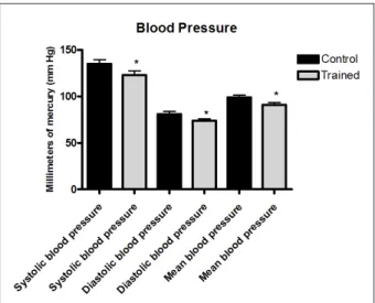
Respiratory retraining and blood pressure - In our sample of hypertensive patients, there was a decrease in blood pressure (Graphic 1). Systolic, mean, and diastolic BP all dropped (135 ± 14.32 mm Hg to 123 ± 14.29 mm Hg, p < 0.05; 99 ± 7.8 mm Hg to 91 ± 7.5 mm Hg, p < 0.05; and 81 ± 8.75 mm Hg to 74 ± 6.1 mm Hg, p < 0. 05, respectively).
Respiratory retraining and cardiovascular autonomic modulation - Heart rate variability improved (Graphic 2). However, no signiicant change was found in resting heart rate (74 ± 8 bpm to 72 ± 9.1 bpm, p > 0.05) and mean RR intervals (1002 ± 3.4 ms to 1004 ± 3.2 ms, p > 0.05). This improvement in heart rate variability was evidenced by an increase in RR interval variation and SDNN index, while the PNN50 index remained unchanged.
Respiratory retraining and thoracicmechanics - After the training period, changes in breathing pattern were noted (Graphic 3). There was a decrease in resting respiratory rate (14.60 ± 1.6 breaths per minute to 10.10 ± 1.1 breaths per minute, p<0.01). No signiicant changes were found in minute volume, which was attributed to the compensatory increase in tidal volume (419.75 ± 60,93 mL to 560.30 ± 60.76 mL, p < 0.01). This tidal volume increase, in turn, was attributed the greater thoracic expansibility (1.9 ± 0.56 cm to 2.33 ± 0.49 cm, p < 0.01), while diaphragmatic expansibility remained unaltered (Graphic 3). In the study sample, a moderate negative correlation (Pearson’s coefficient = 0.5 with p < 0.05) was found between tidal volume and systolic and mean blood pressure (not shown), as shown by the linear regression analysis (Graphic 4).
Graphic 1 - Slow breathing and blood pressure decrease: *p<0,05.
Graphic 2 - Improved HRV: SDNN index (*p < 0.05); RR interval variation (*p < 0.05); mean RR intervals, PNN50 index, and resting heart rate (ns, p > 0.05).
Discussion
This study sought to evaluate the effectiveness of a training program using the slow breathing technique in cardiovascular and blood pressure control in a sample of patients with essential hypertension. We have demonstrated that respiratory retraining may improve cardiorespiratory control and decrease blood pressure in hypertensive patients.
Our data are consistent with the studies by Joseph et al11,
Kaushik et al16, Bernardi et al17, and Goso et al18. These studies
evaluated the efects of a single exercise session using the slow breathing technique in healthy, hypertensive and heart failure patients and showed the beneits of spontaneous respiratory modulation in cardiovascular control and blood pressure reduction. We decided to assess the impact of a respiratory retraining program using the slow breathing technique over one month in hypertensive patients. We believed that, performed on a regular basis, these breathing exercises could exert some psychophysiological efects to change breathing pattern at rest. Despite the small sample size owing to the numerous exclusion criteria adopted in the study, data statistics were determinant. Further studies involving larger numbers of patients are needed and, if possible, with probability sampling to assess the efectiveness of this type of intervention and its impact on the population.
Controlling blood pressure is considered the best strategy to reduce the impact of cardiovascular diseases in hypertensive patients; this may be accomplished by prevention campaigns, risk factor management, early diagnosis, and effective treatment for the disease19. Some scientiic evidence suggests
that changes in breathing control may also be regarded as a risk factor associated with lifestyle in hypertensive patients20.
Graphic 3 - ***P < 0.01; ns p > 0.05 (non-signiicant).
Graphic 4 - Integration of the cardiovascular and respiratory systems: the increase in tidal volume is related to a decrease in mean blood pressure.
owing to reports about the greater prevalence of cardiovascular disease in subjects with sleep breathing disorders21. The
mechanism by which sleep breathing disorders afect blood pressure and hypertension remains unclear22. Nonetheless,
Ehlenz et al23 have shown changes in blood pressure regulation
breathing disorders, is strongly associated with changes in systemic and pulmonary blood pressure24. According to Brooks
et al25, these changes in blood pressure control occur both
during sleep and daytime, thereby suggesting a causal role of sleep breathing disorders in the development and maintenance of hypertension. Other than that, epidemiological studies show that sleep-disordered breathing is highly prevalent among hypertensive patients26,27, and also that 50 to 80% of the
patients who experience such changes are hypertensive20.
In this regard, the causal relationship of respiratory disorders might become confusing or disappear when age, obesity prevalence, smoking status, and alcohol consumption are taken into account in hypertensive patients. However, Grote et al20, in a cross-sectional, controlled study involving 1,190
patients, have shown that sleep breathing disorders are risk factors for hypertension and do not depend on body mass index, age, smoking status, alcohol consumption, cholesterol levels, and oxygen and carbon dioxide partial pressures. Therefore, scientiic data point to the existence of a cause and maintenance relationship of hypertension associated with changes in respiratory control.
Spontaneous respiratory modulation and blood pressure reduction - In patients with essential hypertension, these changes in blood pressure are correlated with changes in breathing patterns, as evidenced in this study by statistical correlation and linear regression. According to Joseph et al11,
the decrease in blood pressure during slow breathing, more precisely less than ten breaths per minute (BPM), is associated with improved barorelex sensitivity, which suggests a change in autonomic balance resulting from an absolute or relative reduction in sympathetic activity or increase in parasympathetic tone. One of the diiculties faced by our study was the lack of barorelex measurement, because this is an expensive method that requires a laboratory specially equipped for this purpose. However, the signiicant clinical value of heart rate variability (HRV) analysis s is well characterized in the literature. It has the advantage to allow a selective and non-invasive assessment of the autonomic function28. In our sample,
blood pressure decrease was accompanied by improvement in heart rate variability, suggesting that this is an efect of the autonomic nervous system’s respiratory modulation. Bernardi et al17 studied the efect of a single session of slow
breathing in healthy subjects and hypertensive patients and found increased baroreflex sensitivity with a consequent reduction in blood pressure and heart rate. In our study, there were no changes in heart rate, a inding that contradicts the acute chronotropic efects reported in the literature17 and
previously detected by us in one patient with acute myocardial infarction and hypertension9. We believe that this may be
due to the lack of a more accurate methodology capable of detecting chronotropic changes. In future studies, it would be recommendable that Holter electrocardiographic recordings be used to better assess these heart rate changes, as well as ABPM (Ambulatory blood pressure monitoring), to analyze the degree of the anti-hypertensive efect aforded by this breathing technique.
Autonomic imbalance in hypertension and breathing - The autonomic nervous system plays a key role in controlling blood pressure and heart rate, and is an important pathophysiological
factor in the development of hypertension29. Sympathetic
hyperactivity has been frequently associated with hypertension, in which the sensitivity of baroreceptor control is impaired, thereby compromising the autonomic function30. This state
of hyperexcitation with increased sympathetic discharges is well characterized in the literature, which also reports the beneits of mental relaxation associated or not with breathing exercises in hypertensive patients16. In essential hypertension,
the sympathetic hyperactivity results not only in arteriolar vasoconstriction, but also in chemoreflex activation, an important mechanism that regulates breathing,since the excitatory state is distributed to all organs and systems31.
Hypertensive patients usually show signs of hyperventilation at rest, suggesting that the cardiovascular and respiratory abnormalities may be related to a common excitatory pattern of the autonomic nervous system that could be modulated by the adoption of a slower breathing pattern11. In this context,
the generalized hyperexcitatory pattern could explain the signs of hyperventilation found in patients with essential hypertension11; and as breathing can be voluntarily controlled,
the long-term practice of a breathing pattern may also induce changes in cardiovascular control.
Heart rate variability is reduced in hypertensive patients, as compared with normotensive subjects, and PNN50 and SDNN indices are lower in the time-domain method, relecting decreased vagal tone (parasympathetic) and increased sympathetic tone, respectively32. In the present study, there
was an improvement in heart rate variability, based on an increase in RR interval variation and the SDNN index, while the PNN50 index did not change signiicantly (p > 0.05). This suggests that respiratory retraining using the slow breathing technique enhanced autonomic function not by increasing the parasympathetic tone, but rather by decreasing sympathetic activity. Our data corroborate the studies by Joseph et al11
and Bernardi et al17, who also noted an improvement in heart
rate variability.
According to Kaushik et al16, the hyperexcitation caused
by increased sympathetic discharges may be modulated by sessions of relaxation and breathing exercises, together or alone. However, in this author’s opinion, breathing exercises and relaxation are likely to modulate the autonomic nervous system through diferent mechanisms that need to be further elucidated. In this study, the hypothesis that breathing exercises may have provided some degree of relaxation to the patients cannot be discarded. Nevertheless, given the negative and significant statistical correlation (p < 0.05) between tidal volume and blood pressure, the modulation of respiratory mechanics is thought to play a key role in this anti-hypertensive efect.
Integration of cardiovascular control and benefits of respiratory retraining - Cardiovascular and respiratory systems have a close relationship regarding their mechanisms of neural control, the physiology of which is not discussed in this paper. It is noteworthy, however, that in this mechanism of cardiovascular and respiratory control, the solitary tract nucleus plays a major role, since it receives aferent inputs from baroreceptors located in the heart and blood vessels, chemoreceptors, and lung stretch receptors33. Accordingly,
References
1. Souza MFM, Timerman A, Serrano CV Jr, Santos RD, Mansur AP. Tendência do risco de morte por doenças circulatórias nas cinco regiões do Brasil de 1979 a 1996. Arq Bras Cardiol. 2001; 77: 562-8.
2. Keys A. The Seven Countries Study: a multivariate analysis of death and coronary heart disease. Cambridge (MA): Harvard University Press; 1980. 3. Diretrizes Brasileiras de Hipertensão Arterial. Sociedade Brasileira de
Hipertensão, Sociedade Brasileira de Cardiologia e Sociedade Brasileira de Nefrologia. Hipertensão. 2002; 6: 1-2.
4. Stamler J, Stamler R, Neaton JD. Blood pressure, systolic and diastolic, and cardiovascular risks: U.S. population data. Arch Intern Med. 1993; 153: 598-615.
5. MacMahon S, Peto R, Cutler J, Collins R, Sorlie P, Neaton J, et al. Blood pressure, stroke, and coronary heart disease: part 1. Prolonged diferences in blood pressure: prospective observational studies corrected for the regression
dilution bias. Lancet. 1990; 335: 765-74.
6. Suzuki S, Ohta T. Non-pharmacological treatment of hypertension in the elderly. Nippon Rinsho. 2005; 63 (6): 1010-5.
7. Schein MH, Gavish B, Herz M, Rosner-Kahana D, Naveh P, Knishkowy B, et al. Treating hypertension with a device that slows and regularises breathing: a randomised, double-blind controlled study. J Hum Hypertens. 2001; 15 (4): 271-8.
8. Grossman E, Grossman A, Schein MH, Zimlichman R, Gavish B. Breathing-control lowers blood pressure. J Hum Hypertens. 2001; 15 (4): 263-9. 9. Pinheiro CHJ, Cézar ID, Marinho MJF, Araújo FCS. Efeitos dos exercícios de
controle respiratório sobre a pós-carga cardíaca em pacientes hipertensos no pós-operatório tardio de revascularização do miocárdio. [Resumo]. In: 11º Encontro de Iniciação à Pesquisa da Universidade de Fortaleza. Fortaleza; 2005.
have common neuroanatomical pathways, which may provide a biological basis for the efects of breathing modulation on cardiovascular control and blood pressure reduction.
In view of this body of evidence, our indings might be explained in many ways. During slow breathing, tidal volume increases to compensate for the reduced respiratory rate and to maintain minute ventilation34,35, which may be responsible
for the autonomic changes by reducing sympathetic activity or activating the Hering-Breuer relex18. According to Bernardi
et al17, this increase in barorelex depends solely on a lower
respiratory rate, rather than on controlled breathing and, thereby, a frequency of six breaths per minute increases barorelex, while a frequency similar to that of spontaneous breathing, such as 15 breaths per minute, fails to do it. Joseph et al11 believe that changes in respiratory amplitude, that is, in
chest expansion, may increase the luctuations in RR intervals, render barorelex more efective, and decrease blood pressure. On the other hand, the increase in tidal volume and activation of the Hering-Breuer reflex, which reduces chemoreflex sensitivity and consequently may highlight barorelex eicacy, decrease blood pressure and sympathetic activity.
These results may be explained also by chemoreflex reduction, the increase of which is known to elevate sympathetic activity and blood pressure37. In the opinion of Bernardi et al17,
slow breathing training enhanced oxygen saturation and the regulation of thypoxic chemorelex, improving exercise capacity in patients with cardiovascular disease. Although respiratory gas exchanges were not measured in our sample, we believe that the increase in tidal volume attributed to greater chest expansion enhanced oxygenation in these patients. We suggest that other studies be made to evaluate this metabolic parameter by analyzing maximal oxygen uptake (VO2) and carbon dioxide production (VCO2). However, our data have shown that respiratory rate reduction does not induce hyperventilation in these patients following the technique, since minute volume remained unchanged and tidal volume underwent a compensatory increase. These data were conirmed by Joseph et al11, because in their study slow breathing did not change
minute volume and end-tidal carbon dioxide (ETCO2). As CO2is maintained within resting ranges, slow breathing is well
tolerated by patients.
Breathing pattern management seems to be a fairly reasonable tool since voluntary stimulation is used to induce relex changes in the cardiovascular system. The existence of common neuroanatomical bases in the modulation of cardiovascular and respiratory control is regarded as the biological foundation for this intervention. However, the exact mechanism of such integration is yet to be established. Slow breathing is not a new method, but rather an ancient Eastern tradition of Yoga38. Thanks to the beneits that yoga
provides patients with cardiovascular diseases, its components (stretching, relaxation, and breathing techniques) sparked interest within the scientific community39. Some studies
have shown improvement in cardiorespiratory control in hypertensive patients who practice Bhramari Pranayama, a yogic technique that involves slow breathing patterns, while techniques that use high-frequency breathing show antagonistic efects40.
Study limitations and inal remarks - Study sample consisted of patients with well-controlled hypertension. The next step would be to analyze how reproducible these indings are in patients with more severe degrees of hypertension with or without the presence of other comorbidities.
Based on our findings, we came to the conclusion that the correction of abnormalities in respiratory control of hypertensive patients improves cardiovascular and respiratory control, acting as an adjunctive for clinical and pharmacological treatment of hypertension. Slow breathing is a straightforward method with no contraindications that ofers a rather valid cost-beneit, improving autonomic balance and respiratory control and lowering blood pressure in patients with essential hypertension. However, further studies with larger series of patients and probability sampling are needed to assess the impact of this intervention on a greater cohort of hypertensive patients.
Potential Conlict of Interest
10. Radaelli A, Raco R, Perfetti P, Viola A, Azzellino A, Signorini MG, et al. Efects of slow, controlled breathing on baroreceptor control of heart rate and blood pressure in healthy men. J Hypertens. 2004; 22 (7): 1361-70.
11. Joseph CN, Porta C, Casucci G, Casiraghi N, Maffeis M, Rossi M, et al. Slow breathing improves arterial baroreflex sensitivity and decreases blood pressure in essential hypertension. Hypertension. 2005; 46 (4): 714-8. 12. World Medical Association (WMA). Declaration of Helsinki. Somerset West:
(South Africa) 1996. In: 48th General Assembly-WMA. 2000 October 6. Acessado em 2006 nov. 10. Disponível em: <http:// www.wma.net>. 13. World Medical Association (WMA). Declaration of Helsinki. Edinburgh
(Scotland) 2000. In: 52nd General Assembly. 2000 October 13. Acessado em 2006 nov 10. Disponível em: <http://www. wma.net>.
14. IV Diretrizes Brasileiras de Hipertensão Arterial. Arq Bras Cardiol. 2004; 82 (supl. 4): 2-22.
15. Joint National Committee on Detection, Evaluation and Treatment of High Blood Pressure (JNC). Seventh Report of Joint National Committee on Prevention, Detection, Evaluation and Treatment of High Blood Pressure: The JNC 7 report. JAMA. 2003; 299: 2560-72.
16. Kaushik RM, Kaushik R, Mahajan SK, Rajesh V. Efects of mental relaxation and slow breathing in essential hypertension. Complement Ther Med. 2006; 14 (2): 120-6.
17. Bernardi L, Porta C, Spicuzza L, Bellwon J, Spadacini G, Frey AW, et al. Slow breathing increases arterial barorelex sensitivity in patients with chronic heart failure. Circulation. 2002; 105: 143-5.
18. Goso Y, Asanoi H, Ishise H, Kameyama T, Hirai T, Nozawa T, et al. Respiratory modulation of muscle sympathetic nerve activity in patients with chronic heart failure. Circulation. 2001; 104: 418-23.
19. Gus I, Harzheim E, Zaslavsky C, Medina C, Gus M. Prevalence, awareness, and control of systemic arterial hypertension in the state of Rio Grande do Sul. Arq Bras Cardiol. 2004; 83 (5): 424-8.
20. Grote L, Ploch T, Heitmann J, Knaack L, Penzel T, Peter JH. Sleep-related breathing disorder is an independent risk factor for systemic hypertension. Am J Respir Crit Care Med. 1999; 160 (6): 1875-82.
21. He J, Kryger MH, Zorick FJ, Conway W, Roth T. Mortality and apnea index in obstructive sleep apnea: experience in 385 male patients. Chest. 1988; 94: 9-14.
22. Wright J, Johns R, Watt I, Melville A, Sheldon T. Health efects of obstructive sleep apnoea and the efectiveness of continuous positive airways pressure: a systematic review of the research evidence. Br Med J. 1997; 314: 851-60. 23. Ehlenz K, Peter JH, Kafarnik H, von Wichert P. Disturbances in volume regulating
hormone system – a key to the pathogenesis of hypertension in obstructive sleep apnea syndrome? Pneumologie. 1991; 45 (Suppl. 1): 239-45.
24. Schneider H, Schaub CD, Andreoni KA, Schwartz AR, Smith PL, Robotham JL, et al. Systemic and pulmonary hemodynamic responses to normal and obstructed breathing during sleep. J Appl Physiol. 1997; 83: 1671-80.
25. Brooks D, Horner RL, Kozar LF, Render-Teixeira CL, Phillipson EA. Obstructive sleep apnea as a cause of systemic hypertension: evidence from a canine model. J Clin Invest. 1997; 99: 106-9.
26. Lavie P, Ben-Yosef R, Rubin AE. Prevalence of sleep apnea syndrome among patients with essential hypertension. Am Heart J. 1984; 108 (2): 373-6. 27. Fletcher EC, DeBehnke RD, Lovoi MS, Gorin AB. Undiagnosed sleep apnea
in patients with essential hypertension. Ann Intern Med. 1985; 103: 190-5. 28. Task Force of the European Society of Cardiology and the North American
Society of Pacing and Electrophysiology. Heart rate variability: standards of measurement, physiological interpretation and clinical use. Circulation. 1996; 93: 1043-65.
29. Julius S. Autonomic nervous system dysregulation in human hypertension. Am J Cardiol. 1991; 67: 3B-7B.
30. Guzzetti S, Piccaluga E, Casati R, Cerutti S, Lombardi F, Pagani M, et al. Sympathetic predominance in essential hypertension: a study employing spectral analysis of heart rate variability. J Hypertens. 1988; 6: 711-7. 31. Somers VK, Mark AL, Abboud FM. Potentiation of sympathetic nerve
responses to hypoxia in borderline hypertensive subjects. Hypertension. 1988; 11: 608-12.
32. Menezes AS Jr, Moreira HG, Daher MT. Análise da variabilidade da freqüência cardíaca em pacientes hipertensos, antes e depois do tratamento com inibidores da enzima conversora da angiotensina II. Arq Bras Cardiol. 2004; 83 (2): 165-8.
33. Spyer KM. Central nervous integration of cardiovascular control. J Exp Biol. 1982; 100: 109-28.
34. Bernardi L, Spadacini G, Bellwon J, Hajric R, Roskamm H, Frey AW. Effect of breathing rate on oxygen saturation and exercise performance in chronic heart failure. Lancet. 1998; 351: 1308-11.
35. Spicuzza L, Gabutti A, Porta C, Montano N, Bernardi L. Yoga and chemorelex response to hypoxia and hypercapnia. Lancet. 2000; 356: 1495-6. 36. Bernardi L, Gabutti A, Porta C, Spicuzza L. Slow breathing reduces
chemorelex response to hypoxia and hypercapnia, and increases barorelex sensitivity. J Hypertens. 2001; 19: 2221-9.
37. Francis DP, Coats AJS. Chemorelex-barorelex interactions in cardiovascular disease. In: Bradley TD, Floras JS, eds. Sleep apnea: implications in cardiovascular and cerebrovascular disease. New York: Marcel Dekker; 2000. p. 33-60.
38. Schell FJ, Allolio B, Schonecke OW. Physiological and psychological efects of Hatha-Yoga exercise in healthy women. Int J Psychosom. 1994; 41: 46-52. 39. Jayasinghe SR. Yoga in cardiac health (a review). Eur J Cardiovasc Prev Rehabil.
2004; 11 (5): 369-75.