INTRODUCTION
Melanocytic choroidal nevi are the most common intraocular tu-mors(1-4), with a prevalence of 6.5% in Caucasian populations(2).
Most choroidal nevi do not undergo malignant transformation throughout the patient’s life, but some can grow during adulthood
(1 in every 160 cases in 5 years)(5) and/or undergo malignant
transfor-mation to choroidal melanoma, with an incidence of approximately
1 in 8000 cases per year(3). Several studies reported risk factors for
the malignant transformation of choroidal nevi as follows: thickness >2 mm, presence of adjacent subretinal fluid, orange pigmenta-tion, nevus margin located ≤3 mm from the optic disc, presence of
associated symptoms(2,4-6), presence of lesions detected via A-mode
ultrasound (US) characterized by medium/low homogeneous
inter-nal reflectivity and the absence of choroidal hollowing(6), and the
absence of retinal drusen(6). The presence of three or more risk factors indicates a >50% probability of tumor growth within 5 years(2,6).
The detection of growth in the early stages is the most reliable predictive factor for lesion transformation into a melanoma. Conside-rably, US has an important role as it allows the accurate measurement of the height of lesions and size comparisons at different time inter-vals (every 6 months to 1 year)(4). One of the disadvantages of US is the inability to detect flat or minimally elevated nevi (<1 mm in height)(7).
Comparisons of choroidal nevus measurements obtained using 10- and 20-MHz
ultrasound and spectral domain optical coherence tomography
Comparação entre a medida de nevus de coroide obtida com 10-MHz e 20-MHz e tomograia de coerência
óptica de domínio espectral
Maírade França Martins1, Kelcia KieFer2, liliane alMeida andrade Kanecadan3, Patricia novita Garcia3, rubens n. belFort3, norMa alleMann3
Submitted for publication: March 9, 2016 Accepted for publication: November 7, 2016 1 Curitiba, PR, Brazil.
2 Vitória, ES, Brazil.
3 Department of Ophthalmology, Universidade Federal de São Paulo, SP, Brazil.
Funding: No specific financial support was available for this study.
Disclosure of potential conflicts of interest: None of the authors have any potential conflict of interest to disclose.
Corresponding author: Norma Allemann. Rua Olimpíadas, 134/ 51 - São Paulo - SP - 04551-000 - Brazil - E-mail: norma.allemann@pobox.com
Approved by the following research ethics committee: UNIFESP (CEEA: 00651412.5.0000.5505). ABSTRACT
Purpose: To compare measurements of lesions clinically diagnosed as choroidal nevi using spectral domain optical coherence tomography (SD-OCT ) and 10- and 20-MHz ultrasound (US).
Methods: This prospective study, which was conducted between May 2011 and December 2011, evaluated eyes diagnosed with choroidal nevus via photographic documentation using 10- or 20-MHz A- and B-mode US (experienced examiner using both the transpalpebral technique and direct contact) or SD-OCT in the enhanced depth imaging mode (performed by a different examiner blinded to the US results). Anteroposterior (AP) and transverse (T ) US sections corresponded to sections adjusted perpendicularly on SD-OCT.
Results: We evaluated 14 eyes from 12 patients (six males, mean patient age= 64.5 years) diagnosed with choroidal nevus. The choroidal nevi of all samples had a melanocytic profile. Moreover, eight nevi were located at the equator, five nevi were located in the posterior pole (peripapillary in one sample), and one nevus shifted from the equator to the periphery. On SD-OCT, the maximum measurable dimension was 9 mm. The lesions in the posterior pole were easier to evaluate, and image acquisition of lesions located more peripherally was possible depending on patient cooperation. The accurate assessment of height was difficult. Baseline dimensions on 10- and 20-MHz US were larger than those determined via OCT. No significant differences in height were observed between US and SD-OCT. All parameters were statistically similar between 10- and 20-MHz US measurements. Conclusions: No significant difference in the AP and T diameters was observed between 10- and 20-MHz US measurements; however, these measurements (AP and T ) were significantly higher than those obtained using OCT. No significant differences in height were observed among the techniques adopted.
Keywords: Choroidal neoplasms/ultrasonography; Nevus, pigmented/pathology; Tomography, optical coherence; Ultrasonography
RESUMO
Objetivo: Comparar as medidas obtidas de lesões diagnosticadas clinicamente como nevus de coroide através da tomografia de coerência óptica de domínio espectral (Spectralis, Heidelberg Engineering, Inc.), ultrassonografia com 10 MHz e de 20 MHz.
Métodos: Estudo prospectivo realizado entre maio e dezembro de 2011, avaliou olhos com diagnóstico de nevus de coroide, utilizando documentação fotográfica, ultras-sonografia com transdutor 10-MHz e 20-MHz A- e B-mode e SD-OCT em modo de EDI, por um examinador diferente para cada técnica. Os cortes realizados perpendiculares entre si, correspondentes ao corte ântero-posterior e latero-lateral à ultrassonografia.
Resultados: Foram avaliados 14 olhos de 12 pacientes (6 do sexo masculino), com média de idade média de 64,5 anos. Todos os nevus tinham um perfil melanocítico. Observou-se 8 nevus no equador, 5 no polo posterior (peripapilar em uma amostra), e 1 deslocado a partir do equador para a periferia. Em SD-OCT, a dimensão máxima mensurável foi de 9 mm. As lesões no polo posterior eram mais fáceis de avaliar e aqui-sição de imagens de lesões mais periféricas era possível, dependendo da colaboração do paciente. A avaliação precisa da altura era difícil. As dimensões usando transdutor 10-MHz e 20-MHz US foram maiores que as encontradas pelo SD-OCT. Não foram observadas diferenças significativas na altura entre métodos SD-OCT e US. Todas as medidas foram estatisticamente semelhantes entre 20-MHz e 10-MHz.
Conclusão: Para o parâmetro AP e T não foi detectada diferença entre as medidas utili-zando US de 10-MHz e de 20-MHz. Porém estas medidas se mostraram significativamente maiores em relação à medida obtida com OCT. Para a altura, não foram detectadas diferenças estatística em relação à técnica utilizada, US 10-MHz e 20-MHz e SD-OCT.
Ma rt i n s MF, e t a l.
Other complementary methods such as fluorescein angiography, autofluorescence imaging, and optical coherence tomography (OCT) are important during the evaluation and follow-up of choroidal nevi(2,8-11). OCT can identify factors that increase the risk of malignant transformation, including subretinal fluid (even when the volume to be clinically detected is small), cystoid macular edema, and changes in the retinal pigment epithelium; however, it does not infer much importance during the diagnosis itself(9,10).
The use of OCT to measure choroidal thickness has been studied
in both normal eyes(12) and those with complications(13-15). The use
enhanced depth imaging (EDI)-OCT is an additional, innovative
diagnostic imaging modality implemented in the SPECTRALIS® OCT
apparatus. The EDI mode is a modality enabling high-resolution OCT imaging of external retinal layers, the choroid, and the lamina cribro-sa. It allows the detection of structural changes “beyond the RPE” in a reproducible manner, which is difficult to achieve using standard OCT. EDI-OCT measures the distance between the RPE and the cho-rioscleral interface to obtain the choroidal thickness.
This study aimed to compare measurements of lesions clinically diagnosed as choroidal nevi using SD-OCT and 10- or 20-MHz US.
METHODS
This prospective study, which was conducted between May and December 2011, evaluated patients from the Ocular Oncology Sector of Universidade Federal de São Paulo, who received a clinical diagno-sis of choroidal nevus as assessed by two experts in ocular oncology using photographic documentation of the lesion (retinography) and ocular US.
The study was approved by the Ethics Committee of the Univer-sity, and all patients read and signed a consent form.
All eyes exhibited optical transparency, and the location of the lesion permitted OCT examination.
US images were obtained using A- and B-mode 10- and 20-MHz
transducers (Ultrascan®,Alcon, Fort Worth,TX, USA). The
examina-tions were performed with the patient in the supine position with standardization of instrument parameters and examination techni-ques. For the 10-MHz transducer, the transpalpebral technique was adopted, which involved the application of gel on the closed eyelid with a gain of 60 dB and depth of 40 mm, to identify the lesion and measure the anteroposterior (AP) and transverse (T) basal diameters and height. Using the 20-MHz transducer, the direct contact technique was adopted under topical anesthesia, and it involved the application of gel over the conjunctiva with a gain of 65 dB to identify the lesion and determine its size. In both techniques, a gain of 75 dB and depth of 30 mm were used to assess the vitreous cavity.
After mydriasis, all eyes were examined using SD-OCT (Spectralis, Heidelberg Engineering, Inc.) in the EDI mode with a 20 × 30 degree field, a linear section with a length of 9 mm, a resolution of 100 sec-tions, and a total of 1536 scans per second in the A-mode to generate an axial resolution of 7 microns, as described previously(15).
Measurements were obtained by both methods (US and OCT) in the same manner. The AP diameter was measured from the optic nerve toward the periphery, the transverse (T) basal diameter was acquired perpendicular to the AP, and height was measured between the internal limiting membrane and the top of the sclera. When it was impossible to see the sclera on OCT due to the shadow caused by the high internal reflectivity of the nevus, an imaginary line was drawn to join the images from the origin of the adjacent sclera, and this line was considered the posterior limit of the lesion (Figure 1).
SD-OCT examinations were performed by examiners blinded to the US examination results on the same date.
To detect differences between measurement protocols for each of the methods employed (10- and 20-MHz US and SD-OCT) and between the AP and T diameters and height, analysis of variance was performed using repeated measures after the measurement of each
dimension in triplicate, corresponding to one measurement on each apparatus used. Differences were also found between the sexes. Age was used as a covariate, and its possible effects were removed from the model. For the multivariate statistical test, Pillai’s trace was calculated, as it is more robust for the assumption of violations in the model, as reported by Olsen (1974). When sphericity could not be assessed using Mauchly’s sphericity test, the degrees of freedom of the univariate tests were adjusted by the lower limit of epsilon (most conservative). Results with p≤0.05 were considered significant, and the alpha value was adjusted by the Bonferroni correction when necessary.
RESULTS
We evaluated 14 eyes from 12 patients (six males) with a mean age of 64.5 years.
Considering the location of the choroidal nevus, eight (57.15%) lesions were located in the equator, five lesions (35.71%) were located in the posterior pole (one with a peripapillary location), one (7.14%) lesion was located in the transition between the equator and peri-phery (7.14%), and all 14 lesions were melanocytic.
Table 1 shows the dimensions calculated for each lesion using distinct methods. The greatest length measurable by OCT was 9 mm. Cases in which the length of the choroidal nevus exceeded 9 mm, measurement of this dimension using OCT was unfeasible.
The choroidal nevi located in the posterior pole were easily eva-luated using OCT. For lesions located in the transition between the equator and periphery, patient cooperation was necessary; thus, the lesion from case 1 was not located.
Height was the most difficult parameter to measure on OCT. For some choroidal nevi, it was possible to determine the origin of the sclera on OCT (Figure 2); however, in others, an imaginary line was drawn to join the images from the origin of the adjacent sclera, and this line was considered as the posterior limit of the lesion (Figure 1).
One choroidal nevus fully covered the papilla, and to define its
size(Figure 3), the following US and OCT measurements were made:
AP basal diameter, the sum of the measurement from the superior pa-pillary margin to the upper limit of the lesion with the measurement from the inferior papillary margin to the lower limit of the lesion; and the T diameter, the sum of the measurement from the nasal papillary margin to the nasal limit of the lesion with the measurement from the temporal papillary margin to the temporal limit of the lesion.
No significant difference in the AP diameter was observed between 10- and 20-MHz US measurements (F=0.165, p=0.696); however, these measurements were significantly larger than those obtained using OCT (F=18.559; p=0.00; Figure 4).
No difference in the T diameter was found between 10- and 20-MHz US measurements (F=0.505, p=0.497); however, these mea-surements were significantly larger than those obtained using OCT (F=38.713, p=0.0002; Figure 4).
No significant differences in height were observed among the techniques (10-MHz US, 20-MHz US, and SD-OCT).
DISCUSSION
US is considered as the most efficient method for assessing the dimensions of a choroidal nevus and identifying tumor growth. A comparison of images obtained using 10- and 20-MHz transducers illustrated that the 20-MHz probe had superior resolution to detect abnormalities in the eye wall and vitreoretinal interface(16).
In our analysis, although no significant difference was observed between the measurements of the choroidal nevus using the 10- and 20-MHz transducers (p<0.05), when the AP and T diameters and height were compared, better resolution was achieved using the
20-MHz probe under direct contact, resulting ina better definition
The 20-MHz transducer used for image acquisition was part of a specific model of ophthalmic ultrasound equipment (Ultrascan, Alcon). Considering that manufacturers use distinct methods for adjusting the image, depth of focus, and resolution in US transducers, the findings presented in this study should be correlated with the equipment model used.
OCTis recently used to identify factors that increase the risk of
malignant transformation(8-10), but it has also proven beneficial for the serial monitoring of choroidal melanocytic lesions; however, it has
restrictions for the measurement of AP and T diameters and height(7).
In one study(7),images of choroidal tumors located in the
poste-rior pole were analyzed using Spectralis OCT in the EDI mode, and the researchers observed that the measurement of tumors with heights of <1 mm was feasible only on OCT because flat lesions were undetectable via US. According to these authors, the limitations of OCT include a maximum limit of measurement (9 mm) and better documentation of lesions located at the limit of the vascular arcades in the posterior pole.
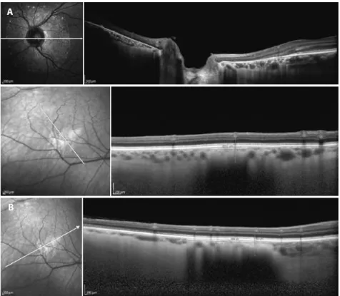
In this study, tumors thinner than 1 mm could be measured by US, whereas OCT measurements were limited to a maximum length of Figure 1. Choroidal nevus located at the posterior pole. Anteroposterior diameter, 6.2 (20-MHZ ultrasound
Ma rt i n s MF, e t a l.
Table 1. Dimensions of choroidal melanocytic lesions diagnosed as choroidal nevi using 10- and 20-MHz US and SD-OCT
ID Eye Location Age (years)
10-MHz US 20-MHz US SD-OCT
(mm) (mm) (mm)
02 OS Posterior pole, peripapillary 28 AP: 15.1 AP: 14.8 AP: 4.73
T: 14.3 T: 14.3 T: 4.69
H: 1.2 H: 1.2 H: 0.59
03 OD Temporal inferior posterior pole 60 AP: 6.0 AP: 10.6 AP: NM
7 h T: 9.0 T: 8.9 T: NM
H: 0.8 H: 1.1 H: 0.15
04 OD Temporal superior posterior pole 60 AP: 8.4 AP: 6.4 AP: 7.86
10 h T: 10.2 T: 8.2 T: NM
H: 1.2 H: 0.9 H: 0.34
05 OS Temporal superior posterior pole 60 AP: 9.0 AP: 6.3 AP: NM
2 h T: 12.2 T: 9.8 T: NM
H: 0.8 H: 1.0 H: 1.16
06 OS Temporal posterior pole 66 AP: 4.4 AP: 4.4 AP: 1.99
4 h T: 3.5 T: 3.9 T: 2.33
H: 1.0 H: 1.0 H: 0.22
07 OS Temporal superior posterior pole 68 AP: 7.0 AP: 5.9 AP: 1.76
1 h T: 9.2 T: 9.4 T: 1.64
H: 1.6 H: 1.4 H: 0.47
08 OD Temporal inferior posterior pole 60 AP: 7.9 AP: 7.7 AP: 2.894
8 h T: 8.8 T: 8.4 T: 2.894
H: 1.8 H: 1.6 H: 0.552
09 OD Superior posterior pole 64 AP: 6.6 AP: 6.2 AP: 5.60
12 h T: 8.8 T: 11.0 T: 6.12
H: 2.0 H: 2.3 H: 1.08
10 OS Temporal posterior pole 61 AP: 5.6 AP: 7.5 AP: 1.576
4 h T: 6.8 T: 6.8 T: 1.577
H: 1.4 H: 1.4 H: 0.524
11 OS Temporal superior posterior pole 73 AP: 7.0 AP: 5.6 AP: 4.23
1 h T: 7.7 T: 6.7 T: 3.42
H: 1.6 H: 1.9 H: 3.42
12 OD Temporal inferior posterior pole 80 AP: 9.1 AP: 9.7 AP: NM
8 h T: 9.7 T: 9.7 T: 6.88
H: 2.4 H: 2.4 H: 0.79
13 OD Temporal superior posterior pole 85 AP: 10.7 AP: 12.7 AP: 3.84
10 h T: 12.1 T: 11.1 T: 4.73
H: 3.0 H: 2.4 H: 1.081
14 OD Posterior pole 65 AP: 8.0 AP: 7.1 AP: 1.55
10 h T: 9.1 T: 11.2 T: 1.38
H: 1.5 H: 1.6 H: 0.41
ID= identification; OD= right eye; OS, left eye; AP= anteroposterior diameter; T= transverse diameter; H= height; NM= not measurable; US= ultrasound; SD-OCT= spectral domain optical coherence tomography.
9 mm. AP and T diameters measured using US (10-MHz and 20-MHz transducers) were significantly larger (p<0.05) than those obtained using OCT. In some cases, US could assess dimensions >9 mm, and identification of the margins of the lesion using OCT was not feasible. It is assumed that US may overestimate measurements, primarily those of flat choroidal nevi, because lesion margins are poorly defined during the examination.
Figure 2. Choroidal nevus (case 14) located at the posterior pole and identiied by spectral domain optical coherence tomography (0.41 mm in height) and a comparison with 20-MHz ultrasound (1.6 mm in height).
Figure 3. A) Choroidal nevus on spectral domain optical coherence tomography. Peripapillary location indica-ting the limit between the choroid and sclera. Height, 0.59 mm. B) Nevus located at the posterior pole at which the limit between the choroid and sclera is not evident. The anteroposterior diameter was 2.894 mm, and the transverse diameter was 1.636 mm.
A
Ma rt i n s MF, e t a l.
Figure 4. Correlation of the size of the choroidal nevus using 10- and 20-MHz ultrasound and spectral domain optical coherence tomography.
Height is the most important variable for the evaluation of benign melanocytic choroidal nevi. The level of difficulty in measuring AP and T diameters was identical using US and OCT, that is, the limits cannot be accurately assessed in all lesions. In OCT, height was the most difficult parameter to measure because the shadow caused by the high internal reflectivity of the nevus hindered visualization of the posterior limit of the lesion.
Height as measured using OCT was not significantly different (p<0.05) from that obtained using 10- or 20-MHz US.
According to one study(7), the posterior limit of the lesion, that is, the sclera, was observed only in melanocytic tumors with heights of <0.9 mm. In this study, the posterior margin of the tumor was obser-ved in two cases in which the height detected using US was >1 mm (1.2 and 1.5 mm, respectively).
Comparative analysis of images obtained using 10- or 20-MHz US and OCT revealed no relationship between increased reflectivity on US and increased technical difficulty in visualizing the posterior scleral limit on OCT, as previously reported(17).
OCT could not assess lesions located peripherally. Considerably, it has been reported that the most suitable images are obtained at the posterior pole(7,18); however, after maximal mydriasis and with patient cooperation, images of nevi located beyond the posterior pole were acquired. These images were of poorer quality, and the maximum range was limited to the equator.
One should also consider that technological advancements may reduce the limitations of OCT in evaluating choroidal lesions, allowing the assessment of peripherally located and thicker lesions and conse-quently the measurement of OCT sections larger than 9 mm.
In the model tested, OCT was unfeasible for evaluating choroidal nevi located beyond the posterior pole, detecting lesions larger than 9 mm in length, and accurately assessing lesion height (thickness).
Thus, it can be concluded that 10- and 20-MHz US were more suitable for serial measurements of choroidal nevi; however, small, flat, or slightly elevated choroidal nevi that are difficult to detect on US and are located in the posterior pole or the middle periphery can also be evaluated using OCT.
CONCLUSIONS
No significant differences in the AP and T diameters were obser-ved between 10- and 20-MHz US measurements; however, these measurements (AP and T) were significantly larger than those obtained using OCT.
No significant differences in height were observed among the techniques adopted (10-MHz US, 20-MHz US, and SD-OCT).
REFERENCES
1. Sumich P, Mitchell P. Choroidal nevi in a white population. Arch Ophthalmol. 1998; 116(5):645-50.
2. Kaiserman I, Kaiserman N, Pe’er J. Long term ultrasonic follow up of choroidal naevi and their transformation to melanomas. Br J Ophthalmol. 2006;90(8):994-8.
3. Singh A, Kayani P, Topham A, Estimating risk of malignant transformation of a choroidal nevus. Ophthalmology. 2005;112(10):1784-89.
4. Johnna MS, Yang TA. Choroidal melanoma: prognosis In: Ryan SJ. Retina. 4th ed. Cali-for nia: Mosby; 2006. p.699-707.
5. Thiagalingam S, Wang JJ, Mitchell. Absent of change in choroidal nevi across 5 years in an older population. Arch Ophthalmol. 2004;122(1):89-93.
6. Shields CL, Furuta M, Berman EL, Zahler JD, Hoberman DM, Dinh DH, et al. Choroidal nevus transformation into melanoma. Arch Ophthalmol. 2009;127(8):981-7. 7. Torres VL, Brognoni N, Kaiser PK, Singh AD. Optical coherence tomography enhanced
depth imaging of choroidal tumors. Am J Ophthalmol. 2011;151(4):586-93.e2. 8. Shields CL, Pirondini C, Biancotto C, Materin MA, Harmon SA, Shields JA.
Autofluores-cence of choroidal nevus in 64 cases. Retina. 2008;28(8):1035-43.
9.Singh AD, Belfort RN, Sayanagi K, Kaiser P. Fourier domain optical coherence tomogra-phic and auto-fluorescence findings in indeterminate choroidal melanocytic lesions. Br J Ophthalmol. 2010;94(4):474-8.
10.Espinoza G, Rosenblatt B, Harbour W. Optical coherence tomography in the evalua-tion of retinal changes associated with suspicious choroidal melanocytic tumors. Am J Ophthalmol. 2004;137(1):90-5.
11. Muscat S, Parks S, Kemp E, Keating D. Secondary retinal changes associated with choroidal naevi and melanomas documented by optical coherence tomography. Br J Ophthalmol. 2004;88(1):120-4.
12. Branchini L, Regatieri CV, Flores-Moreno I, Baumann B, Fujimoto JG, Duker JS. Repro-ducibility of choroidal thickness measurements across three spectral domain optical coherence tomography systems. Ophthalmology. 2012;119(1):119-23.
13. Spaide RF. Age-related choroidal atrophy. Am J Ophthalmol. 2009;147(5):801-10. 14. Maruko I, Iida T, Sugano Y, Oyamada H, Sekiryu T, Fujiwara T, et al. Subfoveal choroidal
thickness after treatment of Vogt-Koyanagi-Harada disease. Retina. 2011;31(3):510-7. 15. Fujiwara T, Imamura Y, Margolis R, Slakter J, Spaide R. Enhanced depth imaging optical coherence tomography of the choroid in highly myopic eyes. Am J Ophthalmol. 2009;148(3):445-50.
16. Mendes MH, Bentijane AJ, Cavalcante AF, Castanheira VR, Cheng CT, Carani JC. Com-parative study of ultrasound images obtained with 10 MHz and 20 MHz probes in the evaluation of the abnormalities of the posterior segment of the globe. Rev Bras Oftalmol. 2009;68(5):291-5.
17. Say EA, Shah SU, Ferenczy S, Shields CL. Optical coherence tomography of retinal and choroidal tumors. J Ophthalmol. 2012; 2012: 385058.
![Figure 1. Choroidal nevus located at the posterior pole. Anteroposterior diameter, 6.2 (20-MHZ ultrasound [US], lower left) and 5.59 mm (optical coherence tomography [OCT], top left); transverse diameter, 11.0 (20-MHZ US, lower right) and 6.12 mm (OCT,](https://thumb-eu.123doks.com/thumbv2/123dok_br/15440406.598312/3.892.203.670.124.824/choroidal-posterior-anteroposterior-diameter-ultrasound-coherence-tomography-transverse.webp)