NMO-DBr
The Brazilian Neuromyelitis Optica Database System
Marco A. Lana-Peixoto1, Lívia Edwiges Talim1,
Alessandra C. Faria-Campos2, Sérgio V.A. Campos2,
Cristiane F. Rocha1, Lucas A. Hanke2, Natália Talim1,
Paulo Henrique Batista2, Carolina R. Araujo1, Rodrigo Kleinpaul1, for the
Brazilian Committee for Treatment and Research in Multiple Sclerosis
ABSTRACT
Objective: To present the Brazilian Neuromyelitis Optica Database System (NMO-DBr), a database system which collects, stores, retrieves, and analyzes information from patients with NMO and NMO-related disorders. Method: NMO-DBr uses Flux, a LIMS (Laboratory Information Management Systems) for data management. We used information from medical records of patients with NMO spectrum disorders, and NMO variants, the latter defined by the presence of neurological symptoms associated with typical lesions on brain magnetic resonance imaging (MRI) or aquaporin-4 antibody seropositivity. Results:
NMO-DBr contains data related to patient’s identification, symptoms, associated conditions, index events, recurrences, family history, visual and spinal cord evaluation, disability, cerebrospinal fluid and blood tests, MRI, optic coherence tomography, diagnosis and treatment. It guarantees confidentiality, performs cross-checking and statistical analysis. Conclusion: NMO-DBr is a tool which guides professionals to take the history, record and analyze information making medical practice more consistent and improving research in the area.
Key words: NMO-DBr, Brazilian Neuromyelitis Optica Database System, neuromyelitis
optica, NMO variants, database.
NMO-DBr: o banco de dados brasileiro em neuromielite óptica
RESUMO
Objetivo: Apresentar o Brazilian Neuromyelitis Optica Database System (NMO-DBr), um sistema de banco de dados que coleta, arquiva, recupera e analisa informações de pacientes com neuromielite óptica (NMO) e doenças relacionadas. Método: NMO-DBr usa o sistema Flux, um LIMS (Laboratory Information Management Systems) para gerenciamento de informações. As informações foram colhidas dos prontuários de pacientes com espectro de NMO e variantes de NMO, estas últimas definidas por quadro neurológico associado a lesões encefálicas típicas à imagem pela ressonância magnética (IRM) ou à soropositividade do anticorpo anti-aquaporina-4. Resultados: NMO-DBr contém dados relativos a identificação, sintomas, condições associadas, eventos índices, recorrências, história familiar, avaliação visual e da medula, incapacidade, exames do líquor e de sangue, IRM, tomografia de coerência óptica (OCT), diagnóstico e tratamento. O sistema assegura confidencialidade, cruza dados e faz análises estatísticas. Conclusão:
NMO-DBr é uma ferramenta que possibilita a prática médica mais consistente e promove a pesquisa na área.
Palavras-chave: NMO-DBr, Brazilian Neuromyelitis Optica Database System, neuromielite óptica, variante de NMO, banco de dados.
Correspondence
Marco A. Lana-Peixoto Rua Padre Rolim 769 / sala 1301 30130-090 Belo Horizonte MG – Brasil E-mail: [email protected]
Received 20 June 2011. Accepted 27 June 2011.
1CIEM MS Research Center, Belo Horizonte MG, Brazil; 2Department of Computer Engineering, Federal University of Minas
Neuromyelitis optica (NMO) is an idiopathic inlam-matory demyelinating disease of the central nervous system (CNS) most frequently characterized by recur-ring attacks of optic neuritis and myelitis. It can be dis-tinguished from conventional multiple sclerosis (MS) on demographic, clinical, neuroimaging, cerebrospinal luid (CSF) and serological grounds1,2.
he characterization of its immunohistopathology, strongly suggesting an autoantibody-mediated patho-genesis3, led to the search and identiication of a speciic autoantibody, the NMO-IgG4, and promptly, to the rec-ognition of aquaporin-4 (AQP4) as the target antigen in the astrocytes foot processes at the blood brain barrier5. Since then, in the last few years, a massive amount of in-formation has been published, deeply changing the un-derstanding of the disease6-14.
As AQP4 is ubiquitous in the CNS magnetic resonance imaging (MRI) of the brain reveals diferent types of le-sions, some of them with unique patterns, in about 90% of the NMO patients10,12-15. Although most of these lesions are asymptomatic, a variety of clinical manifestations may occur, either at the disease onset or during its course16-19.
In addition to the continued expansion of the con-cept of the disease, both on phenotypical and magnetic resonance imaging (MRI) grounds, the recent introduc-tion of optic coherence tomography (OCT) to the inves-tigation of the demyelinating diseases has provided sig-niicant information about the severity of damage to the retinal nerve iber layer and macula distinguishing NMO from MS patients20.
As NMO spectrum disorders is highly prevalent among the demyelinating diseases of the CNS in South-eastern Brazil21 it is expected that a huge amount of in-formation is available resulting in a better character-ization of the disease. he need to manage such a large amount of data is a clear demand for the use of compu-tational tools and a database to collect, store, retrieve, analyze, and ultimately employ in order to boosting re-search and improving medical care.
Herein we present the Brazilian Neuromyelitis Op-Brazilian Neuromyelitis Op-tica Database System (NMO-DBr), a speciic database system which provides medical professionals with a tool to guide history taking, and record examination, dis-ability scoring, as well as results of laboratory tests, MRI and OCT of patients with NMO and NMO-related dis-orders. he data are then conveniently analyzed, and get ready to be employed.
METHOD
Source of data
NMO-DBr was built at CIEM MS Research Center, Federal University of Minas Gerais Medical School, in Belo Horizonte. We used information from patients’
his-tory, physical examination, (CSF) and laboratory work-up, brain and spinal cord imaging, and OCT. All medical records contained signed Informed Consent by patients or next of kin. Local institutional Ethical Committee ap-proval was secured.
All patients with diagnosis of NMO according to Wingerchuk’s revised criteria2 and neuromyelitis optica spectrum disorders (NMOSD)6 were considered. Addi-tional cases used to build the system included [1] pa-tients with recurring or bilateral optic neuritis, or longi-tudinally extensive transverse myelitis (lesions extending over three or more vertebral segments), associated with brain lesions typical of neuromyelitis optica, (even if they were AQP4-antibody negative); [2] patients with no symptoms related to index events, but with at least two of the following: [i] posterior reversible encephalopathy syndrome (PRES), hypothalamic syndrome or brainstem syndrome; [ii] MRI brain lesions typical of neuromyelitis optica; [iii] AQP4-antibody seropositivity.
An index event was deined as a irst attack of either optic neuritis, acute long transverse myelitis, or both si-multaneously (within a time interval no longer than 30 days), with objective worsening of neurologic function, that lasted longer than 24 hours and that stabilized or eventually resolved either partially or completely. A re-currence was deined as the re-appearance of symptoms of a previous index event lasting longer than 24 hours, and separated by an interval longer than 30 days. Re-currences could also have partial or complete recovery. Fatigue alone and transient fever-related worsening of symptoms were not considered a relapse. Symptoms that occurred within a month after the initial symptoms of an attack were considered part of the same episode.
Visual function was evaluated and scored by using the Snellen Test Types and the Snellen’s chart. Best vision was obtained with prescription glasses and pinhole oc-clude. Wingerchuk’s Quantiication of Optic Nerve and Spinal Cord Impairment was also used to score visual dysfunction in addition to motor, sensory and sphincter impairment individually2. General neurologic disability was evaluated by Kurtzke’s Expanded Disability Status Scale (EDSS)22. Ocular fundus indings were recorded ac-cording to those recently described by Green and Cree.23. Brain and spinal cord MRI data included those consid-ered as atypical for MS, and more consistently found in NMO10,12-15,24. Data derived from optic coherence tomog-raphy (OCT) were recorded as the average overall thick-ness of retinal nerve iber layer (RNFL), the thickthick-ness of the RNFL for each retinal quadrant, and the macular volume and thickness20,25-27.
The NMO-DBr computational system
In-Table 1. Basic components of the Brazilian Neuromyelitis Optica Database System (NMO-DBr).
• Patient’s identiication
• Symptoms in the course of disease • Associated conditions
• Characterization of index events • Recurrences
• Family history of autoimmunity • Visual evaluation
• Spinal cord function evaluation • Disability scoring
• Cerebrospinal luid exam • Laboratory work-up results • Brain resonance imaging • Spinal cord resonance imaging • Optic coherence tomography results • Diagnosis
• Treatment of attacks • Prophylactic treatment
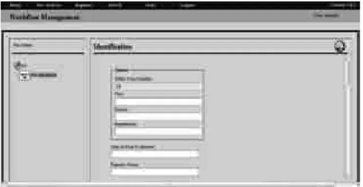
Fig 1. NMO-DBr software. Onset of registration of a new patient.
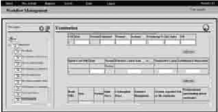
Fig 2. NMO-DBr software. List of symptoms in the course of the disease. The Tree on the left column indicates the task the system is currently executing.
formation Management Systems (LIMS). A LIMS is a complex computational system used to manage labora-tory data with emphasis in quality assurance28,29. In this work we used the Flux LIMS, a worklow based system to implement a medical record system.Flux uses JAVA as programming language, the Web pages were JSP and the J2EE technology was used for code development. Flux is based on the SIGLa system30. Applets are used for the graphical interface. he database was developed with MySQL, a robust, scalable and free DBMS. To deine a worklow, the Together Worklow Editor community edi-tion was used with the XPDL standard. For managing the worklows Flux uses a specialized worklow manager. A worklow manager consists of a set of functions that con-trols the activities of a worklow. he worklow manager maintains a list of activities that have been executed, as well as the order of execution and the activities that are available for execution.
RESULTS
NMO-DBr consists of standardized forms, a database system and thw web-based interface. he system guar-antees conidentiality through the use of passwords for system access as well as intellectual property for all par-ticipants through an approved NMO-DBr Code of Be-havior. Relationship with third parties is also regulated by NMO-DBr regulations.
he program contains items related to patients’ reg-istration and identiication (Fig 1), demographic, clinical and disability status data, as well as the results of labora-tory and imaging tests (Table 1).
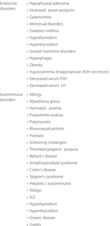
NMO-DBr shows a list of symptoms in the course of the disease (Fig 2). In addition to general symptoms the list contains symptoms related to involvement of the optic nerves and spinal cord, the cerebral cortex, corti-cospinal tract in its course from the frontal motor area through the posterior limb of the internal capsule and the cerebral peduncles, hypothalamus, brainstem and cerebellum (Table 2). he application conveniently dis-plays the list in separate anatomic or functional com-partments. his is followed by clinical characterization of the index events and recurrences regarding date, type of involvement (whether optic neuritis, myelitis or both simultaneously) and degree of recovery (Fig 3). NMO-DBr also includes a dropdown list of endocrine and au-toimmune disorders, identiied in association with NMO (Table 3), and collects the Family history of autoimmu-nity as shown in Fig 4.
Wing-Fig 4. NMO-DBr software. Family history of autoimmunity.
Fig 5. NMO-DBr software. Fields for collecting information on Wingerchuk’s Quantiication of Optic Nerve and Spinal Cord Im-pairment and Kurtzke’s Expanded Disability Status Scale.
Fig 6. NMO-DBr software. Fields for results of cerebrospinal luid analysis and indings of magnetic resonance imaging of the brain and spinal cord.
Table 2. Possible symptoms in the course of neuromyelitis optica.
Type Related symptoms
Optic nerve •
• •
Blurred vision
Scotoma and loss of VF Optic disc edema or atrophy
Spinal cord •
• •
Motor disturbances Sensory disturbances Sphincter/sexual disturbances
Posterior reversible encephalopathy syndrome
•
• •
Impaired consciousness, agitation, convulsion Visual disturbances Diplopia, nystagmus
Hypothalamus •
• • • • •
Hypothermia, fever Orthostatic hypotension Tachycardia
Sleep disturbances Hyperphagia
Endocrine disturbances
Brainstem •
• • • • • • • •
Nausea, vomiting Diplopia, nystagmus Miosis
Ptosis
Facial pain or dysestesia Facial paralysis Hearing loss
Vertigo and vestibular disturbances Ataxia
General symptoms • • • • • • • • •
Headache Pain
Lhermitte’s symptom Uhthof’s symptom Fatigue
Memory disturbances Other cognitive disturbances Anxiety
Depression
Other symptoms •
• • • •
Painful spastic paroxysms Tremor
Myoclonus Chorea Dyskinesia
Fig 3. NMO-DBr software. Fields for the irst index event with date, type and degree of recovery.
erchuk’s Scale for motor, sensory and sphincter impair-ment and Kurtzke’s EDSS (Fig 5) are similarly recorded.
he-molytic complement (CH50) and its fractions (C3 and C4), as well as vitamin B12, folate, methylmalonic acid, and homocysteine serum concentrations . Data on CSF included normal CSF, number of cells per cubic milli-meter; number of neutrophils per cubic millimilli-meter; number of eosinophils per cubic millimeter, concentra-tion of protein (mg/dL), IgG index, and presence of oli-goclonal bands outnumbering those of serum.
he section for collecting data on MRI contained the main findings we have observed in our cohort as well those so far reported by investigators in AQP-4 seroposi-tive patients10,12-15,24. Figure 6 displays the ields for CSF and
MRI data on NMO-DBr. As the most common and typ-ical indings are listed in the software the user has no dii-culty looking for them on patients’ individual exams. Data on thickness of the RNFL and macular volume and thick-ness are then collected in an equally user-friendly way. In the diagnosis section NMO-DBr takes into ac-count the sensitivity variability of the AQP4 antibody assays31-36 and the typical lesions which have been de-scribed on brain MRI10,12-15,24. In addition to choices within the widely accepted NMO spectrum disorders2,6 the system includes patients with formes frustres of NMO, typical lesions of NMO on brain MRI and AQP4-antibody seronegativity; and those who have brainstem syndrome, hypothalamic syndrome, or symptoms of en-cephalopathy in association typical lesions of NMO on brain MRI or AQP4-antibody seropositivity. hese latter diagnostic categories are named after NMO variants by NMO-DBr. A inal diagnostic choice is Not NMO.
The NMO-DBr next section was specifically de-signed to collect data on management of acute attacks and relapse prevention. Treatment of attacks included oral prednisone, intravenous pulse of methylpredniso-lone, plasmapheresis, and IV immunoglobulin. For each coursed of treatment the degree of recovery is recorded. Approach to relapse prevention included the use of oral prednisone, azathioprine, rituximab, intravenous immu-noglobulin, mitoxantrone, micophenolate mofetil and cy-clophosphamide. Data for each of these therapeutic reg-imens is then recorded with its corresponding eicacy.
DISCUSSION
By using a specialized data management system we have been able to create a worklow to specify the NMO patients data format and implement the medical records system in a short amount of time. In this work we used the Flux LIMS, a worklow based system to implement a medical record system. Flux incorporates a worklow management system, making it possible to create and manage customized worklows. For each new application a worklow is deined with its activities, rules and pro-cedures. During the execution, for each worklow cre-ated, the values of attributes deined in a worklow de-scription ile are stored in Flux’s database, allowing them to be managed and retrieved upon request. hese char-acteristics increase system’s lexibility and extend its us-ability to include the needs of multiple types of condi-tions. he resulting NMO-DBr turned to be a fast, easy and friendly to use system. It provides data consistency through automated cross-checking; performs statistical analysis, and guarantees conidentiality for patients and investigators alike.
Recent advances in the immunopathogenesis of NMO have emphasized the deleterious efect of
aqua-Table 3. Endocrine and autoimmune disorders associated with neuromyelitis optica.
Endocrine
disorders • Hypophyseal adenoma• Increased serum prolactin
• Galactorrhea • Menstrual disorders • Diabetes mellitus • Hypothyroidism • Hyperthyroidism
• Growth hormone disorders • Hyperphagia
• Obesity
• Hyponatremia (Inappropriate ADH secretion) • Decreased serum FSH
• Decreased serum LH
Autoimmune
disorders • Allergy• Myasthenia gravis
• Hemolytic anemia • Polyarteritis nodosa • Polymyositis • Rheumatoid arthritis • Psoriasis
• Sclerosing cholangitis • Thrombocytopenic purpura • Behçet’s disease
• Antiphospholipid syndrome • Crohn’s disease
• Sjögren’s syndrome • Hepatitis ( autoimmune) • Vitiligo
• SLE
porin-4 antibody. he lesions are initiated by its binding to AQP-4 in any part of the brain parenchyma, activating complement, and then destroying astrocytes11. As AQP-4 is ubiquitous in the CNS, many diferent manifestations antedating or even in the absence of optic neuritis or my-elitis may occur31-33. On the other hand, serum detection of anti-AQP4 antibody in NMO patients varies widely by diferent assays and in diferent populations34-36. Factors such as the use of corticosteroid or immunossupressant drugs may also interfere with the test sensitivity32.
NMO-DBr application takes into account these facts as it reckons data on neurologic symptoms antedating or not related to the classical NMO index events. he system, ultimately, allows to broaden the spectrum of the disease adding, among its formes frustres or NMO variants, patients who are anti-AQP4 antibody seronega-tive but, on the other hand, have typical NMO lesions on brain MRI in association with either just one of the index events, or with symptoms related to reversible encepha-lopathy, hypothalamus or brainstem syndrome.
he NMO-DBr system is a comprehensive and spe-ciic database to collect, store, retrieve, analyze and sup-port research, ultimately improving medical care in the expanding ield of NMO.
ACKNOWLEDGMENT – The authors would like to thanks Satya Sistemas for participating in this research by making the Flux System available and assisting in the development of the worklow.
REFERENCES
1. Wingerchuk DM, Hogancamp WF, O’Brien PC, Weinshenker BG. The clin-ical course of neuromyelitis optica (Devic’s syndrome). Neurology 1999;53: 1107-1114.
2. Wingerchuk DM, Lennon VA, Pittock SJ, Lucchinetti CF, Weinshenker BG. Revised diagnostic criteria for neuromyelitis optica. Neurology 2006;66: 1485-1489.
3. Lucchinetti CF, Mandler RN, McGavern D, et al. A role for humoral mecha-nisms in the pathogenesis of Devic’s neuromyelitis optica. Brain 2002;125: 1450-1461.
4. Lennon VA, Wingerchuk DM, Kryzer TJ, et al. A serum autoantibody marker of neuromyelitis optica: distinction from multiple sclerosis. Lancet 2004; 364:2106-2112.
5. Lennon VA, Kryzer TJ, Pittock SJ, Verkman AS, Hinson SR. IgG marker of optic-spinal multiple sclerosis binds to the aquaporin-4 water channel. J Exp Med 2005;202:473-477.
6. Wingerchuk DM, Lennon VA, Lucchinetti CF, Pittock SJ, Weinshenker BG. The spectrum of neuromyelitis optica. Lancet Neurol 2007;6: 805-815. 7. Lana-Peixoto MA. Devic’s neuromyelitis optica: a critical review. Arq
Neu-ropsiquiatr 2008;66:120-138.
8. Matà S, Lolli F. Neuromyelitis optica: An update. J Neurol Sci 2011;doi: 10.1016/j.jns.2011.01.002.
9. Bizzoco E, Lolli F, Repice AM, et al. Prevalence of neuromyelitis optica spec-trum disorder and phenotype distribution. J Neurol 2009;256:1891-1898. 10. Ito S, Mori M, Makino T, Hayakawa S, Kuwabara S. ‘Cloud-like enhancement’
is a magnetic resonance imaging abnormality speciic to neuromyelitis optica. Ann Neurol 2009;66: 425-428.
11. Saadoun S, Waters P, Bell AB, Vincent A, Verkman AS, Papadopoulos MC. Intra-cerebral injection of neuromyelitis optica immunoglobulin G and
human complement produces neuromyelitis optica lesions in mice. Brain 2010;133:349-361.
12. Pittock SJ, Lennon VA, Krecke K, Wingerchuk DM, Luchinetti CF, Weinsh-enker BG. Brain abnormalities in neuromyelitis optica. Arch Neurol 2006; 63:390-396.
13. Pittock SJ, Weinshenker BG, Lucchinetti CF, Wingerchuk DM, Corboy JR, Lennon VA. Neuromyelitis optica brain lesions localized at sites of high aquaporin 4 expression. Arch Neurol 2006;63:964-968.
14. Nakamura M, Misu T, Fujihara K, et al. Occurrence of acute large and edem-atous callosal lesions in neuromyelitis optica. Mult Scler 2009;15:695-700. 15. Kim W, Park MS, Lee SH, et al. Characteristic brain magnetic resonance
imaging abnormalities in central nervous system aquaporin-4 autoimmunity. Mult Scler 2010;16:1229-1236.
16. Chalumeau-Lemoine L, Chretien F, Larbi AGS, et al. Devic disease with brainstem lesion. Arch Neurol 2006;63:591-593.
17. Takahashi T, Miyazawa I, Misu T, et al. Intractable hiccup and nausea in neu-romyelitis optica with aquaporin-4 antibody: a herald of acute exacerba-tions. J Neurol Neurosurg Psychiatry 2008;79:1075-1078.
18. Viegas S, Weir A, Esiri M, et al. Symptomatic, radiological and patholog-ical involvement of the hypothalamus in neuromyelitis optica. J Neurol Neurosurg Psychiatry 2009;80:679-682.
19. Baba T, Nakashima I, Kanbayashi T, et al. Narcolepsy as an initial manifes-tation of neuromyelitis optica with anti-aquaporin-4 antibody. J Neurol 2009;256:287-288.
20. de Seze J, Blanc F, Jeanjean L, et al. Optical coherence tomography in neuromyelitis optica. Arch Neurol 2008;65:920-923.
21. Talim LE, Talim N, Couy M, et al. Relative frequency of demyelinating dis-eases in the State of Minas Gerais, Southeastern Brazil. Arq Neuropsiquiatr 2011;69 (Suppl 2):17.
22. Kurtzke JF. Rating neurologic impairment in multiple sclerosis: an Expanded Disability Status Scale (EDSS). Neurology 1983;33:1444-1452.
23. Green AJ, Cree BAC. Distinctive retinal neve iber layer and vascular changes in neuromyelitis optica following optic neuritis. J Neurol Neurosurg Psychi-atry 2009;80:1002-1005.
24. Bichuetti DB, Rivero RL, Oliveira DM, et al. Neuromyelitis optica: brain abnor-malities in a Brazilian cohort. Arq Neuropsiquiatr 2008;66:1-4.
25. Merle H, Olindo S, Donnio A, et al. Retinal peripapillary nerve fiber layer thickness in neuromyelitis optica. Invest Ophthalmol Vis Sci 2008;49: 4412-4417.
26. Ratchford JN, Quigg ME, Conger A, et al. Optical coherence tomography helps diferentiate neuromyelitis optica and MS optic neuropathies. Neu-rology 2009;73:302-308.
27. Naismith RT, Tutlam NT, Xu J, et al. Optical coherence tomography dif-fers in neuromyelitis optica compared with multiple sclerosis. Neurology 2009;72:1077-1082.
28. Hinton MD. Laboratory Management Systems. Marcel Dekker, Inc. New York, 1995.
29. Quo C, WDW. Development of a Laboratory Information System forCancer Colaboration Projec ts. 27th Annual International Conference of the
Engi-neering in Medicine and Biology Society 2005;22:108-112.
30. Melo A, Faria-Campos A, DeLaat DM, Keller R, Abreu V, Campos S. SIGLa: an adaptable LIMS for multiple laboratories. BMC Genomics 2010;11 (Suppl 5):S8. doi:10.1186/1471-2164-11-S5-S8.
31. Sato D, Fujihara K. Neuromyelitis optica without typical opticospinal phenotype. Mult Scler 2010;16:1154-1155.
32. Fujihara K. Neuromyelitis optica and astrocytic damage in its pathogenesis. J Neurol Sci 2011;306:183-187.
33. Tanaka A, Yoshida T, Yamada T, et al. A case of cerebral aquaporinopathy. Mult Scler 2010;16:1252-1254.
34. Waters P, Jarius S, Littleton E, et al. Aquaporin-4 antibodies in neuromye-litis optica and longitudinally extensive transverse myeneuromye-litis. Arch Neurol 2008;65:913-919.
35. Fazio R, Malosio ML, Lampasona V, et al. Antiacquaporin 4 antibodies detection by diferent techniques in neuromyelitis optica patients. Mult Scler 2009;15:1153-1163.