DEVIC’S NEUROMYELITIS OPTICA
A critical review
Marco Aurélio Lana-Peixoto
1Abstract – Devic’s neuromyelitis optica (NMO) is an idiopathic inflammatory demyelinating and necrotizing disease characterized by predominant involvement of the optic nerves and spinal cord. In Asian countries relapsing NMO has been known as opticospinal multiple sclerosis. It has long been debated if NMO is a variant of multiple sclerosis (MS) or a distinct disease. Recent studies have shown that NMO has more frequently a relapsing course, and results from attack to aquaporin-4 which is the dominant water channel in the central nervous system, located in foot processes of the astrocytes. Distinctive pathological features of NMO include perivascular deposition of IgG and complement in the perivascular space, granulocyte and eosinophil infiltrates and hyalinization of the vascular walls. These features distinguish NMO from other demyelinating diseases such as MS and acute demyelinating encephalomyelopathy. An IgG-antibody that binds to aquaporin-4, named NMO-IgG has high sensitivity and specificity. Magnetic resonance imaging (MRI) studies have revealed that more frequently there is a long spinal cord lesion that extends through three or more vertebral segments in length. Brain MRI lesions atypical for MS are found in the majority of cases. Treatment in the acute phase includes intravenous steroids and plasma exchange therapy. Immunosupressive agents are recommended for prophylaxis of relapses.
KEY WORS: Devic’s neuromyelitis optica, opticospinal multiple sclerosis, recurring optic neuritis, transverse myelitis, aquaporin-4, NMO-IgG antibody.
Neuromielite óptica de Devic: revisão crítica
Resumo – Neuromielite óptica ou doença de Devic (NMO) é uma doença inflamatória com desmielinização e necrose envolvendo preferencialmente os nervos ópticos e a medula espinal. Desde sua descrição inicial tem havido controvérsia se a NMO é uma variante da esclerose múltipla (EM) ou se é uma entidade independente. Na Ásia a doença é conhecida como esclerose múltipla óptico-espinal. Recentes avanços tem demonstrado que na maioria dos casos a NMO é recorrente e resulta de alterações inflamatórias por ataque à aquaporina-4, uma proteína localizada nos pés dos astrócitos na barreira hemato-encefálica. Patologicamente a NMO difere da EM pela presença de necrose e cavitação no centro da medula, deposição perivascular de IgG e complemento, infiltração de neutrófilos e eosinófilos, assim como por hiperplasia e hialinização dos vasos. O anticorpo contra a aquaporina-4 (IgG-NMO), detectado no soro dos pacientes, tem alta sensibilidade e especificidade. Imagem por ressonância magnética demonstra lesão medular que se estende três ou mais segmentos vertebrais. Na maioria dos casos há lesões cerebrais atípicas para EM. Corticosteróide venoso em altas doses e plasmaférese são usados no tratamento das fases agudas, enquanto os imunossupressores devem ser usados na profilaxia das recorrências.
PALAVRAS-CHAVE: neuromielite óptica, doença de Devic, esclerose múltipla óptico-espinal, neurite óptica recorrente, mielite transversa, aquaporina-4, IgG-NMO.
1MD, Associate Professor of Neurology and Ophthalmology. Department of Ophthalmology and CIEM Multiple Sclerosis Investigation Center, Federal University of Minas Gerais Medical School, Belo Horizonte, MG, Brazil.
Received 22 January 2008. Accepted 4 February 2008.
Dr. Marco Aurélio Lana-Peixoto – Rua Padre Rolim 769 / Conj 1301 - 30130-090 Belo Horizonte MG - Brasil. E-mail: [email protected]
Devic’s neuromyelitis optica (NMO) or Devic’s disease is a severe idiopathic immuno-mediated inlammatory de-myelinating and necrotizing disease that predominantly involves the optic nerves and spinal cord. Recent immu-nopathological evidences suggest that the target antigen
is aquaporin-4, the dominant water channel in the central nervous system (CNS), located in the astrocyte foot pro-cesses at the blood-brain barrier.
was considered a variant of MS, characterized most com-monly by a monophasic course of bilateral optic neuritis and transverse myelitis occurring concomitantly or with-in a short with-interval, and no evidence of disease outside the optic nerves and the spinal cord. Studies of MS in Asia revealed that compared with MS in Western countries, Asian MS patients had more frequent and severe involve-ment of the optic nerves and spinal cord, and a relatively more rapid progression1-3. These observations led Japanese
authors3-7 to divide their patients who did not meet the
criteria for classical Devic’s disease, into two other differ-ent subtypes of MS. Patidiffer-ents who had a relapsing-remit-ting course with involvement of the optic nerves and spi-nal cord and no clinical evidence of disease either in the
cerebrum or the cerebellum were considered to have
op-ticospinal MS. Those with minor brainstem signs, such as nystagmus and diplopia in addition to the opticospinal in-volvement were included in this group. Patients with signs of multiple involvement of the central nervous system, in-cluding the cerebrum and cerebellum were considered to have conventional MS. It was suggested that classical De-vic’s NMO and conventional MS represent two extremes of a spectrum of the same condition3.
Recent clinical, pathological, immunological and imag-ing studies8-13, however, have suggested that most patients
with NMO have a relapsing course8, the disease has
dis-tinctive histopathological and immunopathogenetic fea-tures9-11, and that most patients have magnetic resonance imaging (MRI) brain lesions which are atypical for MS12. It has also been shown that opticospinal MS and relapsing
NMO are most likely the same disease14.
In addition to the idiopathic and pure form of NMO the co-occurrence of optic nerve disease and transverse my-elopathy has been described in association with a variety of conditions such as connective tissue disorders, bacterial and viral infections, and others. These observations might suggest that NMO would be more appropriately consid-ered as a clinical syndrome rather than a single disease. This review focuses on the historical aspects, epidemi-ology, genetics, pathology and imunopathogenesis, clini-cal features, cerebrospinal luid (CSF) abnormalities, MRI indings, diagnostic criteria and current treatment of De-vic’s NMO.
HISTORICAL OVERVIEw
The association between acute myelitis and optic nerve disorder was irst described by Albutt15 in 1870. Nine
years later Erb16 reported on a 52-year-old man with
re-curring optic neuritis in whom transverse myelitis super-vened three months later. The patient ultimately made a complete recovery from the myelitis and a practically complete recovery of visual function. Seguin17 in 1880 was the irst author to report a case in America. Other cases
with this association were also reported in 1880 by Stef-fan18 and Noyes19. In 1882 Chisholm20 recorded a case in which death occurred only 12 days after the onset drawing attention to the severity of the disease. In the same year,
Dreschfeld21 demonstrated for the irst time in an
autop-sied case the occurrence of inlammatory iniltrates in the optic nerves and spinal cord but no brain abnormalities. He credited Gowers for recognizing that optic neuritis and myelitis were both the result of a common cause.
In 1884 Eugène Devic22 reported the case of a 45-year-old French woman seen at the Hôtel-Dieu Hospital of Lyon because of intractable headache and depression in addition to general asthenia. One month later she devel-oped urinary retention, complete paraplegia and blind-ness, and died few weeks later. Autopsy disclosed severe demyelinating and necrotic lesions extending for 4-5 cm length in the lower thoracic and lumbar spinal cord.
The lesions involved both the white and gray matter and were associated with cellular iniltrates and thicken-ing of the vessels walls. There was demyelination of the optic nerves but gross examination of the brain was unre-vealing. Devic22 presented this case at the First Congress of Internal Medicine in Lyon, and mentioned 16 other cas-es reported in the literature. In his paper Devic proposed the identity of the pathological process involving both the spinal cord and the optic nerves, named the syndrome “neuro-myélite optique” or “neuroptico-myélite”, and dis-cussed its relationship with MS.
Fernand Gault23, a disciple of Devic’s, reviewed in de-tail these 17 cases in his doctoral thesis named “De la neu-romyélite optique aigué” and suggested that they repre-sented a distinct nosological entity. The eponym “Devic’s disease” was suggested by Acchiote24 in 1907.
In 1914 Goulden25 reviewed 51 cases in the literature fulilling the diagnostic criteria as proposed by Devic, and added one case of his own. Beck26 in 1927 reviewed 71 cas-es and dcas-escribed rarefaction of the optic nervcas-es and spi-nal cord, polymorphonuclear iniltrates, extensive demy-elination and necrosis extending over multiple segments of the spinal cord. He emphasized that these pathological abnormalities were distinct from those found in MS.
anal-ysis usually died few months after onset of the disease. He proposed that the lesions progressed through a series of different stages. At onset there are prominent perivas-cular iniltrates of polymorphonuclear cells, leucocytes and plasma cells. Then perivascular foci of demyelination and necrosis ensue to coalesce into larger lesions with ax-onal damage. The spinal cord gray matter may be distinc-tively affected or may be involved by extension of the ad-jacent white matter lesions. Necrotic areas are commonly seen in the spinal cord and less frequently within the op-tic nerves. Finally, glial scars are formed although scarring is less frequent and usually only partial in contrast to typ-ical MS lesions. Scott30 in 1952, commenting on Stanbury’s review, disagreed over his conclusion about the inexora-bly poor outcome of the disease and drew attention to cases with good functional recovery. He described cases with visual loss associated with minimal spinal cord dys-function and even the occurrence of “abortive types” with binocular loss of vision not followed by paraplegia. Simi-lar observations had been previously reported by Walsh31. By 1958 over 300 cases of Devic’s disease had already been reported in the literature and the condition was
consid-ered more frequent than previously thought32.
In Latin America NMO was irst reported by Aluizio
Marques33 in 1943 who described two cases in Rio de
Ja-neiro. The irst patient was a 21-year-old mulattoe female who developed simultaneous bilateral blindness and para-plegia in the course of mumps. Cerebrospinal luid
ex-amination disclosed 17 cells /mm3. Clinical examination
three months later revealed partial recovery of the vi-sion and complete recovery of the motor, sensory and sphincter functions. The second case was of a 45-year-old white woman who presented acute transverse myeli-tis followed a month later by bilateral optic neurimyeli-tis. Ex-amination nine months after the onset of the disease dis-closed no recovery.
The concept of Devic’s NMO has been debated since Devic formulated his famous questions “Why such a pe-culiar localization?” “What is the intimate nature of the process?”. To overcome these unanswered questions many clinical criteria for the diagnosis of the disease have been set forth by different investigators. Initially, the mono-phasic course of bilateral optic neuritis and myelitis as well as the simultaneity or short interval between the
in-dex events were emphasized. In 1894, Gault and Devic23
proposed that the diagnosis should be restricted to cases with bilateral optic neuritis and acute myelitis occurring simultaneously or within a few weeks apart. Other signs of CNS involvement could be occasionally observed. Shi-basaki et al.34 in a study of the differences between Brit-ish and Japanese MS considered as having Devic’s disease patients with both acute bilateral optic neuritis and acute myelitis – with no other signs of involvement of nervous
system - occurring in a monophasic course within an in-terval of four weeks.
Imaging, CSF and pathological features were added to clinical indings in the diagnostic criteria suggested by
Mandler et al.35 in 1994. Additionally to the occurrence
of optic neuritis and myelitis, which could be coinciden-tal or separated by an interval of months to years, pa-tients should not have signs of involvement beyond the optic nerves and spinal cord. Magnetic resonance imaging (MRI) criteria included normal brain and the presence of signs of cavitation in the spinal cord. CSF analysis should reveal decreased serum/CSF albumin ratio; normal IgG synthesis and usually absent oligoclonal bands. Patholog-ical features included necrosis and cavitation of the spinal cord with absent or scant inlammatory iniltrates. There should be signs of demyelination in the optic nerves, with or without cavitation whereas no lesions were to be found in the brain, brainstem and cerebellum.
In 1996 O’Riordan et al.36 reviewed the case records of patients seen at the National Hospital of Neurology and Neurosurgery, Queen Square and Moorield’s Eye Hospi-tal between 1986 and 1994. The disease was deined as a complete acute and severe transverse myelitis associat-ed with acute unilateral or bilateral optic neuritis and no signs of brain involvement beyond the optic nerves. The disease could follow a monophasic or relapsing course. Interestingly in ive of their 12 cases a probable etiolo-gy could be identiied. Two patients had probable “acute disseminated encephalomyelitis” two weeks after a non-speciic infectious illness and MRI showed swelling and abnormal sign extending over many segments of the spi-nal cord. One patient had systemic lupus erythematosus, another mixed connective tissue disease, and a third pul-monary tuberculosis. Six other patients exhibited a vari-ety of organ-speciic autoantibodies, further pointing to an immunological mechanism for the disease.
Starting in 1999 a series of investigations conducted at the Mayo Clinic have changed the very concept of NMO and clariied a number of issues concerning its clinical fea-tures, pathology, immunological mechanisms, and imag-ing8-12,37. In a study of 71 patients with the association of optic neuritis (unilateral or bilateral), acute myelitis and no evidence of clinical disease outside of the optic ner-veor spinal cord Wingerchuk et al.8 showed that the great majority of cases have a relapsing course; that patients with monophasic course have more severe index events; that over one third of patients have CSF pleocytosis with
more than 50 cells/mm3; and that a spinal cord MRI with
dif-ferences from MS. Distinctly from MS, in NMO there are a pronounced perivascular deposition of immunoglobulin and complement, eosinophilic iniltration and prominent vascular ibrosis and hyalinization within the lesions. They concluded that these indings support a major role of hu-moral immunity in the pathogenesis of NMO. The next work in this line of investigation was published by Lennon et al.10 in 2004 who identiied a serum autoantibody mark-er of NMO named NMO-IgG, which can distinguish NMO from MS and other demyelinating disorders. The
follow-ing year Lennon et al.11 demonstrated that the NMO-IgG
selectively binds to aquaporin-4 (AQP-4), the predomi-nant water channel within the CNS located in foot pro-cesses of the astrocytes in the blood-brain barrier. Aqua-porins have been known since the seminal study by Pe-ter Agre in 1988 who discovered the irst waPe-ter channel, termed aquaporin 1 and was awarded the Nobel Prize of Chemistry in 200338. Finally Pittock et al.12 reported the inding of nonspeciic brain MRI lesions in the majority of patients with NMO.
EPIDEMIOLOgY
Studies on epidemiology of NMO are still scanty and the data may be misleading as different authors have used distinct terminology, deinition of the disease, diagnostic criteria and methods of survey.
Cases of Devic’s NMO have been reported in all conti-nents and races although the disease is more prevalent in areas with Black, Asian and Indian populations, where MS prevalence is usually low39,40. In Africa and Asia classical MS is rare2,39-41. In Nigeria, Osuntokun42 reported 95 cases of NMO among 97 cases of MS. In South Africa recurrent NMO was observed in seven of eight black patients with demyelinating disease43. In Asia, low prevalence rates of MS have been reported in Japan2,39-41, China44-46, Taiwan47, Ko-rea48, India49, Malaysia50 and Thailand51. Two recent studies in Japan, quoted by Kira40, reveal prevalence rates of 8-9/10-5, a much higher igure than those revealed by early epide-miological studies52 which suggested rates of 0.7-4.0/10-5.
On the other hand there have been many reports about the higher rate of NMO in Asians than in West-ern populations 2,40,41. In the irst Japanese study Okina-ka1 found that out of the 270 cases of demyelinating dis-ease collected in Japan, there were 175 cases of NMO and 66 cases of conventional MS. Kuroiwa et al.53 in 1975 con-ducted a nationwide survey of demyelinating diseases in Japan and found strict Devic’s disease in 7.6% of the 1084 cases collected. In the MS group analysis of the site of in-volvement disclosed lesions in the optic nerves and spi-nal cord in 80% of the cases, whereas the cerebrum was affected in 43%, the brainstem in 70% and the cerebellum in 47%. In an analysis of 488 cases of demyelinating dis-eases in Asian countries, using the same deinitions and
diagnostic criteria, the authors54 found strict Devic’s dis-ease in 7% of the multiple sclerosis group. In a compari-son study between British and Japanese MS patients, Shi-basaki et al.34 found Devic’s disease in none of 204 British cases, in comparison with 5% in the Japanese group. Ad-ditionally, their study showed that the proportion of the opticospinal form among all cases of MS was 42% in Ja-pan and 6% in Britain.
The higher proportion of strict Devic’s disease among MS cases has also been reported in India55-58 (11%-42%), Ko-rea59 (7%), and Philippines60,61 (13%-20%).
One study of MS in Algonkian and Athapaskan
indig-enous people in Manitoba62, Canada, identiied ives
cas-es with the NMO phenotype among seven cascas-es of MS. Autopsy in one of these patients showed marked demye-lination and necrosis of the spinal cord with inlammato-ry iniltrates of lymphocytes and eosinophils, similar his-topathological features to those more recently described by Lucchinetti et al.9.
The frequency of the opticospinal MS has been
re-ported as 36% of MS cases in Hong Kong46, 43% in
Tai-wan44, and 40% to 57% in Japan,39,63,64 although lower (16%) in northern Japan65. Interestingly opticospinal MS has not
been reported in Mediterranean Arabs66 but is frequent
among MS cases in Arabs from Gulf countries67.
In the Caribbean basin the relative frequency of NMO has been studied in the African descent population of
Martinique68,69. In a population-based study in French
Afro-Caribbeans, using the Wingerchuk et al. original di-agnostic criteria8 Cabre et al.68 found 13 cases (17.3%) of relapsing NMO among 75 cases of demyelinating diseas-es. Recently they described a series of 35 (17%) cases in a cohort of 206 cases of demyelinating diseases in which isolated optic neuritis cases were included69. In
Argenti-na one study70 showed that among 134 cases of deinite
MS 10 (7.5%) fulilled the Wingerchuk et al.’s criteria for NMO. In Brazil Lana-Peixoto et al.71 in a study of 67 con-secutive patients with demyelinating disease found 39 patients with classical MS, 20 with opticospinal MS and eight with strict Devic’s disease, suggesting that the optic nerves and spinal cord are more frequently affected in the Brazilian population than in the Caucasian population of North America and Europe. A more recent Brazilian study from a hospital in Rio de Janeiro72 describes the features of a series of 24 NMO patients (20 women and four men; ages at onset 14 to 55 years, mean 32.8) but the relative frequency of the disease among other demyelinating dis-orders is not mentioned. In their series Blacks comprised 14 cases and 22 patients had relapsing disease.
The age of onset ranges from childhood73,74 to late
was 41 years in the relapsing and 29 years in the mono-phasic group.
One recent study of 17 cases under 18 years of age revealed that the median age in pediatric cases was 4.4 years80. On the other hand cases with late onset have been also reported8,79,81. The eldest reported patient at the on-set of the disease was an 81-year-old woman who
devel-oped NMO one month after inluenza immunization81.
Although the gender distribution is variable in differ-ent series women are affected more frequdiffer-ently than men, usually in a ratio higher than in MS. This trend is most evi-dent in China where a study46 showed female to male ratio as high as 9:146. Female to male ratio was 5:1 in a Brazilian series72; whereas in the Mayo Clinic series8 it varied accord-ing to the clinical type of the disease, beaccord-ing 1:1 in mono-phasic NMO and 5:1 in recurrent NMO. Similarly 28 (93%) of the 35 French-Afro-Caribbeans in Martinique with NMO
were women and had the relapsing type of the disease69.
In the pediatric group female to male ratio was 3.2:180.
gENETICS
In the Japanese population the frequency of HLA-DPB1*0501 allele was found to be signiicantly greater (93%) in patients with opticospinal MS than in healthy controls
(63%) but not in patients with conventional MS (66%)82.
A more recent study83, however, suggested that
op-ticospinal MS is not necessarily associated with the DPB1*0501 allele. The authors determined the frequen-cies of the DRB1*1501, DPB1*0501 and DPB1*0301 in 26 patients with opticospinal MS, 167 with convention-al MS and 156 normconvention-al subjects. All opticospinconvention-al MS pa-tients were negative for DPB1*0301 whereas the fre-quency of the DPB1*0501 allele was similar in optico-spinal MS and conventional MS yet higher than in the healthy controls. In DPB1*0301 positives a frequency of the DPB1*0501 was low but similar in conventional MS and controls. They also found that the DPB1*0301 al-lele was strongly associated to the development of peri-ventricular lesions which were found in 97% of the con-ventional MS patients who were DPB1*0301 positive in contrast with only 16% of those who were DPB1*0301 negative, and 8% of the opticospinal MS patients.
It is somewhat surprising that given the genetic un-derpinning of the disease only ive pairs of familial cases
have been reported in the literature. McAlpine84 in 1938
described two identical twins with severe cervicothorac-ic myelitis and bilateral optcervicothorac-ic neuritis, occurring in one at age 24 and the other at age 26. Both patients died 18 and 26 months after onset. Autopsy showed marked demye-lination of both optic nerves and spinal cord with mild or no inlammatory iniltrates. In one case there was a demy-elinating lesion in the lower medulla, whereas in the other case examination of the brain was unrevealing. Ch´len et
al. 85 reported two sisters aged 10 years and 6 years who developed NMO at ages 3 years and 2 years 9 months respectively. Both presented bilateral optic neuritis
fol-lowed by myelitis ive months later. Another paper86
de-scribed NMO in two elderly Japanese sisters with onset at 59 and 62 years of age. The cases of two other sisters of Spanish –American ancestry who developed NMO at
ages 26 and 28 were also documented87. Recently Bradley
and Mikol88 reported by the irst time a
mother-daugh-ter pair with NMO. Differently from other familial cases these patients developed NMO in different stages of life; the daughter at 29 years of age whereas the mother at 62. An additional feature in these cases was the previous his-tory of thymectomy for myasthenia gravis in the daugh-ter. The occurrence of NMO following thymectomy for
myasthenia gravis has been well documented89.
PATHOLOgY AND IMMUNOPATHOgENESIS The basic structural pathological features of NMO have long been known35, 90, 91. In the acute phase of the disease gross pathology of the spinal cord includes dif-fuse swelling and softening extending over multiple spi-nal segments and occasiospi-nally over the entire extension of the cord. Histopathological examination discloses ne-crosis of both grey and white matter with macrophage iniltration associated with myelin and axonal loss. There is variable perivascular inlammatory iniltration. Late in the course of the disease atrophy and cavitation of the involved spinal cord segments and optic nerves ensue, with marked gliosis and cystic degeneration. In necrotic and perinecrotic areas the walls of the microvessels are thickened and hyalinized.
The structural and immunopathological features of NMO were compared with cases of MS, acute demye-linating encephalomyelitis (ADEM) and acute spinal cord infarction9. It was observed that T-cells and macrophages were present to a variable degree in all of them. Howev-er 52% of the MS cases and none of the ADEM and spi-nal cord infarction cases showed deposition of IgG and activated complement. In MS the pattern of deposition was different from that seen in NMO as it was less pro-nounced and found in degenerating myelin sheaths, along with macrophages and oligodendrocytes in the plaque edge, as opposed to the perivascular pattern described in NMO lesions. In an earlier study Lucchinetti et al.92 identi-ied four immunohistochemical types of MS lesions. The most common type is the “pattern 2” type characterized by deposits of complement and immunoglobulins. How-ever, immune complexes in “pattern 2” MS lesion are char-acteristically located at sites of myelin destruction. No complement activation and immunoglobulin reactivity was seen in acute spinal cord infarctions or ADEM cases. Eosinophils and granulocytes are observed in rare cases of fulminant Marburg MS (4% of MS cases) compared with
56% of NMO cases9. All early active NMO lesions
con-tained eosinophils whereas no eosinophils were present in ADEM or spinal cord infarction cases. Hyalinized ves-sels were present in all NMO cases but absent from MS, ADEM or spinal cord infarction cases9.
The pattern of tissue inlammation in early demye-linating active NMO with a unique perivascular pattern supported a role for humoral autoimmunity in the patho-genesis of the disease. The abnormalities of the perivas-cular region with macrophage iniltrate and massive de-position of complement and immunoglobulin associated with the prominent vascular hyalinization deinitely led the authors to suggest the perivascular space as the pri-mary target site of the pathogenic process.
In addition to the lesion pathological features dif-ferent lines of evidence favor humoral immune mecha-nisms in the pathogenesis of NMO. Firstly, there are strik-ing similarities between NMO and a variant of MOG-in-duced experimental allergic encephalomyelitis (EAE) in Brown Norway rats which develop a marked antibody re-sponse associated with pronounced demyelination main-ly affecting the optic nerves and the spinal cord93. Sec-ondly, NMO patients have a number of circulating auto-antibodies and many of them have autoimmune co-mor-bidities such as Sjögren syndrome, systemic lupus erythe-matosus and mixed connective tissue disease94-103. Final-ly, some treatment peculiarities such as improvement of corticosteroid-refractory acute attacks following plasma
exchange104,105 and a better response with general
immu-nossupression than with standard MS immunomodulato-ry drugs106,107 strongly support the role of autoantibodies in the disease process.
On the basis of the immunopathological observations a cascade reaction initiated by the presence of a periph-eral antibody directed against a perivascular antigen
acti-vating the classical complement pathway was proposed9.
Activated macrophages, eosinophils and neutrophils gen-erate cytokines, proteases and free radicals leading to vas-cular and parenchymal damage. Increased vasvas-cular perme-ability will cause futher parenchymal lesion via secondary ischemia. This mechanism may account for the typical lo-cation of the lesion within the spinal cord. Novel antigens liberated during the destructive process may further ex-tend the immune response (Fig 1).
A large number of patients with NMO have brain le-sions on MRI, most of them asymptomatic, but some are clinically evident12. Two autopsy reports by different in-vestigators108,109 describe the pathological features of brain
lesions in NMO patients. Nakamura et al.108 found marked
tissue destruction, cavities and inlammatory changes typ-ical of NMO in the cerebrum, optic nerves and spinal cord of a 63-year-old patient who had an encephalopathy with a cerebral tumefactive lesion 30 years earlier, followed by repeated attacks of optic neuritis and myelitis . More
re-cently Hengstman et al.109 found typical NMO
histopath-ological abnormalities in brain lesions at autopsy of a 35-year-old woman with NMO and multiple brain lesions on MRI. In some of these lesions there were perivascular in-iltrates with large number of eosinophils. These cases show that symptomatic brain lesions may occur in NMO and that the histopathological features of these brain le-sions are identical to those found in the spinal cord and optic nerve.
The reason for the preferential involvement of the op-tic nerves and spinal cord in NMO remains to be discov-ered. It is probable, however that these structures may harbor unique antigenic characteristics driving the
im-Perivascular antigen Activation of complement pathway
Activated macrophages, eosinophils, neutrophils
Cytokines, proteases, free radicals
Vascular damage
Parenchymal damage
Increased vascular permeability Edema Ischemia Novel antigens liberation
mune response, and through epitope spreading follow-ing lesions in these sites the immune response may
broad-en damaging other cbroad-entral nervous system structures109.
The NMO autoantibody marker
Accumulating evidences of humoral mechanisms in the pathogenesis of NMO prompted the identiication of a speciic IgG autoantibody (NMO-IgG) that
localiz-es to blood-brain barrier10. Indirect immunoluorescence
with a composite substrate of mouse tissue showed that NMO-IgG yields a characteristic immunohistochemical pattern of binding in mouse central nervous system. It was prominent in pia and subpia, and outlined the Virchow-Robin space and microvessels in white and grey matter of the cerebellum, midbrain, and spinal cord. It binds to an antigen in the abluminal face of cerebral microvessels, an area represented by astrocytic foot processes.
Lennon et al.10 tested masked serum samples from 102
North American patients with either NMO, high risk syn-drome for NMO or MS (presented with myelitis and op-tic neuritis but did not meet the Wingerchuk et al.’s8 crite-ria for the diagnosis of NMO), and 22 Japanese patients (11 with opticospinal MS, one with high risk syndrome for MS, ive with classical MS and ive with cerebral infarction). They identiied 33 (73%) of the 45 patients with NMO and 16 (45%) of the 35 with high risk syndrome for NMO who were seropositive for this antibody. Two (9%) of the 22 patients with diagnosis of MS were also seropositive for NMO-IgG. Thus this test showed a sensitivity of 73% and speciicity of 91% to differentiate patients with clinical-ly deinite NMO from those with myelitis and optic neu-ritis but could not the strict criteria for the diagnosis of NMO. When NMO and high risk syndrome are considered together the sensitivity drops to 61.3% and the speciicity to 90.9%. The 56 control patients with miscellaneous au-toimmune and paraneoplastic neurologic disorders were all seronegative. In the Japanese group none of the pa-tients with classical MS or cerebral infarction was sero-positive. Of the 12 patients with opticospinal MS or high risk syndrome for NMO seven (55%) were seropositive. In this group, therefore the sensitivity of the test was 58% and the speciicity 100%.
Since the substrates of the NMO-IgG detection assay were not human but mouse brain tissue, there has been a concern that the non-human substrates could be af-fecting its sensitivity and speciicity11. To clarify this is-sue Japanese investigators110 studied the sensitivity and speciicity of a new test using human AQP4-transfected cells as substrates of the indirect immunoluorescence as-say. They tested 148 sera of patients with NMO, high risk syndrome, MS, clinically isolated syndrome suggestive of MS and miscellaneous diseases. The sensitivity of the as-say, named anti-AQP4 antibody asas-say, increased to 91% for
NMO and 85% for high risk syndrome, and the speciicity was 100% for NMO and high risk syndrome as compared with the assay with non-human antigen. Changes from ini-tial negativity to later positivity over some years have also been observed in some patients with typical clinical and imaging features of the disease111.
Anti-AQP4 antibodies can be also detected in patients with the diagnosis of classical MS who have longitudinally extensive spinal cord lesions on MRI scans. In the Matsuo-ka et al.’s series111 two of 17 (11.8%) of these patients were seropositive, and the antibody was detected only in those with spinal cord lesions extending over 10 vertebral seg-ments in length. The anti-AQP4 antibody assay was con-sistently negative in all classical MS patients with longitu-dinally extensive spinal cord lesions shorter than 10 seg-ments in length. The anti-AQP4 titer had no correlations with disease duration, number of exacerbations or effects of immunotherapies, but had an inverse correlation with both the EDSS score and the progression index111.
Aquaporin-4 water channel
NMO-IgG from NMO-positive patients’ serum was found to bind to distal urine-collecting tubules and to basolateral membranes of the epithelial cells of the gas-tric mucosa. This distribution suggested the water chan-nel protein, aquaporin 4 (AQP4) as the target autoantigen in NMO. Aquaporin 4 is an integral protein of astrocytic plasma membranes and is highly concentrated in the as-trocyte foot processes.
Aquaporins constitue a family of water channels that regulate the transport of water in many organs including the nervous system, kidney, gastrointestinal tract, secre-tory glands, inner ear and muscles112. Aquaporin 4 is a ho-motetrameric integral plasma membrane protein found in electrically excitable tissues including brain and spinal cord, retina, inner ear and skeletal muscles. In general it is not expressed in excitable cells, but is found in support-ing cells (astrocytes and ependyma in the nervous system; Müller cells in the retina; Hensen’s and inner sulcus cells
in the ear113. It is the most abundant aquaporin in
mam-malian brain and is concentrated at the blood-brain barri-er, anchored in the astrocytic foot process membrane by
the dystroglycan complex114. Aquaporin-4 is also present
co-local-ization suggests that water and K+ lux are coupled116. It also co-localizes with the glutamate transporter-1 (GLT-1), which is one of the two main astrocytic excitatory ami-no acid transporters, that prevent excessive accumulation of extracellular glutamate117. The strategic localization of AQP4, together with Kir 4.1 and GLT -1 at the perivascu-lar and subependymal end-feet provides several poten-tial mechanisms by which loss of AQP4 may result both in severe damage of myelin and axons in vulnerable areas, such as the spinal cord and optic nerves, as well as revers-ible edema in other brain regions, such as the hypothal-amus and periventricular structures118. Perivascular AQP4 allows a bi-directional water low between the blood and the brain and has been implicated in the pathogenesis of
cerebral edema119. Aquaporin-4 is up-regulated in
astro-cytes in the setting of hypoxia, brain injury, meningitis and encephalitis, high grade astrocytomas, and around meta-static adenocarcinoma. Aquaporin-4-null mice, which lack AQP4 expression at astrocyte end feet, are relatively re-sistant to the development of brain edema in the setting of hypoosmolarity or stroke120.
The distribution of AQP4-rich areas in the central ner-vous system, especially in the central part of the spinal cord, hypothalamus, periventricular area and periaque-ductal areas is highly compatible with that of NMO le-sions121-123. Recent studies124-126 have demonstrated loss of AQP4 in lesions of the spinal cord in NMO patients but not in MS lesions. On the opposite, AQP4 expression may be increased in and around MS plaques. Misu et al.124 re-ported a case of typical NMO which showed a loss of AQP4 and glial ibrillary acidic protein (GFAP) immunos-tainning especially in active spinal cord lesions. In a
sub-sequent study Misu et al.127 conducted an
immunohisto-chemical analysis of 12 cases of NMO, six of MS, 7 of brain and spinal cord infarction, and eight normal controls. They observed loss of AQP4 in 90% of the acute and chronic NMO lesions, which were more pronounced in the active perivascular lesions where immunoglobulins and comple-ments were deposited. In contrast, AQP4 immunoreactivi-ty was increased in MS lesions. In normal controls and and infarctions AQPO4 was diffusely expressed but the stain-ing in the spinal cord was stronger in the grey matter than in the white matter. In NMO cases the areas surrounding the AQP-4-absent lesions had AQP4 expression similar to that of controls127.
Similarly one study126 demonstrated loss of AQP4 in op-tic nerve and spinal cord lesions of a NMO case as well as increased expression of AQP4 in all active and some inac-tive lesions from seven cases of secondary progressive MS. They also observed higher expression of AQP4 in normal-appearing white matter of MS patients than in controls.
Roemer et al.125 analysed and compared patterns of
AQP4 immunoreactivity in nervous tissues of nine patients
with NMO, 13 with MS, nine with infarcts and ive normal controls. In normal brain, optic nerve and spinal cord the distribution of AQP4 expression resembles the vasculo-centric pattern of immune complex deposition observed
in NMO lesions9. Cerebral white matter showed limited
AQP4 reactivity whereas the reactivity was most intense at glia limitans and subependyma. Within the cortex AQP4 reactivity was concentrated in astrocytic foot processes extending to abluminal surface of blood vessels. With-in the braWith-instem AQP4 predomWith-inated With-in subependymal regions of the fourth ventricule, in a rim or rosette pat-tern. In the spinal cord it was prominent within both grey and white matter in a rim or rosette pattern. In the optic nerve the staining was also in a rim and rosette pattern. Therefore the rim and rosette patterns found in NMO le-sions just represent the normal patterns at sites of AQP4 concentration125.
AQP4 expression in MS lesions correlates with the
stage of demyelinating activity126. In acute MS lesions
AQP4 is diffusely increased in the periplaque white mat-ter of active lesions, whereas chronic inactive lesions are devoid of AQP4. In some MS cases there are foci of in-lammatory iniltrates lacking demyelination, associated with AQP4 loss. Loss of AQP4 is not due to necrosis and cavitation. In NMO two basic pathology patterns can be found. The most prevalent lesion pattern involves the spi-nal cord and optic nerves. Loss of AQP4 occurs in the con-text of vasculocentric immune complex deposition, ac-tive demyelination and vascular hyperplasia with hyalin-ization. These lesions are frequently cavitary and involve both the grey and white matter in the spinal cord. The less frequent lesion pattern is found in the spinal cord and me-dulla extending into the area postrema, and is highly in-lammatory but with no evidence of demyelination. The area postrema has high expression of AQP4 and lacks a blood-brain barrier. It is related to control of osmoregu-lation and brain homeostasis including control of blood low autoregulation, edema and immune regulation.
Although it has been suggested that AQP4 autoanti-bodies cause NMO, probably by inhibiting AQP4,11,110,127-129
other authors130 keep a more skeptical view pondering
CLINICAL fEATURES
Neuromyelitis optica may present either with the si-multaneous occurrence of acute transverse myelitis and optic neuritis or with these index events occurring sep-arately by an indeterminate time interval. Following in-stallation of disease by the presence of both myelitis and optic neuritis, its course may be either monophasic with no further events, or relapsing with additional attacks of either transverse myelitis, optic neuritis or both. In the
Wingerchuk et al.8 series 48 (68%) of the 71 NMO cases
had a relapsing and 23 (32%) a monophasic course. Re-lapsing course is more frequently associated with female sex, older age at onset, longer time interval between index events, less severe motor impairment with the irst my-elitis attack, and the presence of systemic autoimmuni-ty8,79. A irst attack interval longer than six months almost always predicts relapsing disease79. The relapsing course in this Western series is compatible with the characteris-tic polyphasic course of the opcharacteris-ticospinal MS described in Asian countries40,131. Patients with relapsing NMO tend to have a greater number of attacks than relapsing-remitting MS patients in similar follow-up duration132.
In rare cases viral illnesses or immunizations precede the clinical onset. Simultaneous bilateral optic neuritis and myelitis occurred at onset in about one third of the monophasic cases and in none of the relapsing cases in Wingerchuk et al.’s series9. Bilateral optic neuritis and my-elitis occurring within one month usually point to a mono-phasic course8. Optic neuritis and myelitis are equally fre-quent index events at the onset in Caucasians8,133 where-as in patients of African origin, optic neuritis is more fre-quently the presenting symptom of the disease68,69,72. Neu-rologic impairment is usually more severe in the
mono-phasic than in the relapsing group8. Blindness developed
in over 50% of patients of the monophasic group and in 28% in the relapsing group8. Patients with unilateral op-tic neuritis are indistinguishable from those with bilater-al optic neuritis; the visubilater-al loss is usubilater-ally more severe in NMO than in MS and may not be as responsive to high-dose steroid therapy69.
Spinal cord involvement occurs usually in the form of transverse myelitis with paraparesis, bilateral sensory loss and sphincter dysfunction. Radicular pain, paroxys-mal tonic spasms and Lhermitte’s symptom occur in one third of the relapsing cases but are rare or absent in
pa-tients with monophasic NMO8.
Neurologic symptoms indicating disease outside the optic nerves and spinal cord have been observed in about 15% of patients and include symptoms of encephalopathy,
brainstem dysfunction and hypothalamic abnormalities8.
In 10% of the cases NMO affects the hypothalamus and brainstem12, especially the areas around the fourth ventri-cule, nucleus tractus solitarius, area postrema and the
sub-ependymal regions. Brainstem symptoms include vomit-ing, vertigo, hearing loss, facial weakeness, trigeminal neu-ralgia, diplopia, ptosis and nystagmus8,12,132. Some patients have nausea and intractable hiccups121. In one study121 re-spiratory failure occurred in eight (17%) of 47 cases. Most patients with brainstem and hypothalamic symptoms have
brain MRI and positive IG-NMO antibodies132.
Endocrine dysfunction associated with relapsing NMO (Vernant’s syndrome) was irst described by Vernant et al.134 in 1997 who reported eight women from Martinique and Guadeloupe with recurring attacks of transverse myeli-tis and optic neurimyeli-tis associated with endocrine dysfunc-tion. Seven of them had secondary amenorrhea that co-incided with exacerbations of NMO. One postmenopaus-al woman and two others had gpostmenopaus-alactorrhea with hyperp-rolactinemia. Four patients had hypothyroidism, one di-abetes insipidus and three patients were obese and had hyperphagia, probably due to hypothalamic dysfunction. Brain MRI showed gadolinium enhancement of the hypo-thalamic-hypophyseal region in three patients. In a series of nine cases of NMO one patient was incidentally not-ed to have gadolinium enhancement of the hypothalam-ic-hypophyseal region associated with clinical evidence of central hypothyroidism and an elevated prolactin lev-el135. More recently Poppe et al.136 described two patients with relapsing NMO who presented hypersomnolence, hyponatremia and hypothermia in association with brain MRI lesions that selectively involved the hypothalamus. Hyperprolactinemia has also been reported in Asian pa-tients with opticospinal MS137.
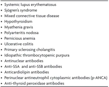
Neuromyelitis optica has been associated with many other autoimmune diseases, and even more frequently with the presence of circulating autoantibodies in the ab-sence of clinical manifestations of their associated
condi-tions (Table 1). Wingerchuk and Weinshenker79 found
au-Table 1. Autoimmune disorders and serum autoantibodies asso-ciated with neuromyelitis optica.
• Systemic lupus erythematosus • Sjögren’s syndrome
• Mixed connective tissue disease • Hypothyroidism
• Myathenia gravis • Polyarteritis nodosa • Pernicious anemia • Ulcerative colitis
• Primary sclerosing cholangitis • Idiopathic thrombocytopenic purpura • Antinuclear antibodies
• Anti-SSA and anti-SSB antibodies • Anticardiolipin antibodies
toimmune diseases in 19 (36%) of 57 patients with relaps-ing NMO and in only one (4%) of 23 patients with mono-phasic NMO. Interestingly patients with systemic lupus erythematosus or Sjögren’s syndrome who never devel-oped optic neuritis or myelitis are NMO-IgG seronegative whereas those with optic neuritis or myelitis are usually seropositive138,139. Patients with myathenia gravis who un-dergo thymectomy may develop NMO probably by dys-regulation of B-cell autoimmunity in myasthenia, exacer-bated by thymectomy140.
Typically NMO has a worse outcome than MS8,68,69,79.
Notwithstanding the index events are more severe in the monophasic than in relapsing NMO, in the long run the outcome is worse in relapsing disease as patients tend to accumulate neurologic impairment with repeating re-lapses. Relapses tend to occur early, in clusters and at un-predictable intervals8. Within ive years of disease onset 50% of patients in the relapsing group are unable to walk without assistance and 32% die from respiratory failure secondary to acute cervical spinal cord lesion. The 5-year survival rate was 90% in the monophasic group and 68% in the relapsing group8. Predictors factors for poor out-come include presence of other autoimmune disorder, high frequency of attacks during the irst two years of dis-ease, and poor motor recovery following the index event 79. The prognosis is still worse in patients of African origin
than in whites. In the Afro-Caribbean series69 63% of the patients had EDSS score ≥ 6 at the inal examination, and 33% died after a mean time of 8.7 years. In the Brazilian series72 most of the white patients had a low EDSS score, but six of the 12 melanodermic patients died after a mean disease duration of 12 years. In children the prognosis may be more favorable than in adults. An analysis of 17 chil-dren (onset before age 18 years) with NMO showed that after a median disease duration of three years 16 patients
were ambulatory without need for assistance80.
Neuromyelitis optica and infections
Viral and bacterial diseases preceding or occurring in temporal association with NMO have been described. In
the Mayo Clinic series8 a nonspeciied antecedent viral
event occurred in 30% of the patients in the monophasic and 23% in the relapsing group. In other studies the infec-tious agent could be identiied. In the irst reported case
in Brazil the disease occurred in the course of mumps33.
Acute infectious mononucleosis preceded NMO for three
weeks in a 29-year old man who had a complete recovery141
and cases of varicella-zoster virus infections in associa-tion with NMO were reported by some investigators142-145.
We had the opportunity to examine a 65-year-old wheelchair-bound woman with paraparesis and sphincter disturbances who developed severe optic neuritis 16 years after the onset of the myelitis. Spinal cord MRI disclosed
a cervicothoracic lesion whereas the CSF showed three
lymphocytes/mm3 and a protein content of 77 mg%.
ELI-SA and Western blot test for HTLV I-II were positive in both the serum and CSF. The patient made no recovery.
Another interesting viral association with NMO re-ferred to our MS Center occurred in a 39-year old man who was HIV-seropositive for three years. He suddenly developed severe paraparesis, dysesthesia in the lower limbs, a T10 sensory level and sphincter disturbances. Se-quential optic neuritis occurred on the right and then on the left three and six months later. Spinal cord MRI dis-closed a lesion extending over the entire thoracic cord. Laboratory work-up was unrevealing except for HIV se-ropositivity. A previous case of NMO in a
HIV-seroposi-tive patient had been reported in an African woman146 in
whom a spinal cord MRI scan disclosed four gadolinium-enhancing lesions on T6, T7, T10, and T12 levels.
Neuromyelitis optica has also been observed in pa-tients with pulmonary tuberculosis9,36,72,147-149 and syphi-lis150,151. Three of the 24 patients in the Brazilian series72 had pulmonary tuberculosis.
We documented a rather unique association in a 34-year-old mulattoe man who experienced bitemporal loss of the visual field and headache. His visual acuity evolved to hand movement in both eyes in 3 days. Brain MRI showed a gadolinium-enhanced lesion in sellar re-gion and thickening of the optic chiasm. Biopsy of the lesion disclosed “non-speciic granulomatous reaction”. His vision improved after a course of IV methylprednis-olone. Fourteen months later he developed lumbar pain, severe paraparesis and bladder dysfunction. Spinal cord MRI showed a T2- hyperintense lesion in thoracic and lumbar levels. Cerebrospinal luid examination revealed
35 WBC/mm3 (38% eosinophils), a protein content of 60
mg%, and a strong positive immunoreaction for schisto-somiasis mansoni. The patient had full recovery after cor-ticosteroid treatment152.
CEREbROSPINAL fLUID
In most NMO patients CSF analysis exhibits some ab-normalities. In the Mayo Clinic series8 pleocytosis (>5
WBC/mm3) was present in 79% of the patients and was
greater than 50 WBC/mm3 in 35%. Cell count varies
broad-ly and can reach igures over 2000 cells/mm3. This
strik-ing abnormality had long been observed153. Neutrophils
are commonly found, and even the presence of eosino-phils can be noted154. Protein content and some cytokines as interleukin (IL)-17 and IL-8, and the numbers of IL-5 and IL-6, IgG and IgM secreting cells are increased154,155.
CSF features may be helpful in distinguishing NMO
from MS. Pleocytosis greater than 50 WBC/mm3 rarely
pa-tients with NMO8. Similarly, IgG index which is usually ele-vated in MS is normal in patients with NMO156. An addition-al difference is the higher levels of matrix metaddition-alloprotein-
metalloprotein-ase-9 in the CSF of MS as compared with NMO patients157.
MRI AbNORMALITIES
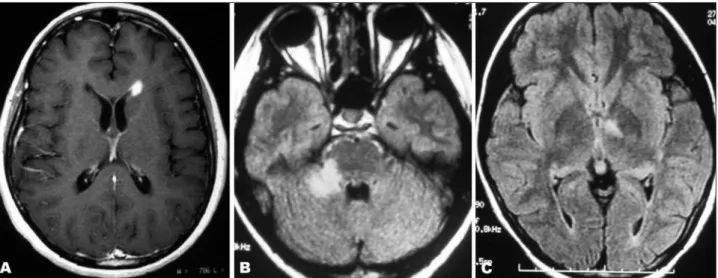
Neuroimaging has expanded the very concept of De-vic’s NMO deining the typical features of the spinal cord lesion (Fig 2) and disclosing asymptomatic cerebral lesions in the majority of patients (Fig 3). Furthermore, it provides clinicians with the most reliable element for the diagnosis of NMO – the longitudinally-extensive transverse myeli-tis lesion – distinguishing it from MS. Imaging of the spi-nal cord typically shows a gadolinium-enhanced lesion extending through several vertebral segments, and in the acute phase, marked swelling of the cord 36,158,159. Cavity-like longitudinally-extensive lesions are seen in cases with relatively severe disease, and cervical lesions may extend to lower medulla 160. In late stages of the disease spinal cord atrophy will ensue. On the opposite, spinal cord le-sions in MS patients usually do not extend beyond two vertebral segments in length, do not occupy the entire cord transverse area, and are not associated with cord swelling or atrophy161-163.
One study122 correlated the MRI features of
optico-spinal MS with the NMO-IgG seropositivity. Longitudi-nally-extensive spinal cord lesions (LESCL) were found in all but one of the 12 NMO-IgG-positive opticospinal MS patients and in 57% of the NMO-IgG-negatives. The only NMO-IgG-positive patient without LESCL had extensive spinal cord atrophy suggesting the previous existence of a longitudinally-extensive lesion. A similar observation was
reported in another study164 which detected the presence
of anti-AQP4 antibodies in 25 of 28 patients with
LES-CL and in 12 of 17 opticospinal MS patients with long at-rophy of the spinal cord. Although the LESCL are more frequently seen in opticospinal MS than in conventional
MS some authors165-167 have found them in one-fourth of
Asian patients with conventional MS relecting the great-er susceptibility of these populations to more sevgreat-ere spi-nal cord damage168.
Fig 2. MRI scans of the spinal cord in patients with neuromyelitis op-tica. (A) Swollen cervical spinal cord with a longitudinally-extensive lesion mainly involving the central area of the cord. (B) Longitudinal-extensive irregular lesion from T4 to T9 levels. (C). Axial imaging of thoracic cord showing central pattern of involvement. (D) Extensive atrophy of the thoracic spinal cord in a late stage of the disease.
Longitudinally-extensive spinal cord lesions in anti-AQP4-positive patients predominantly involve the up-per-to-middle thoracic cord with a predominant central grey matter pattern. Even short lesions in AQP4 anti-body-positive patients tend to show this central involve-ment preference. On the other hand, in AQP4 anti-body-negative opticospinal MS patients the lesions are usually extremely long extending from the upper cervi-cal cord to the middle thoracic cord and have a holocord pattern. Even in cases with short spinal cord lesions this holocord pattern can be observed. Finally, in anti-AQP4 antibody-negative conventional MS patients both short spinal cord lesions and LESCL most frequently affect the cervical cord and present a peripheral pattern of involve-ment. This hetereogeneity of the anti-AQP4 antibodies in patients with LESCL may relect differences in their patho-genesis. In anti-AQP4 antibody-positive cases there may be predominance of humoral mechanisms, whereas in pa-tients with LESCL and absence of anti-AQP4 antibodies
T-cell mediated immune mechanisms may predominate168.
It was long held that the presence of brain lesions at onset of disease outside the optic nerves ruled out the diagnosis of Devic’s NMO. However, the recent
de-velopment of the Wingerchuk et al.’s diagnostic criteria8
prompted a new look at this issue12. A review of 60 cases satisfying these criteria, except for the absolute criteri-on of lacking symptoms beycriteri-ond the optic nerves and spi-nal cord, and the supportive criterion of having a normal brain MRI at onset, disclosed MRI brain abnormalities in 60% of the patients. IgG-NMO antibodies were detect-ed in 41 (68%) patients. In 30 patients the initial brain MRI was normal although half of them developed brain ab-normalities in subsequent scans. Most of the lesions were small and nonspeciic, not fulilling the Barkof’s diagnos-tic criteria for MS. Some of the lesions were hemispheric and conluent extending to subcortical areas; others in-volved the hypothalamus, thalamus, region around the fourth ventricle, and the cerebral peduncle or cerebel-lum. Six patients (10%) had MS-like lesions and four ful-illed Barkof’s criteria for MS.
In a subsequent study Pittock et al.123 described the brain MRI indings in eight out of 89 (9%) NMO patients who had lesions in the hypothalamus and in areas sur-rounding the third and fourth ventricles which have high expression of aquaporin-4. All but one of these patients had symptoms not related to optic nerve and spinal cord involvement. They showed that although most brain le-sions found in NMO patients are nonspeciic, lele-sions in the hypothalamus and brainstem are typical of the dis-ease. Lesions in these structures in NMO patients had al-ready been described by others134,136,159.
In Brazil, Domingues et al.169 in 2004 drew attention to the presence of brain lesions on MRI scans of a child with
NMO. They reported the case of a 10-year-old mulattoe boy with recurring optic neuritis and myelitis. His brain MRI disclosed multiple periventricular lesions, including a large one with a conluent pattern extending to the pa-rieto-occipital subcortical area. The spinal cord lesion ex-tended throughout the cord, and the CSF examination
re-vealed marked pleocytosis (1675/mm3, with predominance
of neutrophis). Papais-Alvarenga et al.72 had also observed the presence of nonspeciic brain lesions in their series.
THE SPECTRUM Of DEVIC’S NEUROMYELITIS OPTICA
Recent advances in the understanding of the immuno-pathogenetic mechanisms of NMO and the discovery of the NMO-IgG as a marker of the disease have provided evidences suggesting that the disease may encompass a number of different phenotypic expressions sharing com-mon immunological and pathological grounds.
The traditional view of Devic’s NMO as a monophasic condition in which bilateral optic neuritis and myelitis oc-cur simultaneously or within an interval of few weeks with no evidence of brain lesions just deines a narrow band of a broad spectrum of clinical expressions which may be de-termined by the inluence of different genetic factors. At present time, the spectrum of Devic’s NMO includes pa-tients whose clinical features can be categorized into a number of phenotypic subtypes such as:
(1) Classical or strict Devic’s NMO – This group is char-acterized by monophasic bilateral optic neuritis and acute myelitis with clinically estimated lesions conined to the optic nerves and spinal cord and no brain lesions on MRI outside the optic nerves. In Asian countries the term “De-vic’s disease” is strictly used to identify these cases 131.
Wingerchuk et al.8 have demonstrated that patients with
unilateral optic neuritis are indistinguishable from those with bilateral optic neuritis and the interval over which patients develop the index events has no diagnostic sig-niicance. Rather, patients with NMO can be classiied into a monophasic and a relapsing type.
(2) Relapsing NMO – Patients in this group develop repetitive attacks of unilateral or bilateral optic neuritis and myelitis with clinically estimated main lesions con-ined to the optic nerves and spinal cord. Interestingly, in the same year (1894) Devic22 reported the case of his
sin-gle patient with a monophasic course, he and Gault23
For a long time Japanese authors3-7,41,49,53 have identiied patients with clinical features of relapsing NMO as having “opticospinal MS”, emphasizing that they differ from con-ventional MS patients in a number of features (Table 2).
More recently one study160 showed that 28% of the
opti-cospinal MS patients have a “pure optiopti-cospinal MS” sub-type as they had normal brain MRI except for lesions in the optic nerves and spinal cord in repeated examinations for a minimal follow-up of ive years.
(3) Relapsing NMO with asymptomatic brain lesions on MRI not meeting diagnostic criteria for MS – These pa-tients comprise most of the NMO cases in both Eastern and Western populations12,160. Brain MRI lesions are large-ly nonspeciic and usuallarge-ly involve the brainstem and the hypothalamus or may present as large tumefactive and coalescent lesions in the cerebral hemispheres12. Most of these patients are seropositive for NMO-IgG.
(4) Relapsing NMO with asymptomatic brain lesions on MRI meeting diagnostic criteria for MS – These pa-tients are usually seropositive for NMO-IgG and comprise
10% of the cases of relapsing NMO12 . They may represent
the extreme portion of the spectrum of conditions as de-scribed by Shibasaki et al.3 in 1974, which ranges from clas-sic Devic’s disease to conventional MS.
(5) Relapsing NMO with symptomatic brain lesions – Patients with NMO may present symptoms of cerebral in-volvement such as consciousness disturbances, agitation, emotional liability, in addition to brainstem signs as eye movement disorders, facial weakness, nausea and dysar-tria. Histopathology examination of these lesions reveal the typical changes described in the spinal cord of NMO patients9. In one autopsy-proven case the disease started with signs of encephalopathy due to a brain tumefactive lesion, followed by repeated relapses of optic nerve and spinal cord involvement109.
(6) Relapsing NMO with autoimmune diseases – Over
a third of NMO patients have either symptoms of other autoimmune conditions or seropositivity for other circu-lating autoantibodies8.
(7) Isolated recurring optic neuritis or isolated recur-rent acute myelitis (high-risk syndromes for NMO) – Pa-tients with either isolated recurrent optic neuritis or re-curring isolated longitudinally-extensive spinal cord dis-ease are in high risk of developing the second index event that characterizes the fully-developed disease. The
con-dition is known as forme fruste or limited forms of NMO.
NMO-IgG antibody was detected in 25% of the patients with recurrent optic neuritis and 52% of the patients with recurrent transverse myelitis; 46% when both conditions
are considered together10. Anti-AQP4 antibody assay with
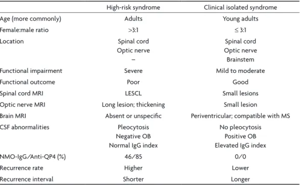
human antigen yielded a sensitivity of 85% and speciic-ity of 100% in these cases 110. Isolated transverse myelitis and isolated optic neuritis may be either high-risk syn-dromes for NMO or clinically isolated synsyn-dromes herald-ing MS. Table 3 depicts the differences between high-risk syndromes for NMO and clinically isolated syndromes which may convert to MS.
In one study of 72 patients with recurrent optic neu-ritis Pirko et al.170 found that the 5-year conversion rate to NMO was 12.5% and to MS, 14.4%. Among 5 patients with two or more lesions consistent with MS on brain MRI, two converted to MS and none to NMO, while among 11 pa-tients without such lesions, none converted to MS and two converted to NMO. Conversion to NMO occurred earlier than conversion to MS, more often in women than in men and in those with a higher relapse rate. Visual out-come was worse in NMO group.
DIAgNOSIS
In 1999 Wingerchuk et al.8 reviewed 71 cases of NMO
examined at the Mayo Clinic between 1950 and 1993. The cases fulilled either the “strict criteria” for diagnosis of NMO (bilateral optic neuritis and myelitis occurring with-in two years of one another without symptomatic disease outside of the optic nerveand spinal cord) or “not meet-ing strict criteria” (unilateral optic neuritis or develop-ment of a second index event over a period greater than two years). They observed that there was no difference between the two groups regarding demographics, clinical or paraclinical features and outcome. Therefore this dis-tinction makes no sense. On the other hand there are im-portant differences between the monophasic and relaps-ing groups regardless of the index event interval and if op-tic nerve involvement is unilateral or bilateral. Based on the clinical, laboratory and imaging data from both groups they designed a set of diagnostic criteria for NMO (Table 4) which includes three absolute criteria (optic neuritis, myelitis and absence of clinical evidence of disease out-side of the optic nerve and the spinal cord), and six
sup-Table 2. Differential features of relapsing NMO (opticospinal mul-tiple sclerosis) as compared with conventional mulmul-tiple sclerosis.
• • • • •
• •
• • • • •
Preponderance in non-white populations Higher preponderance in females Higher age at onset
Greater disability (higher EDSS scores)
Longitudinally-extensive spinal cord lesions (≥vertebral seg-ments in length)
Lower proportion of secondary progressive disease
Disability determined by relapses rather than by progression of the disease
Lower number of brain MRI lesions
Brain MRI lesions usually do not meet criteria for MS CSF with greater pleocytosis and higher protein content CSF may have neutrophils and eosinophils
portive criteria (three major and three minor criteria). Di-agnosis requires the presence of all absolute criteria and either one major or two minor supportive criteria.
Recently Wingerchuk et al.132 revised their previous di-agnostic criteria for NMO not to exclude patients with
neurologic symptoms implicating lesions outside the op-tic nerves and spinal cord and those whose brain MRI le-sions meet MS imaging criteria (Table 5). In a new analy-sis of 96 NMO patients including the detection of NMO-IgG antibodies they found that the 1999 diagnostic
cri-Table 3. Differences between high-risk syndrome for neuromyelitis optica and clinically isolated syndromes her-alding multiple sclerosis.
High-risk syndrome Clinical isolated syndrome Age (more commonly) Adults Young adults
Female:male ratio >3:1 ≤ 3:1
Location Spinal cord
Optic nerve –
Spinal cord Optic nerve Brainstem Functional impairment Severe Mild to moderate
Functional outcome Poor Good
Spinal cord MRI LESCL Small lesions Optic nerve MRI Long lesion; thickening Small lesion
Brain MRI Absent or unspeciic Periventricular; compatible with MS CSF abnormalities Pleocytosis
Negative OB Normal IgG index
No pleocytosis Positive OB Elevated IgG index
NMO-IgG/Anti-QP4 (%) 46/85 0/0
Recurrence rate Higher Lower
Recurrence interval Shorter Longer
LESCL, longitudinally extensive (≥3 vertebral segments) spinal cord lesion; OB, oligoclonal bands.
Table 4. Wingerchuk et al.’s 1999 diagnostic criteria for neuromyelitis optica8.
Diagnosis requires all absolute criteria and one major supportive criterion or two minor supportive criteria Absolute criteria
1. Optic neuritis 2. Acute myelitis
3. No evidence of clinical disease outside of the optic nerve or spinal cord Supportive criteria
Major
1. Negative brain MRI at onset (does not meet criteria for MS)
2. Spinal cord MRI with signal abnormality extending over ≥3 vertebral segments 3. CSF pleocytosis of >50WBC mm3 OR >5 neutrophils/mm3
Minor
1. Bilateral optic neuritis
2. Severe optic neuritis with ixed visual acuity worse than 20/200 in at least one eye 3. Severe, ixed, attack-related weakness (MRC grade ≤ 2) in one or more limbs
Table 5. Wingerchuk et al.’s revised diagnostic criteria for neuromyelitis optica132.
Deinite neuromyelitis optica Optic neuritis
Acute myelitis
At least two of three supportive criteria