Arq Neuropsiquiatr 2004;62(2-B):543-546
RECURRENT NEUROM YELITIS OPTICA WITH DIFFUSE
CEN-TRAL NERVOUS SYSTEM INVOLVEM ENT
Case report
Renan B. Domingues
1, Gustavo W. Kuster
2, Flávio Lanes
3, Dagoberto Callegaro
4ABSTRACT - Several demyelinating disorders can affect children. The differential diagnosis between these diseases is usually an arduous task. Diagnostic criteria have been proposed for some of these disorders, however most of them have not yet been clini-cally and prospectively validated. Here we present a case of a ten year-old boy with recurrent bilateral optic neuritis and spinal cord involvement. Clinical and cerebrospinal fluid data have fulfilled diagnostic criteria for Devic´s neuromyelitis optica (NMO). The dif-ferential diagnosis with multiple sclerosis (MS) has become troublesome since not only optic nerves and spinal cord were involved. In one of the relapses a left hemiparesis with facial involvement was registered. Magnetic resonance imaging was also compati-ble with MS. This case illustrates that CNS demyelinating disorders can fulfill diagnostic criteria for more than one demyelinating disease, making the clinical judgment an important tool in the management of these patients.
KEY WORDS: neuromyelitis optica, Devic´s disease, multiple sclerosis.
Neuromielite óptica recorrente com envolvimento difuso do sistema nervoso central: relato de caso
RESUMO - Diversas doenças desmielinizantes podem ocorrer em crianças, sendo muitas vezes o diagnóstico diferencial entre elas difícil. Critérios diagnósticos têm sido propostos para algumas destas entidades, entretanto nenhum deles pode ser considerado definitivo. O objetivo deste trabalho é apresentar o caso de um paciente de 10 anos de idade, com quadro recorrente de neurite óptica bilateral e mielopatia. Os dados clínicos e liquóricos preencheram critérios para o diagnóstico de neuromielite óptica de Devic. O diagnóstico diferencial foi especialmente difícil em relação à esclerose múltipla, pois não apenas os nervos ópticos e medu-la foram acometidos, visto que em um dos surtos registrou-se hemiparesia, com acometimento facial. A ressonância magnética foi também compatível com esclerose múltipla. Este caso ilustra que pacientes com doenças desmielinizantes do SNC podem preencher critérios diagnósticos para mais de uma delas, o que torna o julgamento clínico uma ferramenta ainda importante na abordagem e condução clínica destes casos.
PALAVRAS-CHAVE: neuromielite óptica, doença de Devic, esclerose múltipla.
1Professor Adjunto, Doutor, Escola de M edicina da Santa Casa de M isericórdia, Vitória, ES Brasil (EM ESCAM );2M édico Interno, EM ESCAM ;3M édico Radiologista,
M ULTISCAN, Vitória ES, Brasil;4M édico Assistente, Doutor, Serviço de Neurologia do Hospital das Clínicas da Faculdade de M edicina da Universidade de São Paulo
(HCFM USP), São Paulo SP, Brasil.
Received 7 August 2003, received in final form 26 November 2003. Accepted 9 January 2004.
Dr. Renan Domingues - Avenida Nossa Senhora da Penha 595/1208 - 29055-131 Vitória ES - Brasil. E-mail: [email protected]
Neuromyelitis optica (NMO) (Devic´s syndrome) is an asso-ciation of optic neuritis with myelitis.The neuropathological fea-tures and the clinical evolution of NM O suggest that this is a distinct disease. Several other diseases such as multiple sclero-sis (M S), collagen diseases, and infections can present w ith myelitis and optic neuritis1. As the result of the difficult
dis-tinction between NM O and other diseases sharing the same clinical features some diagnostic criteria have been proposed, however, none of them have been prospectively validated so far1,2.
The evolution of NMO can be monophasic or recurrent.The prog-nosis is usually worse than in MS. The relapses can be confined
in the optic nerve or spinal cord, however, they can be found in different areas of central nervous system (CNS)2. In such
cas-es the differential diagnosis w ith M S can become more trou-blesome because clinical diagnostic criteria for M S and NM O may be superposed.
Here w e report the clinical and diagnostic features of a patient w ith recurrent NM O w ho fulfilled diagnostic criteria for both M S and NM O.
CASE
respiratory symptoms, or diarrhea have been reported. The patient had no previous history of neurological disorders, and had received regular immunizations against B hepatitis, tuberculosis, tetanus, diphtheria, pertussis, measles, mumps, rubella, and poliomyelitis.
One week after symptoms have begun he was brought to our attention. No abnormalities were found on general examination. The patient was fully oriented. He had no problems w ith fluency, com-prehension, and repetition.Visual acuity was severely affected on both sides. Fundoscopic examination disclosed bilateral optic atrophy. He had full extraocular movements. Facial sensation and musculature were intact. Swallow ing was normal. Arms strength and tone w ere nor-mal, but there was a severe weakness of his legs (+ + + /4+ ), with spas-ticity. Babinski sign was present bilaterally. Sensory examination revealed spinal cord level at T4.
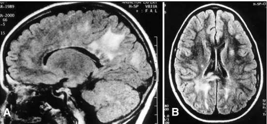
A brain magnetic resonance imaging (M RI) has show n more than nine T2-hyperintense lesions, most of them in periventricular white matter, but also w ith juxtacortical and pericallosum region involve-ment (Figs 1A e 1B). Neither gadolinuim-enhancing nor infratento-rial lesions were seen. Spinal MRI has shown multiple hyperintense T2 lesions in the upper cervical region, predominating in the dorsal region. A large T2 hyperintense lesion with mild mass effect was seen extending from the low cervical level through the conus medularis (Fig 2) There was not gadoliunium enhancement in spinal cord lesions. Cerebrospinal fluid (CSF) analysis has revealed pleocytosis (165 cells/mm3), with polymorphonuclear predominance (65%), protein and
glucose concentration were 165 and 63 mg⁄dl, respectively. No oligo-clonal bands were found by agarose gel electrophoresis or isolelet-ric focusing. Antinuclear antibodies, anti-SSA, anti-SSB, hepatitis B surface antigen, hepatitis C antibodies, p-ANCA, anticardiolipin anti-bodies w ere all negative. IgM antianti-bodies against HSV, VZV, CM V, and EBV w ere not found. Complement levels w ere w ithin normal values. The hypothesis of NM O, acute disseminated encephalomyelitis (ADEM ), or M S were initially raised and high doses of methylpred-nisolone (500 mg a day for five days) followed by ciclophosphamide (500 mg, once) w ere given. After a few days a complete recovery of spinal signs and partial visual recovery were seen. About a month
la-544 Arq Neuropsiquiatr 2004;62(2-B)
ter a new attack was registered w ith isolated visual involvement. Methylprednosolone was given again, and the patient was maintained with prednisone 40 mg a day with further reduction of prednisone and azathiprine introduction (2-3 mg/Kg/day). Visual acuity at this
Fig 1A. Sagittal Fast-FLAIR M RI scan shows multiple hyperintense lesions, some of them coalescent, in the periventricular white matter extending to the pericallosum region, with minimal mass effect. B. Axial Fast-FLAIR brain images shows sev-eral hyperintense white matter lesions, in the corona radiata and corpus callosum splenium.
Fig 2. Fast Spin Echo Heavy T2-Weighted sequence shows an extensive
hyper-intense lesion involving the spinal cord from the low cervical level (not shown) to the conus with mild mass effect.
time was 20/40 at left and he was able to count fingers at 30 cm of distance w ith his right eye. Three months later a new attack was reg-i st er ed . A t t h reg-i s t reg-i m e a l ef t h em reg-i p a r esreg-i s w a s r eg reg-i st er ed . Methylprednosolone (1000 mg a day for three days) and intravenous immunoglobulin (IVIG, 0.4 g/day for five days) w ere given. A com-plete recovery was seen except for the visual acuity deficit. Prednisone (40 mg a day) and azathiprine (2-3 mg/Kg/day) w ere maintained. Several months later there was a new relapse after an attempt to reduce prednisone. Paraparesis plus bilateral optic w orsening w ere registered. This new attack was treated again w ith methylpred-nisolone plus IVIG. The preventive schedule was altered and present-ly the patient is using subcutaneous glatiramer acetate 20 mg a day, mitoxantrone, 5 mg/m2, every three months, and oral prednisone 20
mg/day. Since this schedule was introduced no other attack has been registered. Blood cell counts have been ordered monthly and echocar-diography has been performed every three months, in order to assess mitoxantrone side effects.
DISCUSSION
Diagnostic criteria for NM O have been recently proposed by Wingerchuck and col.2. According to such criteria,
diagno-sis requires three absolute criteria: 1) optic neuritis, 2) acute myelitis, and 3) no evidence of clinical disease outside the optic nerve or spinal cord; as w ell as at least one of the follow ing major supportive criteria: 1) negative brain M RI at onset, 2) spinal cord M RI with signal abnormality extending over 3 ver-tebral segments, 3) CSF pleocytosis of > 50 WBC/mm3or
> 5neutrophils/ mm3, or tw o of the follow ing minor
support-ive criteria: 1) bilateral optic neuritis, 2) severe optic neuritis, 3) severe, fixed, attack-related weakness in one or more limbs.
Our patient has initially presented w ith optic neuritis, myelitis, had pleocytosis w ith polymorphonuclear predomi-nance, and M RI spinal cord abnormalities. Three absolute cri-teria, two major supportive cricri-teria, and three minor support-ive criteria were present. Laboratory tests have excluded oth-er diseases, such as collagen and vascular diseases, auto-anti-bodies syndromes, and infections. Therefore, the signs and symptoms initially displayed by our patient are consistent w ith the diagnosis of NM O according to Wingerchuck’s and col. criteria.
The possibility of other demyelinating CNS diseases w ere also raised. ADEM usually follow s an infection or a vaccine. There were no histories of both.Also,ADEM is usually monopha-sic and our patient had a multiphamonopha-sic disease. Although recur-rent ADEM can occur, the recurrences are usually registered in the first six months and our patient had new attacks through the first 18 months of disease3,4. Diffuse sclerosis was ruled
out since this is a rapidly progressive disease w ith w hite mat-ter lesions w ith mass effect4,5.
The differential diagnosis w ith M S is more difficult in this case. M S can be found in children and usually presents w ith attacks reflecting white matter involvement6-8. Only after years
Arq Neuropsiquiatr 2004;62(2-B) 545
of disease a secondary progressive stage is usually seen. Some patients have a primary progressive disease but a relapsing-remitting course is the rule. M RI criteria for M S diagnosis require three of the follow ing: 1) one gadolinium-enhancing or nine T2 hyperintense lesions, 2) one infratentorial lesion, 3) at least one jutacortical lesion, and 4) at least three periven-tricular lesions9. Recommended diagnostic criteria for M S
w ere recently proposed by M cDonald and col10. According to
these criteria, if there are two attacks compatible with MS, doc-umented by objective evidence of two lesions separated in time and necessarily separated in space may be sufficient to make an MS diagnosis solely on clinical grounds. Our patient had objective evidence of more than tw o lesions separated in space, such as optic neuritis, spinal cord lesion, and left hemi-paresis. M RI findings w ere compatible w ith Barkhof and col. criteria, since there were more than nine T2 enhancing lesions, being more than three periventricular, and at least one juxta-cortical lesion.
NM O can have a more diffuse brain involvement and relapsing course. In the series of Wingerchuck and col. there were five patients with recurrences not confined to optic nerves or spinal cord.Two of them had facial numbness, two had ver-tigo, and one had cerebellar tremor. These authors have per-formed M RI studies in 28 patients with NM O. Brain parenchy-ma was norparenchy-mal in 22, but three (11%) have satisfied the cri-teria for M S diagnosis.
Our case points to the difficult differential diagnosis of recur-rent demyelinating CNS diseases. Clinical criteria have provid-ed a more uniform clinical approach to these patients11.
How ever, they still require future refinements because some overlapping can still occur. It is possible that more overlap-ping sit uat ions can present ly be document ed because M cDonald’s and cols. criteria are more sensitive than Poser’s criteria. In M cDonald’s and col. criteria it is stressed that there should be no better explanation than M S for the clinical pic-ture to define M S diagnosis10. In our patient w e believe that
NMO is a better explanation for the whole clinical picture than M S. This conclusion was strongly based on clinical judgment since clinical criteria for both NM O and M S w ere fulfilled.
The diagnostic problems can have therapeutic implica-tions. NM O is a distinct disease w ith more severe prognosis than M S. There are few studies addressing NM O treatment. The combination of prednisone and azathioprine have reduced the attacks frequency in an uncontrolled series12,13. Plasma
treatment.
REFERENCE
1. Cree BAC, Goodin DS, Hauser SL. Neuromyelitis optica. Semin Neurol 2002;22:105-122.
2. Wingerchuck DM, Hongcamp WF, O’Brien PC, Weinshenker BG. The clinical course of neuromyelitis otpica. Neurology 1999;53:1107-1114. 3. Dale RC, Souza C, Chong WK, Cox TCS, Harding B, Neville BGR. Acu-te disseminaAcu-ted encephalomyelitis, multiphasic disseminaAcu-ted encepha-lomyelitis and multiple sclerosis in children. Brain 2000;123:2407-2422. 4. Fontaine B. Les formes frontières de sclérose em plaques. Rev Neurol
(Paris) 2001;157:929-934.
5. Dupel-Pottier C. Critères diagnostiques des formes frontiers de sclérose em plaques. Rev Neurol (Paris) 2001;157:935-943.
6. Silva A, Sá MJ. Esclerosis múltiple de inicio juvenil. Rev Neurol 1999;28:1036-1040.
7. Balássy CS, Bernet G, Wöber-Bigöl C, et al. Long-term MRI observa-tions of childhood-onset relapsing-remmiting multiple sclerosis. Neu-ropediatrics 2001;32:28-37.
8. Pinhas-Hamiel O, Barak Y, Siev-Ner I, Achiron A. Juvenile multiple scle-rosis: clinical and prognostic characteristics. J Pediatrics 1998;132:735-737. 9. Barkhof F, Filippi M, Miller DH, et al. Comparison of MR imaging cri-teria at first presentation to predict conversion to clinically definitive multiple sclerosis. Brain 1997;120:2059-2069.
10. McDonald IW, Compston A, Edan G, et al. Recommended diagnostic criteria for multiple sclerosis: guidelines from the international panel on the diagnosis on multiple sclerosis. Ann Neurol 2001;50:121-127. 11. Tintoré M, Rovira A, Río J, et al. New diagnostic criteria for multiple
sclerosis. Neurology 2003;60:27-30.
12. Mandler RN, Ahmed W, Dencoff JE. Devic´s neuromyelitis optica: a prospective study of seven patients treated with prednisone and aza-thioprine. Neurology 1998;51:1219-1220.
13. de Seze J, Stojkovic T, Ferriby D, et al. Devic´s neuromyelitis otpica: clin-ical, laboratory, MRI and outcome profile. J Neurol Sci 2002;197:57-61.