Excessively crying infants are more common among
children of parents with restless legs syndrome
José Carlos Pereira JrI, Márcia Pradella HallinanII, José Hugo de Lins PessoaI
DOI: 10.5935/MedicalExpress.2015.06.04
I Faculdade de Medicina de Jundiaí, Departamento de Pediatria, São Paulo, Brazil
II Universidade Federal de São Paulo, UNIFESP, Departamento de Psicobiologia, Instituto do Sono, São Paulo, Brazil
OBJECTIVE: We have frequently observed that infants presenting with excessive crying and fussing, or colic at night have parents with Restless Legs Syndrome. Our objective was to determine if these infants are more likely to have parents with Restless Legs Syndrome (Willis-Ekbom Disease).
METHODS: We interviewed 67 families with infants and children, in search of a history of excessive crying and fussing during their irst four months of life. Their parents were investigated for Restless Legs Syndrome.
RESULTS: Among the 134 interviewed parents, 39 (29%) had Restless Legs Syndrome. Among the 96 children, 37 (38%) presented excessive crying and fussing. Of these, 28 (76%) had at least one parent with Restless Legs Syndrome. Among the 59 children without excessive crying and fussing only 14 (24%) had at least one parent with Restless Legs Syndrome. The association between events (children of parents with vs. without Restless Legs
Syndrome) was measured by the phi coeicient (0.510), indicating a more than trivial association. The estimated association was 75.7 vs. 27.7, Odds Ratio = 10 at 95% conidence interval, 3.82-26.15).
CONCLUSION: Children with excessive crying and fussing were more likely to have at least one parent with Restless Legs Syndrome. The present evidence is insuicient to conclude that infantile excessive crying and fussing is equivalent to a a probable diagnosis of parental Restless Legs Syndrome. However, they provide information as well as the necessary motivation to undertake more extensive studies of infants with excessive crying and fussing.
KEYWORDS: Restless legs syndrome; Childhood restless legs syndrome; Colic; Infant.
Pereira-jr JC, Pradella-Hallinan M, Lins-Pessoa JH. Excessively crying infants are more common among children of parents with restless legs syndrome. MedicalExpress (São Paulo, online). 2015;2(6):M150604.
Received for Publication on August 5, 2015; First review on August 22, 2015; Accepted for publication on September 23, 2015
E-mail: [email protected]
■
INTRODUCTIONExcessive crying and fussing (ECF) - infantile colic - among infants younger than 4 to 5 months of age is not rare. It is believed that as many as 40% of infants cry or fuss primarily during the late afternoon and evening often to such an extent as to elicit concern from their parents; almost one-third of these infants exhibit a behavior similar to what appears to be a condition of intense distress.1-3 Healthcare providers are often tasked with
reassuring these families that their child’s condition is not serious and that it is a self-limiting phenomenon that will resolve itself spontaneously within a few months. It is important to teach these parents how to cope effectively with the condition because their anxiety may exacerbate
their infants’ colic. Infants with ECF are difficult to soothe,
although a few practical measures may be of some
assistance; however, parental support is the mainstay
of managing the condition.1-3 Most of these infants will
stop exhibiting ECF by 3 to 4 months of age, and the vast
majority will stop exhibiting it before 6 months of age.1,2 When we consider the diagnosis of excessive crying
and fussing in an infant, it is important that no causes
or diseases underlying the infant’s distress are found and that the infant is both healthy and well nourished;
otherwise, the diagnosis of “idiopathic” colic cannot be
made.1-3 There is only limited agreement among experts
regarding how much crying or fussing is considered
excessive; the average duration of crying during the first 3 months of life varies from 42 minutes to 2 hours per
day.2 As defined by Wessel a diagnosis of “idiopathic” colic is appropriate for an infant who is healthy and well fed
WED/RLS. Both manifest themselves primarily during the
evening and cause sleep disturbances, and both diseases are characterized by complaints or discomfort and, in the case of adults, discomfort which primarily affects the legs. Additionally, many infants behave as if they are experiencing
discomfort in a particular part of the body. Both ECF and
WED/RLS patients constantly move their legs. Furthermore,
we have frequently encountered parents diagnosed with WED/RLS (the parents) who have stated that their children
had ECF when they were 3 to 4 months of age. Thus, we
conducted this prevalence study to determine whether ECF at night is more common among infants of parents with WED/RLS than among infants of parents without WED/
RLS, or verify the possibility that colic in small infants is
related to presence of WED/RLS in their parents.
■
PATIENTS AND METHODS GeneralOver a period of 8 months (June/3/2014 to February/26/2015), we interviewed 67 families (134 parents, 96 children) who presented to our private office due to concerns regarding their infants, who were 10
months of age or younger.
All the interviews were performed when both
parents of the 67 families were present in their child’s
interview and in the enquiries about WED/RLS in
themselves, as described below.
For diagnostic purposes, eight infants had previously
received a trial dosage of domperidone prescribed by their respective attending neonatologists to exclude occult
gastroesophageal reflux as an alternative diagnosis to ECF. When each infant reached 4 months of age, his or her information was collected for this study, specifically
information regarding increased crying or fussiness during the evening and night. Infants who were younger than 4 months at the time of study enrollment were
followed until 4 months of age, during which time their
behavioral characteristics were carefully observed and
recorded. Patients who were older than 4 months of age,
with a maximum of 10 months of age, at the time of their
first interview were enrolled in the study; their data were
collected retrospectively.
Excessive crying and fussing
Parents of infants with or without ECF were questioned regarding ECF in both older children and siblings of the infants enrolled in the study; a diagnosis of ECF (crying or fussing or both) was established when
infants did not sleep continuously for at least 9 hours each
night (sleep interruption was ignored when the infants were awoken for breast or bottle feedings but quickly returned to sleep after being fed). The minimum amount of time
a week, and for longer than 3 weeks.1 Not all infants with
colic fulfill these criteria, although many cry and fuss more
than other babies and elicit concern from their parents.1-3
One characteristic of infants with ECF relates to sleep: it has been demonstrated that these infants sleep less than their unaffected counterparts.4,5 Thus, ECF may be considered
a pediatric sleep disturbance, and many theories have
been postulated regarding the possible pathophysiology
underlying infantile colic; however, no specific causes have been identified as yet.1-3
A different, ailment that affects many people is the sensorimotor disease known as Willis-Ekbom Disease, or
Restless Legs Syndrome (WED/RLS).6 It is believed that 5
to 10% of the population may suffer from WED/RLS.6 The
lifetime prevalence of WED/RLS among Brazilian adults
is 6.4%, although data pertaining to Brazilian pediatric
population are lacking.7 In population-based studies from
the United Kingdom, the United States, and Turkey, the
prevalence of WED/RLS among children was approximately
2 to 4%, wheres the prevalence of the moderate to severe
WED/RLS among children was 0.5 to 1%.8,9 manifestation of
the problem is diagnosed based on a patient’s self-reported
history; therefore, it may be impossible to determine if a
child who is unable to articulate his or her symptoms suffers from WED/RLS.9-11 Because some children may behave as
if they are having discomfort in their legs, experts have
hypothesized that they may be prone to develop WED/RLS.
As such, the diagnoses of “possible” and “probable” WED/
RLS have been created to describe this type of behavior among children who are unable to express their symptoms
in their own words. As the terms may indicate, probable
WED/RLS is more likely to occur than possible WED/RLS. The prevalence of childhood WED/RLS has been studied primarily among older children who can describe their
symptoms,10,11 which means that little is known regarding
WED/RLS among younger children.11 Numerous studies
have documented that WED/RLS is characterized by an underlying genetic predisposition; various genes have been linked to the WED/RLS phenotype.6,9 The family histories
of the majority of patients with WED/RLS are consistent with an autosomal dominant pattern of inheritance. When one considers the possibility of WED/RLS in a pediatric
patient, it is helpful to determine if one of the child’s parents
has the disease.10 WED/RLS is a typical “case-description”
disease, as it is diagnosed when 5 criteria are met by the patient.6,9 The criteria are as follows: 1) The patient must
be either resting or inactive (lying down or sitting); 2) The patient feels an urge to move his/her legs, an urge that is
usually accompanied by paresthesias deep inside the legs or pain; 3) The symptoms are relieved by movement; 4) The symptoms occur exclusively or predominantly during the evening and night; and 5) The symptoms are not the result of any other medical or behavioral condition.6,9
considered positive for ECF (crying or fussing or both) was
2 hours, occurring between 7 PM and 6 AM, for at least 6
weeks(both conditions not necessarily continuous) during
the infant’s first 4 months of life.
Inclusion criteria were as follows: (i) 54 infants who
were healthy and well nourished; (ii) 42 children, siblings of the above mentioned infants, if their parents were certain regarding their behavior during their 4 first months of life
(iii) a gestational age of at least 38 weeks and a weight of
at least 2,800 grams at birth; (iv) an Apgar score equal to
or greater than 8 at 5 minutes of life.
Exclusion criteria were as follows: (i) children older than 10 months of age upon their initial consultation; (ii) infants who were not well-appearing and had a history of poor feeding; (iii) adopted children (iv) children with
gastroesophageal reflux (v) three siblings because their
parents were uncertain regarding their behavior during that period.
Restless legs syndrome
The first step in diagnosing adult WED/RLS in our
study entailed the delivery of a handout to each set of parents
that included a written explanation regarding WED/RLS, its symptoms, its precipitating conditions, and its manifestations
in individuals with it. The handout was written so that individuals without a medical background could understand
its contents. After the parents read the handout, one or two
face-to-face interviews with only the parents were conducted to make a reliable diagnosis of WED/RLS. To ensure that no parent was misdiagnosed with WED/RLS all 5 criteria for the diagnosis of WED/RLS were necessary to make the diagnosis of adult WED/RLS;9 the only exception to this rule applied to three parents receiving selective serotonin reuptake inhibitors (SSRIs) for a mild depression condition. Cases in which a secondary cause of WED/RLS was suspected were excluded; We considered only patients who had clear symptoms (even mild) of the disease for an average of 3 times a month or more during the previous year as potential patients with WED/RLS. We did not attempt to determine the severity of the WED/RLS
of our “parent” patients. The attempt to diagnose possible or
probable WED/RLS in any of the children enrolled in the study
were made supplementary to the main study, because our aim
was exclusively to determine whether ECF is more common among the children of parents with WED/RLS than among the children of parents without WED/RLS.
Statistical analysis
The significance of the association between ECF and
parental WED/RLS was assessed by means of the chi-square test with continuity correction. The degree of association
between both factors was measured by the phi coefficient
and the odds ratio evaluated the magnitude between the difference of infants or children born of parents with and without WED/RLS.
This project was submitted to and approved by the Committee of Ethics on Research at Faculdade de
Medicina de Jundiaí (case number 166/2014). Formal
written informed consent was obtained from all couples participating in the study.
■
RESULTSOf the 134 parents (67 couples) interviewed, 39 (29%) had a past medical history indicative of WED/RLS. In 4 couples, both parents suffered from WED/RLS; 95 parents did not have the disease. Of the parents with WED/RLS, 21 were women, a result expected given the prevalence of
women with the condition in the general population. The majority of adults with WED/RLS suffered from either mild or moderate disease. Of the parent patients enrolled in our
study, only 6 were aware that they suffered from the disease.
Four parents had severe WED/RLS and were interested in treatment with pramipexole. Although mild side effects
occurred in one patient, all 4 experienced improvement in their symptoms within the first few days of treatment.
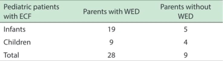
Of the 96 infants and children studied (54 infants, 42 children), 37 (38.5%) had ECF as defined above, and 59 did not, as shown in Table 1. Of the 37 infants and children with ECF, 28 (75.7%) had at least 1 parent with WED/ RLS, as seen in Table 2. Of the 59 infants and children who did not have ECF, 45 of them (76%) were born to couples in which none of the parents had WED/RLS, whereas the remaining 14 (23.7%) were born to couples in which at least one parent had WED/RLS, as shown in Table 3. Of the 42 infants and children with at least one parent with WED/ RLS, 28 (66, 6%) had ECF.
The significance of the association between infantile
colic and parental WED/RLS was assessed by means of the
chi-square test with continuity correction. A significant chi
squared (χ2 = 22.87, p < 0.05) association occurred. The
degree of association between factors was measured by
the phi coefficient (f = 0.510), indicating a more than trivial
association. Infants and children with ECF were more likely to have at least one parent with WED/RLS than those who
did not (75.7 vs. 27.7; OR: 10; confidence interval (95%): 3.82-26.15). Therefore, we conclude that an association between ECF, or infantile colic, and parental WED/RL was
observed.
When infants with ECF eventually slept, their
parents frequently reported that their sleep was restless.
Additionally, many of the infants continued moving their
legs while being breastfed or bottle fed.
Eight babies had previously received domperidone;
none had experienced improvement in their symptoms, and
six experienced worsening symptoms following treatment. One mother reported that she believed her baby was
Table 1 - Distribution of the pediatric patients studied accordingly their age and in relation to be present or not excessive crying and fussing (ECF)
Pediatric patients Number Positive for ECF
Negative for ECF
Infants
(up to 10 months) 54 24 30
Children
(23 to 69 months) 42 13 29
Total 96 37 59
Table 2 - Distribution of the pediatric patients with excessive crying and fussing (ECF) in relation to their parents with or without Willis-Ekbom disease (WED)
Pediatric patients
with ECF Parents with WED
Parents without WED
Infants 19 5
Children 9 4
Total 28 9
Table 3 - Distribution of the pediatric patients without excessive crying and fussing (ECF) in relation to their parents with or without Willis-Ekbom disease (WED)
Pediatric patients
without ECF Number
Parents without WED
Parents with WED
Infants 30 24 6
Children 29 21 8
Total 59 45 14
ECF during her second day of domperidone treatment; we noted dramatic restlessness and movements that appeared to primarily involve the inferior limbs. The infant returned to baseline following the cessation of domperidone. The parents of infants without ECF more frequently reported that their children’s sleep was restful and that these infants spent less than one hour crying or fussing for no more than
6 weeks during their first 4 months of life. During feeding,
these infants’ leg movements were minimal.
■
DISCUSSIONThe data collected in this study suggest that infantile ECF is more common among children of parents with WED/ RLS than among children of parents without WED/RLS. The
numbers obtained by our study are statistically significant
and indicate a more than trivial association.
However, bias may have affected the resultsof our investigation because it was performed by clinicians who gained some notoriety (in the city where they work) as health professionals with an interest in sleep medicine.
The design of our study has some weaknesses, one of
which was pointed out above; two others are: (i) the same
interviewer (JCPJr) performed all the inquiries, which
means that the investigator was not blinded with respect to the results of the infants and parents in the studied group; (ii) some children were studied prospectively and others retrospectively.
However, we emphasize that is a clinical pilot study;
and as is the case in some initial and pilot studies our interest was to verify if what we have observed deserves
or not a better planned “blinded” clinical study. And we
do accept that health providers who do not specialize in sleep medicine may observe different values from these ones we obtained.
The percentage of parents (young adults) in our
study with WED/RLS (29%) was clearly higher than that
present in the general population(5-10%);6,7 the apparent
explanation lies in the fact that we were evaluating a
specific population, namely children with ECF who had
sleep disturbances; because our results indicate that ECF appears to be more common among children of parents
with WED/RLS, we may have diagnosed a greater number
of subjects with WED/RLS than are to be found in the general population.
In contrast to ECF, the pathophysiology of WED/ RLS is somewhat well understood, although much of the evidence is circumstantial. The most significant attribute
of the disease is that almost all patients show remarkable improvement in their symptoms when treated with dopaminergic agonists.4This finding prompted the notion
that the hypofunction of some dopaminergic system may be central to the pathophysiology underlying WED/RLS.9,11
The “iron theory” represents an additional explanation for
the disease’s underlying pathophysiology because patients who had been previously deprived of iron may show improvement in their symptoms following treatment with iron.6,9 Numerous theories regarding the pathophysiology
of WED/RLS have been formulated, although they have not
been experimentally proven.12-16 WED/RLS is a severe cause
of sleep disturbance for many affected patients.9
Although based on circumstantial evidence,
one theory regarding the pathogenesis of WED/ RLS asserts that the disease manifests secondary to disequilibrium between the thyroid axis and dopaminergic (neurohormone dopamine) neurons of the tuberoinfundibular pathway;12-15 and it may also be
the consequence of a hypofunction of the dopaminergic (neurotransmitter dopamine) cell group area 11 of the hypothalamus.16 The neurohormone dopamine, in some
patients, may not modulate the hormone thyrotropin; consequently, enhanced sensitivity of the somatosensory
receptors may result from increased thyroid hormone activity; the neurotransmitter dopamine (from area
11 of the hypothalamus) may be unable, in prone patients, to modulate the plethora of action potentials
parents and studies in which pediatricians collaborate with
specialists in sleep medicine. Additionally, a longer
follow-up period may provide information regarding how infants with ECF develop particularly concerning WED/RLS. The parents of older children diagnosed with WED/RLS should be questioned regarding ECF when these children were small infants. Polysomnography studies of infants with ECF could be compared with similar studies of infants without ECF to identify any possible difference in the sleep patterns
of the two groups. Because iron deficiency may be a trigger for WED/RLS symptoms, it would be interesting to evaluate whether this deficiency in infants causes more frequent
ECF when compared with infants with normal iron values.
■
CONCLUSIONOur findings suggest that infants with ECF in the
evening and night are more likely to have parents with WED/RLS than parents without WED/RLS. Because Willis-Ekbom disease appears to have an underlying
genetic predisposition, we believe that our findings may be of interest to individuals within the fields of both sleep
medicine and pediatrics.
■
AUTHOR CONTRIBUTIONSPereira Jr JC conceived and designed the study, was also responsible for the face-to-face interviews, the writing of the manuscript, and researches for the pertinent
literature. Márcia Pradella-Hallinan M. and Lins Pessoa JH. were responsible for the revision of the manuscript and provided important notes to it.
■
CONFLICT OF INTERESTAuthors declare no conflict of interest regarding
this project.
BEBES COM CHORO E AGITAÇÃO EXCESSIVOS TÊM MAIS COMUMENTE PAIS COM SÍNDROME DE PERNAS INQUIETAS
OBJETIVO: Temos frequentemente observado que infantes
que apresentam choro excessivo e agitação ou cólicas noturnas têm pais com Síndrome de Pernas Inquietas. Nosso objetivo foi determinar se estes infantes são mais propensos a terem pais com a Síndrome de Pernas Inquietas.
MÉTODOS. Foram entrevistadas 67 famílias com infantes
e crianças em busca de uma história de choro excessivo e agitação durante os primeiros 4 meses de vida. Seus pais foram investigadas para Síndrome de Pernas Inquietas.
RESULTADOS: Dentre os 134 pais entrevistados, 39
neurons, action potentials that would be interpreted as
paresthesias by the somatosensory cortex.12-15 Infants
with ECF may have immature dopaminergic neurons
until they are 3-4 months old. Therefore, they may
experience discomfort in their legs while trying to
sleep, which would likely result in increased crying
and fussiness. Infants of parents with WED/RLS may show a stronger mismatch between their thyroid axes and the dopaminergic regulatory mechanisms of the hypothalamus and would therefore experience more unpleasant symptoms compared to other infants.
The finding that some of the infants with ECF showed
worsening symptoms when treated with domperidone is notable because it suggests that notwithstanding the fact that this dopamine antagonist that does not cross the blood-brain-barrier it exacerbates the symptoms of WED/ RLS;17 the only significant dopaminergic pathway that
may be affected by the antagonism of domperidone is the tuberoinfundibular pathway.13 It is important to remember
that the neurohormone dopamine is not only a modulator of prolactin but is also a modulator of thyrotropin.13 The
observation that domperidone exacerbates the symptoms of WED/RLS represents one important clinical fact regarding the disease because it challenges the hypothesis that WED/RLS symptoms are generated inside the blood-brain-barrier.18-19 It has been observed previously that
patients with hyperthyroidism who are not WED/RLS
sufferers may have symptoms of the disease, including both
an unpleasant sensations in the legs and an urge to move;
however, when they are cured of their hyperthyroidism,
these symptoms disappear.20,21
We do not believe that the evidence generated by
this study is sufficient to conclude that infantile ECF is equivalent to WED/RLS. Indeed, this should be looked upon as a pilot study, a clinical investigation that aimed only to
determine whether ECF is more common among infants of parents with WED/RLS than among infants of parents
without WED/RLS; however, we believe that our findings may have raised several important questions. First, it is
possible that some infants with ECF experience symptoms that mimic WED/RLS symptoms. It is also possible that the same disequilibrium involving thyroid hormone and dopamine that may underlie the pathophysiology of WED/ RLS is also present in infants with ECF. This mismatch
may disappear within the first few months of life, after
the dopaminergic neurons of the hypothalamus of these
infants mature. Moreover, this mismatch may only be
temporary as are some cases of WED/RLS associated with pregnancy.15 However, we believe that additional studies
must be undertaken before these hypotheses may be considered valid.
The extent to which the data we obtained in this
study are confirmed will likely depend on the results of
(29%) tinham doença Willis-Ekbom. Entre as 96 crianças avaliadas 37 (38%) apresentaram choro excessivo e agitação. Destas, 28 (76%) apresentaram pelo menos um dos pais com Síndrome de Pernas Inquietas. Entre as 59 crianças sem choro excessivo e agitação, apenas 14 (23, 7%)
apresentaram pelo menos um dos pais com a Síndrome de Pernas Inquietas. A associação entre os eventos (crianças de pais com ou sem Síndrome de Pernas Inquietas) foi medida
pelo coeficiente phi (0,510), indicando uma associação mais
do que trivial. As crianças com choro excessivo e agitação mostraram-se mais propensas a ter pelo menos um dos pais
com a doença Willis-Ekbom (75,7 vs. 27,7, “Odds Ratio” = 10, com intervalo de confiança de 95%, 3,82-26,15).
CONCLUSÃO: A evidência gerada por este estudo não é
suficiente para concluir que o choro infantil excessivo
e agitação é equivalente a um diagnóstico provável da
doença Willis-Ekbom parental. No entanto, eles fornecem informações, bem como a motivação necessária para
empreender estudos mais extensos sobre bebês com choro excessivo e agitação.
PALAVRAS-CHAVE: Síndrome de pernas inquietas, cólicas;
colic; Síndrome infantil de pernas inquietas.
■
REFERENCES1. Barr RG, Fujiwara T. Crying in Infants. In Rudolph CD, Rudolph AM, Lister GE, First LR, Gershon AA editors: Rudolph’s Pediatrics 22 ND edition. McGraw Hill, New York, 2011. Pages 318-321.
2. Turner TL, S Palamountain. Infantile colic: Clinical features and diag -nosis. In: M Augustyn (ed).UpToDate. UpToDate; 2015.
3. Turner TL, S Palamountain. Infantile colic: Management and outcome. In M Augustyn (ed). UpToDate. UpToDate; 2015.
4. James-Roberts IS, Conroy S, and Hurry J. Links between infant crying and sleep-waking at six weeks of age. Early Child Dev. 1997;48(1-2):143-52.
5. Weissbluth M. Sleep and colic. In Sheldon SH, Ferber R, and Kryger MH, editors. Principle and Practice of Pediatric Sleep Medicine. Elsevier Saunders, Philadelphia 2005. Pages 113-25.
6. Ekbom Jr K, Ulfberg J. Restless legs syndrome. J Intern Med. 2009;266(5):419-31.
7. Eckeli AL, Gitaí LL, Dach F, Ceretta H, Sander HH, Passos AD, et al. Prevalence of restless legs syndrome in the rural town of Cassia dos Coqueiros in Brazil. Sleep Med. 2011;12(8):762-7.
8. Picchietti D, Allen RP, Walters AS, Davidson JE, Myers A, Ferini-Strambi L. Restless legs syndrome: prevalence and impact in children and adolescents--the Ped REST study. Pediatrics. 2007;120(2):253. 9. Pichietti DL. Restless legs syndrome/Willis-Ekbom disease and peri
-odic limb movement disorder in children. In: Chervin R (ed). UpToDate. UpToDate; 2015.
10. Pereira Jr. JC, Pradella-Hallinan M, Alves RC. Childhood restless legs syndrome. Medicalexpress. 2014;1(3):116-22.
11. Walters AS. Simple sleep related movement disorders of childhood including benign sleep myoclonus of infancy, rhythmic movement disorder, and childhood restless legs syndrome and periodic limb
movements in sleep. Sleep Medicine Clinics. 2007;2(3):419-32.
12. Pereira JC Jr., Pradella-Hallinan M, Lins Pessoa JH. Imbalance between thyroid hormones and the dopaminergic system might be central to the pathophysiology of restless legs syndrome: a hypothesis. Clinics. 2010;65(5):548-54.
13. Pereira JC, Hallinan MP. Willis-Ekbom disease (Restless Legs Syn -drome) Pathophysiology: The Imbalance between Dopamine and Thyroid Hormone Theory. J Sleep Disorders Ther. 2013;2:139. 14. Pereira JC Jr., Silva Neto JL, Pradella-Hallinan M. Restless legs syndrome
in subjects with knee prosthesis: evidence that symptoms are gener-ated in the periphery. Clinics. 2011;66(11):1955-9.
15. Pereira JC Jr, Rocha e Silva IR, Pradella-Hallinan M. Transient Willis-Ekbom disease (restless legs syndrome) during pregnancy may be caused by estradiol-mediated dopamine overmodulation. Med Hy-potheses. 2013;80(2):205-8.
16. Clemens S, Rye D, Hochman S. Restless legs syndrome: revisiting the dopamine hypothesis from the spinal cord perspective. Neurology. 2006;67(1):125-30.
17. Rios Romenets S, Dauvilliers Y, Cochen De Cock V, Carlander B, Bayard S, et al. Restless legs syndrome outside the blood-brain barrier-exacerbation by domperidone in Parkinson’s disease. Parkinsonism Relat Disord. 2013;19(1):92-4.
18. Allen RP (2012) Restless legs syndrome (Willis-Ekbom disease) and periodic limb movements. In Morin CM, and Espie CA editors: The Oxford Handbook of Sleep and Sleep Disorders. Oxford University Press, Inc; New York, 707-25.
19. Pereira JC. Are symptoms of restless legs syndrome generated in the periphery of the nervous system or are they born centrally? J Neurosci Rural Pract. 2013;4(1):1-2.
20. Tan EK, Ho SC, Eng P, Loh LM, Koh L, Lum SY, et al. Restless legs symptoms in thyroid disorders. Parkinsonism Relat Disord. 2004;10(3):149-51. 21. Tan EK, Ho SC, Koh L, Pavanni R. An urge to move with L-thyroxine: