* Corresponding author.
E-mail: rcsvenez@unifran.br (R.C.S. Veneziani); sergioambrosio@unifran.br (S.R. Ambrósio). A R T I C L E I N F O
Article history:
Received 25 September 2013 Accepted 9 December 2013
Keywords: Salvia officinalis Streptococcus mutans Manool
RP-HPLC method Antimicrobial action Dental caries
A B S T R A C T
In this paper we screened the dichloromethane extract from the aerial parts of Salvia officinalis L., Lamiaceae, against a representative panel of microorganisms that cause caries, conducted a bioassay-guided fractionation to establish themselves the most active metabolite (manool) and determined the Salvia officinalis fraction with the manool highest concentration to be used to activate an ingredient in oral care products such as toothpastes and mouthwashes. Both manool and S. officinalis extract showed very promising minimal inhibitory concentration values (between 6.24 and 31.36 µg.ml-1) and time kill curves against the primary causative agents of dental caries (Streptococcus mutans) revealed that, at twice its minimal bactericidal concentration (12.48 µg.ml-1), manool required 6 h to completely kill the bacteria. Salvia officinalis extract at twice its minimal bactericidal concentration (31.36 µg.ml-1) needed 12 h. The results achieved with Salvia officinalis extract motivated us to develop and validate an analytical RP-HPLC method to detect and determine manool in this extract. The validation parameters were satisfactorily met and evaluated allows us to consider the developed method suitable for use in different labs. In conclusion, our results evidenced that the manool-rich S. officinalis extract can be considered an analytically validated alternative to develop novel and effective antimicrobial agents against the main bacteria responsible for dental caries.
© 2013 Brazilian Society of Pharmacognosy. Published by Elsevier Editora Ltda. All rights reserved.
Original Article
RP-HPLC analysis of manool-rich
Salvia officinalis
extract and
its antimicrobial activity against bacteria associated with
dental caries
Monique R. Moreira, Ariana B. Souza, Maísa A. Moreira, Thamires C. Bianchi,
Luiza J. Carneiro, Fernanda T. Estrela, Raquel A. dos Santos, Ana H. Januário,
Carlos H.G. Martins, Sérgio R. Ambrosio
*, Rodrigo C.S. Veneziani
*Núcleo de Pesquisas em Ciências Exatas e Tecnológicas, Universidade de Franca, Franca, SP, Brazil
Introduction
Despite the modern strategies based on rational drug design and combinatory chemistry, phytomedicines and active substances isolated from plants remain important sources of pharmaceutical products (Calixto, 2000). The emergence of a more sophisticated market for these products has pressed
for regulation and testing, which is a great opportunity for biodiversity-rich countries like Brazil (Desmarchelier, 2010; Seidl, 2002).
herbaceous plant cultivated worldwide, is usually employed in flavoring and seasoning food. Its botanical name refers to its medicinal properties: Salvia is derived from the Latin word “salvare” and means “to cure”; officinalis medicinal means (Miura et al., 2002). The antimicrobial potential of this herb is well established (Bozin et al., 2007; Delamare et al., 2007; Horiuchi et al., 2007a; 2007b) and, a few years ago, a review of the literature on the use of medicinal plants to treat dental diseases in Brazil pointed out that S. officinalis is one of the most employed often species. Therefore, this herb is also a potential candidate to devise novel medicines to be applied in dentistry (Oliveira et al., 2007).
In recent years, our group has concentrated on the antibacterial activity of secondary plant metabolites against strains responsible for pathologies like human oral caries, periodontitis, and endodontic lesions (Carvalho et al., 2011; De Andrade et al., 2011; Porto et al., 2009; Souza et al., 2011a; 2011b). Bearing in mind the potential use of S. officinalis in dentistry, in this paper we screened the dichloromethane extract from the aerial parts of S. officinalis (SOD) against a representative panel of microorganisms that cause caries. We conducted a bioassay-guided fractionation, to establish themselves the most active metabolite (manool, MO, 1), and determine the Salvia officinalis
fraction with the highest MO concentration (labeled SODH2). We assessed the in vitro antimicrobial properties of MO and SODH2 against the main cariogenic bacteria and developed a validated RP-HPLC (Reversed Phase High Efficiency Liquid Chromatography) analytical method to detect and quantify MO in SODH2 according to the guide RE 899/2003 (Anvisa, 2003).
Plant material, bioguided-assay fractionation and manool isolation
Aerial parts (1.0 kg) of Salvia officinalis L., Lamiaceae, were purchased at Nutriervas (SP, Brazil). The identification was carried out by Prof. Milton Groppo and a voucher specimen (SPFR 15178) was deposited in the herbarium of the Departamento de Biologia, Universidade de São Paulo, Ribeirão Preto, SP, Brazil. The plant material was then pulverized, and exhaustively extracted with dichloromethane 5 l at room temperature. This procedure furnished 45.5 g crude extract (SOD) after solvent evaporation. SOD was suspended in 300 ml methanol/H2O (9:1) and filtered; the soluble fraction was partitioned with
n-hexane (400 ml four times) and dichloromethane (400 ml, four times), which yielded the n-hexane-soluble (10.6 g) and the dichloromethane-soluble (6.7 g) fractions (SODH and SODD, respectively). The MIC (Minimal Inhibitory Concentration) values of SODH and SODD were determined against S. mutans, the primary causative agent of dental caries (Chung et al., 2006). Because SODH was the most effective fraction, it was subjected to vacuum chromatography over silica gel 60H (500 g; Merck, art. 7736) (Pelletier et al., 1986) using n-hexane and ethyl acetate increasing amounts of the eluent (1500 ml each fraction). After solvent evaporation, this procedure afforded seven fractions (SODH1-SODH7), which were also assayed against S. mutans. Fraction SODH2 (2.89 g) was the most active antibacterial extract; thus, about 2.00 g of this fraction was partitioned by column chromatography classic over silica gel 60 (100 g; Merck, art. 7734) using n-hexane/ethyl acetate 9:1 as eluent. Fifty fractions were collected and further combined into six new fractions (SODH2.1 to SODH2.6). SODH2.3 (152 mg) was the most active against
S. mutans.1H and 13C NMR analyses identified the diterpene
manool (1) the main constituent of SODH2.3.
Bacterial strains and antimicrobial testing
The MIC and MBC (Minimal Bactericidal Concentration) values were determined by the broth microdilution method using 96-well microplates, in triplicate (Porto et al., 2009). The Streptococcus salivarius strains (ATCC 25975), Streptococcus sobrinus (ATCC 33478), Streptococcus mutans (ATCC 25275),
Streptococcus mitis (ATCC 49456), Streptococcus sanguinis (ATCC 10556), and Lactobacilluscasei (ATCC 11578) were obtained from the American Type Culture Collection (ATCC).
The tested samples (MO and SODH2) were dissolved in dimethyl sulfoxide (DMSO; Synth) at 1 mg.ml-1, followed by
dilution in tryptic soy broth (Difco), which gave the final DMSO content of 5% (v/v) and concentrations ranging from 400.00 to 0.19 mg.ml-1 for MO and from 2000.00 to 0.98 mg.ml-1 for SODH2.
The inoculum was adjusted for each organism, to yield a cell concentration of 5 × 10⁵ colony forming units (CFU) per ml,
according to the guidelines of the Clinical Laboratory Standards Institute (CLSI, 2006). One well was inoculated included, to control the adequacy of the broth for organism growth. One non-inoculated well, free of antimicrobial agent, was employed also, medium to ensure sterility. CHD at concentrations ranging from 0.11 to 7.36 mg.ml-1 was used as control positive; DMSO at
5% (v/v) concentration was the negative control. The microplates (96-wells) were sealed with plastic film and incubated at 37°C,
1
Materials and methods
Reagents and standards
Acetonitrile (chromatographic grade) was supplied by Mallinkrodt Baker Inc. (Phillipsburg, NJ, USA). Water was purified using Milli-Q-plus filter systems (Millipore, Bedford, MA, USA). The internal standard methyl ent -kaur-16(17)-en-19-oate (IS) was synthesized by diazomethane methylation of ent -kaur-16(17)-en-19-oic acid isolated from Mikania hirsutissima,
for 24 h. After this period, resazurin (30 ml) in aqueous solution (0.02%) was added to the microplates, to indicate microorganism viability (Porto et al., 2009). Before resazurin was added, an aliquot of the inoculum was aseptically removed from each well with no apparent microorganism growth and plated onto tryptic soy agar supplemented with 5% sheep blood. The plates were incubated as described previously, in order to determine the MBC. The fractions submitted to the bioguided-assay were tested only against S. mutans.
Time-kill curves
Time-kill assays were performed in triplicate, according to the procedure described by D’Arrigo et al. (D’Arrigo et al., 2010); both MO and SODH2 were tested against S. mutans. Tubes containing MO concentrations at the end of 6.24, 12.48, and 18.72 µg.ml-1 (respectively one, two, and three-times the MBC
of MO for S. mutans) and 15.68, 31.36, and 47.04 µg.ml-1 (one
respectively, two, and three-times the MBC of SODH2 for S. mutans) were inoculated with the microorganism tested, to give an initial bacterial density of 5 × 10⁵ CFU ml-1, and incubated
at 37°C. To determine microorganism viability, samples were removed at 6, 12, and 24 h of incubation, followed by dilution in sterile fresh medium, if necessary. The diluted samples (50 μl) were spread onto tryptic soy agar plate supplemented with 5% sheep blood, incubated at 37°C, and counted after 48 h. Time-kill curves were constructed by plotting the log10 CFU ml versus time. The positive (CHD, at its MBC of 3.69 µg.ml-1)
and the negative (suspension of S. mutans without added MO or SODH2) controls were also assayed in triplicate.
Cytotoxicity assay
We employed the XTT assay to evaluate the effect of both MO and SODH2 on cell viability using the primary human fibroblast cell line obtained from Coriell Cell Repositories (New Jersey, USA). Cells were trypsinized and seeded in a 96-well plate at a concentration of 104 cells/well in DEMEM plus HAM-F10
(1:1, v/v) medium (Sigma, St. Louis, USA) supplemented with 20% fetal bovine serum (Life Technologies, California, USA). After 24 h of incubation at 37ºC, cell cultures were treated for further 24 h with different concentrations of MO or SODH2 (7.8 to 500.0 µM) dissolved in DMSO (1%). Cell viability was assessed with the Cell Proliferation Kit II (Roche, Mannhein, Germany), according to the manufacturer’s instruction. Absorbance of the orange formazan product was read at 490 nm, reference wavelength at 620 nm, in a microplate reader Sunrise (Tecam, Männdorf, Switzerland). Cell viability is expressed as the percentage of the viability achieved with the negative control. Doxorubicin at 3.0 µg.ml-1 was used as positive control.
RP-HPLC method development and validation
The LC (Liquid Chromatograph) consisted of a two-pump system Liquid Chromatography Shimadzu model LC-20A Prominence equipped with a SIL-20A auto sampler, a CTO-20A column oven, a CBM-CTO-20A communications bus module, an in-line degasser DGU-20A3, and a photodiode array detector (SPD-M20A). The LC solution software (version 1.25) was used
for data processing. Analyses were conducted on a Shim-pack CLC-ODS column (25 cm × 4.6 mm i.d, 5 µm; Shimadzu).
To estimate the optimal conditions to develop the method, preliminary experiments were conducted using the gradient scouting run evaluation described by Snyder and Dolan (Snyder and Dolan, 1996). After the final adjustments, the optimized conditions included an isocratic elution system consisting of 90% acetonitrile in water and 0.1% acetic acid (v/v), at a flow rate of 1.0 ml.min-1. The temperature of the column was set at
40°C; absorbance was read at 201 nm.
Preparation of sample solutions
The sample solutions were prepared by dissolving 4.82 mg of SODH2 and 0.75 mg of methyl ent-kaurenoate (IS) in 5 ml of acetonitrile, followed by filtration through a UNIFLO 25/0,2 PTFE syringe filter (Whatman/Schleicher & Schuell, Maidstone, UK).
Selectivity
Spectral purity of the MO peak obtained for all the samples was evaluated by DAD spectral data recorded in the ascending, upper, and descending regions; their respective regions were compared with the MO obtained peaks in the calibration curve. All the spectra matched perfectly, confirming that no other compounds co-eluted with MO.
Linearity
Standard MO solutions (500.0, 400.0, 300.0, 200.0, 100.0, 80.0, 40.0, and 20.0 µg.ml-1) were prepared in acetonitrile; 20-µl
aliquots of these solutions were injected into the HPLC equipment, in triplicate. The data were used to construct the calibration curve for MO concentration and were submitted to regression analysis. The correlation coefficient was calculated using Microsoft Excel®.
Limits of detection (LOD) and quantification (LOQ)
LOD and LOQ values were determined on the basis of the standard deviation of the response (σ) and of the slope of the calibration curve (S), using the expressions LOD = 3.3σ/S and LOQ = 10σ/S.
Precision
The intraday precision was evaluated by analyzing six samples; the interday precision was assayed on two consecutive days. The concentration of MO in SODH2 was determined, to calculate the relative standard deviation (RSD). For the interday precision, the mean values of MO were concentrations compared by t test (p < 0.05) using GraphPad Prism®.
Accuracy
The accuracy of the method was determined by spiking known contents of MO into the SODH2 at low, medium, and high levels (respectively 25, 50, and 100% of the MO content determined for SODH2), in triplicate. Then, solutions were prepared as described previously and analyzed (Ribani et al., 2004).
Robustness
the operational factors related to the chromatographic method-mobile phase flow rate, column oven temperature, percentage of organic solvent (%B) in the mobile phase, detection wavelength, and volume of sample injected (Table 1) were selected. Next, variation levels for each of these operational factors (Table 1) were determined using the method described in “Materials and Methods” at the beginning of the subsection “RP-HPLC method development and validation”. These levels were either represented the level (−1), when the factors were in the negative limit, or level (+1), when the above factors were the positive limit; these levels were divided into eight experiments, the shown in the Plackett–Burman design (Vander Heyden et al., 2001) (Table 2). Two dummy factors had to be included, to reach a saturated design. Each experiment was carried out for the pre-determined limit of −1 and +1 each factor variation. The response y was obtained at the end of each experiment (Table 2).
The responses selected for this study were the peak area and relative retention time (rrt) of the target component detected in SODH2 (MO). On the basis of the eight responses and considering the responses selected, the estimated effect was calculated for each factor using the equation following:
where E is the estimated effect of the selected response x; x is the peak area or rrt of MO; ∑y(+) is the sum of positive responses at level; ∑y(−) is the sum of responses at thenegative level; N is the number of experiments. To improve the interpretation of the results from the robustness study, the effect estimated Ex was converted using the equation RSD % = (S/X)·100, in which RSD % is the relative standard deviation, S is the value of Ex, and X represents the mean of the responses of y, considering the different responses and factors.
Results and discussion
Analysis of Table 3 reveals lower MIC values for MO as compared with SODH2 for all the tested bacteria, which was expected. According to some authors (Gibbons, 2008; Rios and Recio, 2005), extracts like SODH2 are promising in the search of new anti-infection agents at much higher concentrations (up to 100 μg.ml-1) than required for pure those metabolites
such as MO (up to 10 μg.ml-1). Therefore, our results attest to
the importance of both plant-derived MO and SODH2 in the search for new effective antimicrobial agents.
Antimicrobial compounds have different action mechanisms demonstrated (Greenberg et al., 2008). Several authors have emphasized that diterpenes are an important class of plant metabolites for the search of new antibacterial agents; however, the mechanism(s) responsible for this property have not yet been very well elucidated (Urzúa et al., 2008). Urzúa et al. (2008) have suggested that these metabolites
Factors Abbreviation Units Limits Level (-1)
Level
(+1) Nominal
Mobile phase flow
rate
Flow ml.min-1 ± 0.1 0.9 1.1 1.0
Column oven temperature
Temp. °C ± 5 35 45 40
Percentage of organic
solvent (%B) in the
mobile phase
%B % ± 1 89 91 90
Detector wavelength
λ nm ± 5 196 206 201
Volume of injected
sample Inj.
µl ± 1 19 21 20
Table 1
Factors and levels investigated in the robustness test.
Experiment number
Factors
Response
A B C D E F G
Flow Temp Dummy 1
%B λ Inj.
vol.
Dummy 2
1 +1 +1 +1 -1 +1 -1 -1 y1
2 -1 +1 +1 +1 -1 +1 -1 y2
3 -1 -1 +1 +1 +1 -1 +1 y3
4 +1 -1 -1 +1 +1 +1 -1 y4
5 -1 +1 -1 -1 +1 +1 +1 y5
6 +1 -1 +1 -1 -1 +1 +1 y6
7 +1 +1 -1 +1 -1 -1 +1 y7
8 -1 -1 -1 -1 -1 -1 -1 y8
Table 2
Plackett–Burman design for seven factors and eight experiments (Vander Heyden et al., 2001).
Bacteria (ATCC)
MIC (MBC) in µg.ml-1
CHDa MO SODH2
S. mutans (25275) 0.92 (3.69) 6.24 (6.24) 15.68 (15.68)
S. salivarius (25975) 0.92 (0.92) 24.96 (49.92) 31.36 (31.36)
S. sanguinis (10556) 3.69 (3.69) 12.48 (12.48) 15.68 (15.68)
S. sobrinus (33478) 0.92 (0.92) 24.96 (24.96) 31.36 (31.36)
S. mitis (49456) 3.69 (3.69) 12.48 (12.48) 15.68 (15.68)
L. casei (11578) 0.92 (0.92) 12.48 (12.48) 31.36 (62.72)
aChlorhexidine dihydrochloride, positive control.
5% DMSO solution, Negative control did not affect the growth of the microorganisms.
Table 3
promote bacterial lysis and disruption of the cell membrane. According to these authors, the structural features that promote the efficient antibacterial activity include a lipophilic structure, capable of insertion into the cell membrane, and one strategically positioned hydrogen-bond-donor group (HBD; hydrophilic group), which interacts with the phosphorylated groups on the membrane. A careful observation of the results depicted in Table 3 reveals that MO, which contains the structural requirements proposed by Urzúa et al., display very promising MIC values (Gibbons, 2008, Rios and Recio, 2005). Based on these considerations, our results give support to the suggested mechanism of action (Urzúa et al., 2008).
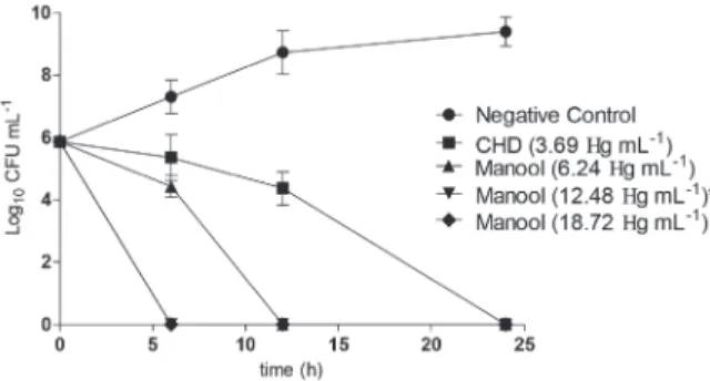
To better understand the observed antimicrobial activity, we conducted time-kill studies of MO and SODH2 using MBC their respective values; we determined how long MO and SODH2 took to completely kill S. mutans, one of the primary causative agents of dental caries. Fig. 1 shows that CHD (positive control) was able to completely kill the S. mutans inoculum at the lowest MBC (3.69 µg.ml-1) after 24 h. At 12.48 µg.ml-1 (twice its
MBC value), MO required 6 h to completely kill the bacteria; SODH2 at twice its MBC (31.36 µg.ml-1) needed 12 h. These
results are remarkable, considering the short period relatively that the active ingredients of oral care products remain in the oral cavity. In addition, neither MO nor SODH2 were cytotoxic to the human fibroblast cell line tested at the concentrations. Therefore, MO and SODH2 are important and selective
plant-Figure 1 - Time-kill curves obtained for manool (A) and SODH2 (B) against Streptococcus mutans using CHD and 5% DMSO solution as positive and negative controls, respectively.
Figure 2 - Typical SODH2 chromatogram obtained at established conditions (isocratic elution system of 90% acetonitrile in water with 0.1% acetic acid at a flow rate of 1.0 ml.min-1, column temperature of 40°C, and detection at
201 nm). Insert: UV spectrum of MO.
derived products with potential application in the control of caries disease.
Considering the increasing importance of plant-derived extracts in the global pharmaceutical market (Desmarchelier, 2010; Seidl, 2002), the results achieved with SODH2 motivated us to develop and validate an analytical RP-HPLC method to detect and determine MO in this extract. Fig. 2 illustrates a typical chromatogram of SODH2; the UV spectral purity confirmed the selectivity of the method.
We calculated the linearity of the plot concentration vs peak area for MO and obtained a correlation coefficient (r2) of 0.9971
(Table 4). The linear regression equation was:
where y is the detector response (in absorbance units) and
x is the MO concentration (in µg.ml-1). As mentioned before,
the LOD and the LOQ values were calculated on the basis of the response (σ = 1111.778) and of the slope of the calibration
curve (S = 29119.333) and corroborated the sensitivity of the developed method (Table 4).
We determined the intraday precision of the method by analyzing the chemical marker (MO) in six SODH2 samples; we assayed the interday precision on two consecutive days. All the RSD values for MO concentrations were less than 5%, and the means obtained on different days did not differ statistically (p < 0.05) (Table 4). To determine the accuracy of the method and after the addition of known contents of MO (25, 50 and 100%), the RSD values for all concentrations obtained, in triplicate, (Table 4) were also less than 5%, indicating that the results were adequate for the purposes analytical of the developed method (Anvisa, 2003).
The robustness test examines sources that potentially are subject to variations when one or a set of responses inherent to the method is being evaluated. To examine these sources, it is necessary to insert a number of factors into the validation procedure and deliberate on selected and
Parameters rrta rrt RSDb (%) E
x on rrt RSD (%) Concentration (µg.ml-1) Concentration RSD (%) RSD (%)
LOQ 0.382
LOD 0.126
Precision
Intraday 0.594 0.008 174.362 0.067
Interday 0.594 0.010 174.028 0.217
Accuracy
Low 0.593 0.009 212.737 0.018
Medium 0.593 0.019 254.138 0.032
High 0.593 0.021 333.639 0.046
Robustness
Flow 0.116 -19.621
Temp. -4.353 2.134
%B -2.511 4.306
λ -0.010 -50.590
Inj. -0.038 10.289
aRelative retention time calculated using the IS. bRelative standard deviation.
Table 4
Analytical parameters of LOD, LOQ, precision, accuracy, and robustness obtained for the developed method.
Acknowledgments
We thank Fundação de Amparo à Pesquisa do Estado de São Paulo, CNPq and CAPES for funds and grants. Our thanks are also expressed to Professor Milton Groppo for plant identification.
Authors contributions
FTE and MAM acquired and processed the plant material and performed the isolation of manool. MRM prepared the manool rich S. officinalis extract. AHJ, ABS and TCB conducted the HPLC-DAD studies. RAS, LJC and CHGM were responsible for the antimicrobial and cytotoxicity assays. SRA and RCSV designed the study, supervised the laboratory work and contributed to critical reading of the manuscript. All the authors have read the final manuscript and approved the submission.
R E F E R E N C E S
Akhondzadeh, S., Noroozian, M., Mohammadi, M., Ohadinia, S., Jamshidi, A.H., Khani, M., 2003. Salvia officinalis extract in the treatment of patients with mild to moderate Alzheimer’s disease: a double blind, randomized and placebo-controlled trial. J. Clin. Pharm. Ther. 28, 53-59.
Anvisa 2003. Guia para validação de métodos analíticos e
bioanalíticos (ed A. N. d. V. S.-. Anvisa). Diário Oficial da União, Brasília.
relatively mild variations. In general, these variations aim to define the possibility of the given oscillation when conducting the method on instruments of other brands or transferring it to another laboratory. Following the methodology described in the robustness parameter (Materials and methods, subsection RP-HPLC method development and validation), we considered each response and all the factors to calculate the effects (Ex), which were converted using RSD %. In the case of MO, the RSD (%) of each response pointed out that the factors “wavelength detector”, “mobile phase flow rate”, and “volume of injected sample” were sensitive to the peak area responses with mean values of 50, 19, and 10%, respectively. The RSD values for the other responses were equal or lower than 10% in the case of the remaining factors. Bearing the developed experimental design and the number of variables, data may be acceptable limit within the range of 20% (Vander Heyden et al., 2001). The high RSD value of the detector wavelength relative to the peak area responses (50%) is due to the sharp UV spectrum profile of MO (Fig. 2). Fig. 2 shows that small variations in the wavelength elicit pronounced changes in the absorbance. The overall assessment of the results obtained during the RP-HPLC analysis allows us to consider the developed method suitable for use in different labs.
Bozin, B., Mlmica-Dukic, N., Samojlik, I., Jovin, E., 2007. Antimicrobial and antioxidant properties of rosemary and sage (Rosmarinus officinalis L. and Salvia officinalis L., Lamiaceae) essential oils. J. Agric. Food. Chem. 55, 7879-7885.
Calixto, J.B., 2000. Efficacy, safety, quality control, marketing and regulatory guidelines for herbal medicines (phytotherapeutic agents). Braz. J. Med. Biol. Res. 33, 179-189.
Capasso, R., Izzo, A.A., Capasso, F., Romussi, G., Bisio, A., Mascolo, N., 2004. A diterpenoid from Salvia cinnabarina inhibits mouse intestinal motility in vivo. Planta Med. 70, 375-377.
Carvalho, T.C., Simao, M.R., Ambrosio, S.R., Furtado, N.A., Veneziani, R.C., Heleno, V.C., Da Costa, F.B., Gomes, B.P., Souza, M.G., Borges dos Reis, E., Martins, C.H., 2011. Antimicrobial activity of diterpenes from Viguiera arenaria against endodontic bacteria. Molecules 16, 543-551. Chung, J.Y., Choo, J.H., Lee, M.H., Hwang, J., 2006. Anticariogenic
activity of macelignan isolated from Myristica fragrans (nutmeg) against Streptococcus mutans. Phytomedicine 13, 261-266.
CLSI 2006. Clinical Laboratory Standards Institute. Methods for Dilution Antimicrobial Susceptibility Tests for Bacteria That Grow Aerobically; Aproved Standard. Clinical and Laboratory Standards Institute.
D’Arrigo, M., Ginestra, G., Mandalari, G., Furneri, P.M., Bisignano, G., 2010. Synergism and postantibiotic effect of tobramycin and Melaleuca alternifolia (tea tree) oil against Staphylococcus aureus and Escherichia coli. Phytomedicine 17, 317-322. De Andrade, B.B., Moreira, M.R., Ambrosio, S.R., Furtado,
N.A.J.C., Cunha, W.R., Martins, C.H.G., Veneziani, R.C.S., 2011. Evaluation of ent-kaurenoic acid derivatives for their anticariogenic activity. Nat. Prod. Commun. 6, 777-780. Delamare, A.P.L., Moschen-Pistorello, I.T., Artico, L.,
Atti-Serafini, L., Echeverrigaray, S., 2007. Antibacterial activity of the essential oils of Salvia officinalis L. and Salvia triloba L. cultivated in South Brazil. Food Chem. 100, 603-608. Desmarchelier, C., 2010. Neotropics and natural ingredients for
pharmaceuticals: why isn’t South American biodiversity on the crest of the wave? Phytother. Res. 24, 791-799.
Gibbons, S., 2008. Phytochemicals for bacterial resistance - Strengths, weaknesses and opportunities. Planta Med. 74, 594-602.
Greenberg, M., Dodds, M., Tian, M., 2008. Naturally occurring phenolic antibacterial compounds show effectiveness against oral bacteria by a quantitative structure-activity relationship study. J. Agric. Food Chem. 56, 11151-11156.
Horiuchi, K., Shiota, S., Hatano, T., Yoshida, T., Kuroda, T., Tsuchiya, T., 2007a. Antimicrobial activity of oleanolic acid from Salvia officinalis and related compounds on vancomycin-resistant enterococci (VRIE). Biol. Pharm. Bull. 30, 1147-1149. Horiuchi, K., Shiota, S., Kuroda, T., Hatano, T., Yoshida, T.,
Tsuchiya, T., 2007b. Potentiation of antimicrobial activity of aminoglycosides by carnosol from Salvia officinalis. Biol. Pharm. Bull. 30, 287-290.
Khalil, R., Li, Z.G., 2011. Antimicrobial activity of essential oil of Salvia officinalis L. collected in Syria. Afr. J. Biotechnol. 10, 8397-8402.
Kontogianni, V.G., Tomic, G., Nikolic, I., Nerantzaki, A.A., Sayyad, N., Stosic-Grujicic, S., Stojanovic, I., Gerothanassis, I.P., Tzakos, A.G., 2013. Phytochemical profile of Rosmarinus officinalis and Salvia officinalis extracts and correlation to their antioxidant and anti-proliferative activity. Food Chem. 136, 120-129. Miura, K., Kikuzaki, H., Nakatani, N. 2002. Antioxidant activity
of chemical components from sage (Salvia officinalis L.) and thyme (Thymus vulgaris L.) measured by the oil stability index method. J. Agric. Food. Chem. 50, 1845-1851.
Oliveira, F.Q., Gobira, B., Guimarães, C., Batista, J., Barreto, M., Souza, M., 2007. Espécies vegetais indicadas na odontologia. Rev. Bras. Farmacogn. 17, 466-476.
Pelletier, S.W., Chokshi, H.P., Desai, H.K., 1986. Separation of diterpenoid alkaloid mixtures using vacuum liquid chromatography. J. Nat. Prod. 49, 892-900.
Porto, T.S., Rangel, R., Furtado, N., De Carvalho, T.C., Martins, C.H.G., Veneziani, R.C.S., Da Costa, F.B., Vinholis, A.H.C., Cunha, W.R., Heleno, V.C.G., Ambrosio, S.R., 2009. Pimarane-type diterpenes: antimicrobial activity against oral pathogens. Molecules 14, 191-199.
Ren, Y.H., Houghton, P.J., Hider, R.C., Howes, M.J.R., 2004. Novel diterpenoid acetylcholinesterase inhibitors from Salvia miltiorhiza. Planta Med. 70, 201-204.
Ribani, M., Bottoli, C.B.G., Collins, C.H., Jardim, I.C.S.F., Melo, L.F.C., 2004. Validation for chromatographic and electrophoretic methods. Quim. Nova 27, 771-780.
Rios, J.L., Recio, M.C., 2005. Medicinal plants and antimicrobial activity. J. Ethnopharmacol. 100, 80-84.
Seidl, P.R., 2002. Pharmaceuticals from natural products: current trends. An. Acad. Bras. Cienc. 74, 145-150.
Snyder, L.R., Dolan, J.W., 1996. Initial experiments in high-performance liquid chromatographic method development I. Use of a starting gradient run. J. Chromatogr. A 721, 3-14. Souza, A.B., De Souza, M.G.M., Moreira, M.A., Moreira, M.R.,
Furtado, N.A.J.C., Martins, C.H.G., Bastos, J,K., Santos, R.A., Heleno, V.C.G., Ambrosio, S.R., Veneziani, R.C.S. 2011a. Antimicrobial evaluation of diterpenes from Copaifera langsdorffii oleoresin against periodontal anaerobic bacteria. Molecules 16, 9611-9619.
Souza, A.B., Martins, C.H.G., Souza, M.G.M., Furtado, N.A.J.C., Heleno, V.C.G., Sousa, J.P.B., Rocha, E.M.P., Bastos, J.K., Cunha, W.R., Veneziani, R.C.S., Ambrósio, S.R., 2011b. Antimicrobial activity of terpenoids from Copaifera langsdorffii Desf. against cariogenic bacteria. Phytother. Res. 25, 215-220. Urzúa, A., Rezende, M.C., Mascayano, C., Vasquez, L., 2008.
A structure-activity study of antibacterial diterpenoids. Molecules 13, 882-891.
Vander Heyden, Y., Nijhuis, A., Smeyers-Verbeke, J., Vandeginste, B.G.M., Massart, D.L., 2001. Guidance for robustness/