Evaluation of radiation dose in voiding cystourethrography
in children*
Avaliação das doses de radiação em uretrocistografia miccional de crianças
Leonardo Vieira Travassos1, Márcia Cristina Bastos Boechat2, Eloá Nunez Santos3, Sérgio Ricardo de Oliveira4, Marcos Otaviano da Silva4, Antonio Carlos Pires Carvalho5
OBJECTIVE: To evaluate dose-area product, skin entrance dose and doses from fluoroscopy and radiography in voiding cystourethrography studies of pediatric patients. MATERIALS AND METHODS: Procedures performed in 37 patients by four physicians of the institution were evaluated. Measurements were performed with an equipment including an ionization chamber directly coupled to the x-ray tube window and an electrometer (Diamentor) connected to a computer for data collection. RESULTS: Some procedures heterogeneity was observed and guidelines for good radiographic techniques were not followed. On average, 11 radiographies are performed for each study, with extended fluoroscopy time delivering a higher average final dose than those reported in the literature. CONCLUSION: The adoption of radiography with high kilovoltage technique and restricted utilization of fluoroscopy can result in a significant reduction of doses during this procedure, considering that the major contribution to the final dose comes from fluoroscopy.
Keywords: X-rays; Dosimetry; Cystourethrography; Fluoroscopy; Pediatric radiology.
OBJETIVO: Analisar o produto dose-área, a dose de entrada na pele do paciente e as doses relativas à fluoros-copia e às radiografias em exames de cistouretrografia miccional em crianças. MATERIAIS E MÉTODOS: Foram avaliados os procedimentos em 37 pacientes, realizados por quatro médicos do serviço. As medições foram realizadas com um equipamento composto de uma câmara de ionização acoplada diretamente à saída do tubo de raios X e um eletrômetro (Diamentor) ligado diretamente ao computador, para a coleta dos da-dos. RESULTADOS: Foi observada alguma heterogeneidade na realização do procedimento, que não segue padrão de técnica radiográfica. São realizadas em média 11 radiografias por exame, usando tempo longo de fluoroscopia, com dose média final mais alta que a encontrada em referências da literatura. CONCLUSÃO: A adoção da técnica de alta quilovoltagem nas radiografias e o uso restrito da fluoroscopia podem propor-cionar importante redução das doses durante a realização deste procedimento, porque o maior contribuinte para as altas doses verificadas foi a utilização da fluoroscopia.
Unitermos: Raios X; Dosimetria; Uretrocistografia; Fluoroscopia; Radiologia pediátrica.
Abstract
Resumo
* Study developed at Instituto Fernandes Figueira da Funda-ção Oswaldo Cruz (IFF/Fiocruz) and Faculdade de Medicina da Universidade Federal do Rio de Janeiro (UFRJ), Rio de Janeiro, RJ, Brazil.
1. Physicist, Scholarship holder, Instituto Fernandes Figueira da Fundação Oswaldo Cruz (IFF/Fiocruz), Rio de Janeiro, RJ, Brazil.
2. PhD, Head for Unit of Radiology at Instituto Fernandes Fi-gueira da Fundação Oswaldo Cruz (IFF/Fiocruz), Rio de Janeiro, RJ, Brazil.
3. Specialist, Pediatric Radiologist at Unit of Radiology – Ins-tituto Fernandes Figueira da Fundação Oswaldo Cruz (IFF/Fiocruz), Rio de Janeiro, RJ, Brazil.
4. Masters, Fellow PhD degree, Fundação Oswaldo Cruz (Fiocruz), Rio de Janeiro, RJ, Brazil.
5. PhD, Associate Professor of Radiology at Faculdade de Medicina da Universidade Federal do Rio de Janeiro (UFRJ), Rio de Janeiro, RJ, Brazil.
of the urinary tract and, according to reports in the literature, represents 30% to 50% of fluoroscopy studies performed in chil-dren(3,4). The main indications for this
im-aging method are: evaluation of repeated urinary infections, vesicoureteral reflux and congenital abnormalities of the blad-der and of the urethra.
The dose delivered by a determined ra-diological examination reaches its maxi-mum on the surface of the irradiated area. One can determine the radiation dose inci-dent on the exposed areas by using the Diamentor, a device that is coupled to the x-ray tube that allows the monitoring of accumulative radiation emitted by the x-ray tube during the examination, and which will not interfere on the procedure perfor-mance. Based on the entrance-surface air
Travassos LV, Boechat MCB, Santos EN, Oliveira SR, Silva MO, Carvalho ACP. Evaluation of radiation dose in voiding cystourethrography in children. Radiol Bras. 2009;42(1):21–25.
tionable benefits. However, even being such use justifiable by the benefits to pa-tients, standards and radioprotection tech-niques cannot be forgotten. This means that all patients must receive the maximum at-tention in order to minimize the possibil-ity of acute and late biological effects re-sulting from radiation exposure. Thus, if radioprotection of patients exposed to ion-izing radiations is important, it is moreover so, with pediatric patients(1,2).
Cystourethrography also referred to as voiding cystourethrography, is a radiologi-cal contrast-enhanced study for evaluation
Mailing address: Dr. Antonio Carlos Pires Carvalho. Rua José Higino, 290, ap. 401, Tijuca. Rio de Janeiro, RJ, Brazil, 20520-202. E-mail: [email protected]
Received October 31, 2008. Accepted after revision November 14, 2008.
INTRODUCTION
unques-kerma rate, one determines the dose-area product (DAP) in gray (Gy) and area unit, and the entrance-skin dose (ESD) in Gy. Variations in the fluoroscopy time during examination as a function of the equipment utilized, the radiologist´s skill, the degree of cooperation from the patient, character-istics of the region under investigation and dimensions of exposed areas, are factors influencing the total dose in the procedure. Considering these factors and the fol-lowing variables, (a) anatomic differentia-tion, (b) discrepancy among techniques uti-lized to obtain pediatric radiographic im-ages and (c) different doses to which pa-tients are exposed, the European Commu-nity has issued the European Guidelines on Quality Criteria for Diagnostic Radio-graphic Images in Paediatrics(5), defining
the quality criteria of images for most of these procedures.
The Instituto Fernandes Figueira of Fundação Oswaldo Cruz (IFF/Fiocruz) is a third-level public maternal and child hos-pital, in which 90% of patients are children. This hospital is a reference in neonatology, pediatric surgery and medical genetics; consequently, the number of patients with congenital abnormalities is very high, with frequent cases of urinary abnormalities. In 2001, the implementation of a program for quality control assurance was initiated in the Division of Radiology with the ap-proval of the Committee for Ethics in Re-search with Humans of IFF/Fiocruz (Re-port No. CAAE-000042/0008-02). Con-sidering the predominantly pediatric profile of the institution, the present study is aimed at analyzing the radiation doses to which children are exposed during voiding cystourethrography.
MATERIALS AND METHODS
The examinations were performed in the period between March 2008 and Sep-tember 2008 in the Division of Radiology at IFF/Fiocruz, by four radiologists with at least eight years of experience with pedi-atric patients. The equipment utilized was PrestilixTM telecommanded unit with image intensifier (General Electric Medical Sys-tems; Milwaukee, USA).
The dose values were obtained by cou-pling a DiamentorTM direct readout
ioniza-tion chamber model M4-KDK (PTW; Freiburg, Germany), to the exit of the x-ray tube. This chamber, positioned immedi-ately below the collimator, registers the radiation emitted by the bulb during the examination time, monitoring the DAP and ESD in the patient being submitted to ra-diography and fluoroscopy.
Thirty seven children, 16 (43%) girls and 21 (57%) boys, with ages ranging from one day to 16 years, were submitted to the examination. The patients were divided into four age groups: infants (less than one year), 1–4 years, 5–9 years and 10 or more years, and were evaluated for radiation doses to which they were exposed during the investigation with radioscopy and radi-ography.
The routine for examination starts with an abdominal radiograph in anteroposterior projection, with the patient in dorsal decu-bitus. The next step is a careful genital an-tisepsis and introduction of a catheter through the urethra to the interior of the bladder. Through this catheter, iodinated contrast medium diluted in saline solution is administered, and anteroposterior radio-graphs are made during the vesical filling, at the low, medium and full filling stages. These images will allow the evaluation of a possible vesicoureteral reflux, of the con-tents and of the bladder wall. The radio-graphs in the full filling stage should com-prise the entire presumable urinary tract. After the bladder filling phase, the voiding study is performed with anteroposterior radiographs in the case of girls, and with lateral or oblique, in the case of boys. Af-ter the voiding is completed, anAf-teroposte- anteroposte-rior abdominal radiography is performed to evaluate the degree of vesical emptying and the presence or not of vesicoureteral reflux.
RESULTS
One half the children were one year old or less at the time the examinations were
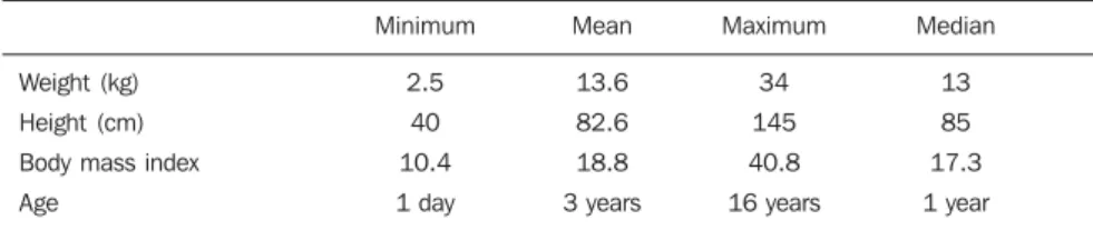
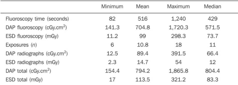
performed. Their weight ranged from 2.5 kg (two children) to 34 kg, and height ranged from 40 cm (two children) to 1.45 m. The body mass index (BMI) was calcu-lated and ranged from 10.4 to 40.8. It is important to observe that the majority of the children were below the ideal weight (21 children with BMI < 18.5). Only one child presented obesity (BMI = 40.8), two were overweight (BMI = 29.1 and 26) and all the others presented BMI ≤ 24 (Table 1). Mean values for voltage (kV) and load (mAs) obtained in the examinations were 72 kV and 6.1 mAs, respectively. The num-ber of exposures ranged between 6 and 18, the average fluoroscopy time was 516 sec-onds, corresponding, on average, to 85% of total dose. Total DAP ranged from 154 to 1,865 cGy.cm2 and total ESD ranged from 17 to 321 mGy (Table 2).
Mean total DAP for radiography and fluoroscopy was separately evaluated for each age group, noticing that in the high-est age group, the utilization of larger films and fluoroscopy fields contributes for a larger irradiated area, and consequently, to an increase in the dose (Table 3).
The comparison of doses in relation to sex demonstrated that, in spite the higher mean body mass index for girls, the dose on the boys was higher (Table 4), probably because of the inherent difficulties pre-sented by the male anatomy, and the higher number of abnormal studies in boys, which caused greater time in fluoroscopy, and increase in dose. A little over half of the studies, 21 (57%), were considered abnor-mal, with a subtle predominance (13) among boys.
The comparison by examiner (radiolo-gist) showed small differences in the way the examinations were conducted, and highlights the extension of the fluoroscopy time by all the examiners as per Table 5. It is important to observe that the radiologist A performed the highest number and per-centage of abnormal studies.
Table 1 Biophysical profile of patients.
Weight (kg) Height (cm) Body mass index Age
Minimum
2.5 40 10.4 1 day
Mean
13.6 82.6 18.8 3 years
Maximum
34 145 40.8 16 years
Median
Table 2 Fluoroscopy time, number of exposures, and observed dose values.
Fluoroscopy time (seconds) DAP fluoroscopy (cGy.cm2)
ESD fluoroscopy (mGy) Exposures (n)
DAP radiographs (cGy.cm2)
ESD radiographs (mGy) DAP total (cGy.cm2)
ESD total (mGy)
Minimum
82 141.3
11.2 6 12.5
2.3 154.4
17
Mean
516 704.8
99 10.8 89.4 14.7 794.2 113.5
Maximum
1,240 1,720.3
298.3 18 391.5
54 1,865.8
321.2
Median
429 571.5
73.7 11 66.4
12 804.4
83.3
DAP, dose-area product; ESD, entrance skin dose.
Table 3 Values observed by age group.
Age group
< 1 year 1–4 5–9
≥ 10
Patients (n)
12 16 6 3
Average exposures
(n)
10 9 11
9
DAP radiographs (cGy.cm2)
34.8 98.4 95.5 247.8
% Total DAP
7% 11% 12% 19%
Mean fluoroscopy time
(seconds)
461 486 531 929
DAP fluoroscopy (cGy.cm2)
463 809 731 1,065
% Total DAP
93% 89% 88% 81%
DAP, dose-area product.
Table 4 Values observed by sex.
Sex
Female Male
Exposures (n)
10 11
Weight (kg)
15.6 12.1
DAP (cGy.cm2)
776 808
DAP fluoroscopy (cGy.cm2)
680 747
Mean fluoroscopy time (seconds)
463 554
ESD (mGy)
92 130
DAP, dose-area product; ESD, entrance skin dose.
Table 5 Values observed by examiner (radiologist).
Examiner
A B C D
Exposures (n)
8 to 18 6 to 15 8 to 11 7 to 16
Average (n)
13.5 9.5
9 11
DAP (cGy.cm2)
939 471 712 896
ESD (mGy)
104 95 95 144
DAP, dose-area product; ESD, entrance skin dose.
DISCUSSION
Costa et al.(6–8) have introduced
video-fluoroscopy to evaluate the swallowing dynamics which requires constant fluoros-copy for detail observation, and dosed the radiation utilized in such procedure in adults, with a mean fluoroscopy time of seven minutes in two similar radiological units, and have observed that, although the doses were within the acceptable range of values in one of them, there was a signifi-cant difference between the values in each
equipment. One of these units produced a DAP five times higher than the other (804 and 4,101 cGy.cm2), in spite of similar se-lected parameters for mAs and kV. These authors have considered that a DAP/minute around 100–120 cGy. cm2 was appropriate, and have concluded that there was a prob-lem in the second equipment, which coin-cidentally, was deactivated few months later. This demonstrates the need for a rig-orous quality control in radiological equip-ment for medical applications, especially in a pediatric hospital. Lacerda et al.(9) have
n
10 6 9 12
Abnormals
8 4 4 5
Studies Fluoroscopy time (seconds)
Mean
470 362 471 525
Minimum
218 82 190 245
Maximum
1,240 1,080 777 1,180
observed that in Brazilian hospitals with older equipment and perhaps inappropriate operational conditions, sometimes the irra-diated field was larger than the selected film because of error in the manual collimation, a fact also observed by Azevedo et al.(10).
Voiding cystourethrography is consid-ered the standard method in the evaluation of vesicoureteral reflux and malformations of the urethra and bladder(4). For being
correct the application of eventually high doses which many times are caused by the radiologist habit of trying to produce a di-agnosis by means of fluoroscopy, forgetting the radiation dose delivered to the patient. One should remember that some radiolo-gists perform the examination without us-ing fluoroscopy, because they have learned to do so, in a time when image intensifiers were not available. Nicholson et al.(11) have
described dose reduction with the utiliza-tion of addiutiliza-tional filters, and emphasized that the removal of the anti-scattering grid and the use of an additional 0.7 mm steel filter alone will reduce the fluoroscopy dose by 75% to 80%.
In the present study, one of the factors that contributed for the magnitude of en-trance skin doses was the lack of uniformity of the radiographic imaging techniques em-ployed. Some radiologists/examiners leave the technical factors in the automatic mode, while others set kV and mA of fluoroscopy and mAs at the radiography mode. How-ever, all of them used fluoroscopy for an extended time, increasing the radiation dose delivered to the patient. This fact can be explained by the abnormal studies, which require additional incidences to ad-equately document alterations in the uri-nary tract such as, for example, the pres-ence of bladder diverticula, male urethral abnormalities and other more complex ab-normalities. Considering that the fluoros-copy time ranged from 82 to 1,240 seconds (mean, 516 seconds), the authors can sug-gest a radical reduction of this time. The utilization of high-kilovoltage and low-milliampere techniques, as well as fluoros-copy only for positioning the patient and observing the most remarkable alterations, will certainly result in an important dose reduction. The observation of total ESD ranging from 17 to 321.2 mGy (the fraction relative to fluoroscopy ranged from 11.2 to 298.3 mGy), of total DAP ranging from 154 to 1,865 cGy.cm2 (with fluoroscopy accounting for 141.4 to 1,720.3 cGy.cm2) and the number of exposures ranging from 6 to 18, suggests that changes in the habits of the radiologists/examiners will result in dose reduction.
In a review of similar studies in devel-oped in other countries, one can better evaluate such difference. Livingstone et
al.(12) have evaluated cystourethrography
studies of an adult Indian population, with a thickness of up to 25 cm, and have ob-served that the mean fluoroscopy time was 2.7 minutes, ranging up to 6.5. The aver-age number of radiographs/exposures was 8.6, mean total DAP was 376 cGy.cm2 (ranging from 43 to 926 cGy.cm2) and ESD, between 1.32 and 32.5 mGy, with an average of 11 mGy.
Persliden et al.(13) have evaluated
cysto-urethrography studies in two pediatric hos-pitals in Sweden, and have found DAP values between 8 and 246 cGy.cm2 in a hospital with conventional equipment, and between 4 and 254 cGy.cm2 in a hospital with a digital equipment. In this latter hos-pital, the fluoroscopy time ranged from 0.1 to 1.6 minute, and in the other one, equipped with a conventional system, between 0.4 and 5.1 minutes. In the present study, in one study, the fluoroscopy time reached more than 20 minutes, perhaps justified by the problems of the patient affected by spine deformity, with severe scoliosis causing extreme difficulty in the manipulation of the region for images acquisition.
On the other hand, Ruiz et al.(14) and
González et al.(15), in Spain, have found
high dose values, up to 300% above those reported by other authors, and have sug-gested measures for dose reduction, ob-serving that voiding cystourethrography correspond to 2.4% of studies performed in the pediatric population.
Osibote & Azevedo(16) and Azevedo et
al.(17) had already observed the lack of
stan-dardization of radiographic techniques in Brazilian general and pediatric hospitals, certainly a contributing factor for dose variations in found in the present study. Azevedo et al.(17) have demonstrated that,
although the doses calculated for chest ra-diography in the same institution are even lower than those reported in reference ar-ticles, they could be still lower, if an appro-priate collimation were utilized, and if studies requisitions were more strict. Kyriou et al.(18) have compared the dose
and the radiographic technique adopted by pediatric and general hospitals, observing increased doses in procedures performed by non-specialists.
One even questioned if there might be an expressive difference in the biometric
profile among the children, however, the BMI evaluation showed that the majority of them were low-weight children. Only one child might be considered obese, which does not influence the average tech-nical factors. However, considering what occurred in the fluoroscopy study, where similar equipment presented very different doses, leading to the finding of a serious problem in one of them which led to its deactivation, one may question if some technical non-conformity might be happen-ing, and thus justify the high doses ob-served in the present study.
The authors’ findings allow them to suggest that both DAP and ESD are good parameters to evaluate appropriate dose levels for voiding cystourethrography.
Higher patients or those in the higher age ranges require larger films to document the images, which in certain instances should include the whole collecting system from the bladder to the kidneys. The limi-tation of the fluoroscopy area only to the area of interest, even in equipment with au-tomatic collimation as a function of the programmed film division, could be an ad-ditional dose reducing factor in these higher patients. It was found that the equip-ment makes divisions only “in the vertical sense”, meaning that one 30 x 40 film will always have images with 30 or 40 cm in height, if the manual collimation is not made, which increases the exposed area. At the first years of life, the patients tend to be more active during the examination and, consequently, end up staying longer under fluoroscopy, which also contributes to the higher final dose. In one of the patients included in this age group, fluoroscopy accounted for 97.85% of total DAP.
Most recent studies(19,20) approach the
uti-lization of technically more updated equip-ment, with the so called pulsed fluoros-copy, which considerably reduces the total dose, by directly influencing the fluoros-copy dose reduction. Ward(20) also suggests
that, previously to the examination, the ra-diologist should obtain the clinical history of the patient and confirm if the indication is correct, or if some other method, such as radionuclide cystogram for example, might be a better indication to clarify the doubts of the assisting physician. Persliden et al.(13)
have observed that in the hospital with con-ventional imaging equipment, where the child undergoes ultrasonography before being submitted to voiding cystoure-thrography and the latter is indicated only when the first study is abnormal, the final dose and the number of radiographs are higher.
CONCLUSIONS AND SUGGESTIONS
The dose delivered to the patient in voiding cystourethrography is high, with fluoroscopy being its primary contributor. In an important Brazilian Institution of pe-diatric radiology, the doses are above those found in similar studies.
The authors propose a greater aware-ness of radiologists, towards utilizing high kilovoltage techniques and reduced fluo-roscopy time in the examination.
REFERENCES
1. Lacerda MAS, Khoury HJ, Silva TA, et al. Radio-proteção, dose e risco em exames radiográficos nos seios da face de crianças, em hospitais de Belo Horizonte, MG. Radiol Bras. 2007;40:409– 13.
2. Lacerda MAS, Silva TA, Khoury HJ, et al. Ris-cos dos exames radiográfiRis-cos em recém-nascidos internados em um hospital público de Belo Ho-rizonte, MG. Radiol Bras. 2008;41:325–9. 3. Al-Imam OA, Al-Nsour NM, Al-Khulaifat S.
Which is the best way of performing a micturat-ing cystourethrogram in children? Saudi J Kid-ney Dis Transpl. 2008;19:20–5.
4. Sulieman A, Theodorou K, Vlychou M, et al. Ra-diation dose measurement and risk estimation for paediatric patients undergoing micturating cysto-urethrography. Br J Radiol. 2007;80:731–7. 5. European Comission. European guidelines on
quality criteria for diagnostic radiographic images in paediatrics. [cited 2008 Oct 25]. Available from: ftp://ftp.cordis.europa.eu/pub/fp5-euratom/docs/ eur16261.pdf
6. Costa MMB, Nova JLL, Carlos MT, et al. Video-fluoroscopia – um novo método. Radiol Bras. 1992;25:11–8.
7. Costa MMB. Uso de bolo contrastado sólido, lí-quido e pastoso no estudo videofluoroscópico da dinâmica da deglutição. Radiol Bras. 1996;29: 35–9.
8. Costa MM, Canevaro LV, Azevedo ACP, et al. Valores típicos do “produto dose-área” obtidos durante o estudo videofluoroscópico da degluti-ção. Radiol Bras. 2003;36:17–20.
9. Lacerda MAS, Silva TA, Khoury HJ. Assessment of dosimetric quantities for patients undergoing X-ray examinations in a large public hospital in Brazil – a preliminary study. Radiat Prot Dosim-etry. 2008;132:73–9.
10. Azevedo ACP, Osibote AO, Boechat MCB. Sur-vey of doses and frequency of X-ray examinations on children at the intensive care unit of a large
reference pediatric hospital. Appl Radiat Isot. 2006;64:1637–42.
11. Nicholson RA, Thornton A, Akpan M. Radiation dose reduction in paediatric fluoroscopy using added filtration. Br J Radiol. 1995;68:296–300. 12. Livingstone RS, Koshy CG, Raj DV. Evaluation of work practices and radiation dose during adult micturating cystourethrography examinations performed using a digital imaging system. Br J Radiol. 2004;77:927–30.
13. Persliden J, Helmrot E, Hjort P, et al. Dose and image quality in the comparison of analogue and digital techniques in paediatric urology examina-tions. Eur Radiol. 2004;14:638–44.
14. Ruiz MJ, Vañó E, González L, et al. Dose-area product values in frequently performed complex paediatric radiology examinations. Br J Radiol. 1996;69:160–4.
15. González L, Vañó E, Ruiz MJ. Radiation doses to paediatric patients undergoing micturating cystourethrography examinations and potential reduction by radiation protection optimization. Br J Radiol. 1995;68:291–5.
16. Osibote AO, Azevedo ACP. Estimation of adult patient doses for common diagnostic X-ray ex-aminations in Rio de Janeiro, Brazil. Phys Med. 2008;24:21–8.
17. Azevedo ACP, Osibote AO, Boechat MCB. Pae-diatric x-ray examinations in Rio de Janeiro. Phys Med Biol. 2006;51:3723–32.
18. Kyriou JC, Fitzgerald M, Pettett A, et al. A com-parison of doses and techniques between special-ist and non-specialspecial-ist centres in the diagnostic X-ray imaging of children. Br J Radiol. 1996;69: 437–50.
19. Ward VL, Strauss KJ, Barnewolt CE, et al. Pedi-atric radiation exposure and effective dose reduc-tion during voiding cystourethrography. Radiol-ogy. 2008;249:1002–9.