DOI: 10.1590/S1516-14392011005000053
*e-mail: [email protected]
A New Possible Atomic Arrangement for the Carbon Atom
in the B-Sites of Ab-Type Carbonate Apatite
Jorge Correa de Araujoa, Elizabeth Lima Moreirab, Valeria Conde Alves Moraesb,
Ana Paula Duarte Moreirac*, Glória Dulce de Almeida Soaresc
a
Rio de Janeiro State University – UERJ, CEP 24435-000, São Gonçalo, RJ, Brazil
b
Brazilian Center for Physics Research – CBPF, CEP 22290-180, Rio de Janeiro, RJ, Brazil
c
COPPE and Polytechnic School, Federal University of Rio de Janeiro – UFRJ,
CEP 21941-972, Rio de Janeiro, RJ, Brazil
Received: March 17, 2011; Revised: July 21, 2011
The crystal structure of a synthetic AB-type carbonate apatite sample was analyzed by Rietveld refinement including a model with carbon atom not fixed in the B-site of the apatite structure. Only one constraint was applied to this model: the fractional occupancies of the atoms in the CO3 ion plus PO4 ion were equal 1.0 per phosphate site with six sites per unit cell. Rietveld refinement of the crystal structure with space group P63/m results in cell parameters a = 9.3583(1) Å and c = 6.9226(5) Å; Z =1; Rwp = 0.0824 and 9.5 wt. (%) of carbonate in this structure. The use of simple geometry formulas showed that the C atom is not located at the center of the equilateral triangle of oxygen O2, O3 and O3’, but to a distance 0.18 Å of this triangle. The results seem to indicate a new 3-D crystal structure of the carbonated apatite in PO4 groups.
Keywords: rietveld, refinement, carbonate, crystal structure
1. Introduction
Calcium phosphates are currently employed as bone substitute in several clinical applications due their similarity with bone1. The
crystal structure of hydroxyapatite, (HA - Ca10(PO4)6(OH)2), allows several ionic substitutions. The incorporation of carbonate ions into the HA structure improves its biological properties as it becomes closely to the composition of the mineral part of bone tissue. For instance, a carbonate ion can replace two different sites in HA lattice: the OH ion (A-site or channel site) and the PO4 site (B-site), increasing apatite solubility.
A deep study in the calcium phosphate structure is important to understand in vivo response. However, the crystal structure of carbonate apatite remains unclear due to uncertainties in the exact localization of the CO3 ions2. As noticed by Fleet and Liu (2003)3
changes in a (= b) and c unit cell parameters of carbonate apatite
(CO3-apatite) were observed, comparing with cell parameters of a pure hydroxyapatite. Suetsugu et al.4 analyzed the structure of
A-type carbonate apatite with partial occupancy in B sites and observed that a parameter took a middle point of the pure HA and
the A-type CO3-apatite powder reference, while c parameter was
larger in comparison with the pure HA. In our previous work5, the
X-ray diffraction profile of the AB-type CO3-apatite synthetic powder showed a contraction of the a lattice parameter and an enlargement
of c lattice parameter. The decreasing of the occupancy factor (O.F.)
of P sites is another partial evidence of the replacement of CO3 ions in the structure of the apatite2.
Elliot6 showed that the planes containing CO
3 triangles are nearly
parallel to the c axis, when the carbonate ions replace OH groups
(A-site) in the channels. When the carbonate ions replace the PO4 groups, the planes with CO3 triangles make a 37 ± 4° angle with c-axis, and the triangle occupies the sloping face of the tetrahedron.
However, according to Wilson et al.7, different locations of the CO 3
ion in the AB- type CO3-apatite can be obtained, probably due to differences in apatite synthesis.
Ivanova et al.2 has analyzed carbonated hydroxyapatite powder
samples by X-ray diffraction and has observed that the carbonate ions randomly occupy two adjacent faces of the PO4 tetrahedron, parallel to the c-axis, indicating that three oxygen atoms O1, O2 and O3 (split
in two O3) of the PO4 tetrahedron make up the coordination for the two carbon atoms labelled C1 and C2. The discrepancies amidst the data of different authors seem to indicate that carbonate triangles may occupy any of the tetrahedral faces, but in each specific case, they can be found to stick to a certain face2.
In this paper, XRD data of an AB-type CO3-apatite sample was analyzed by Rietveld refinement8, using a different approach in which,
the CO3 ion is allowed to move freely during the refinement, in order to determine the C position and the oxygen coordination in PO4 group.
2. Materials and Method
2.1. AB-type carbonated apatite sample (CO
3-apatite)
CO3-apatite was synthesized by wet precipitation method using calcium nitrate (Ca(NO3)2·4H2O), diammonium hydrogen phosphate ((NH4)2HPO4) and ammonium carbonate ((NH4)2CO3) solutions as precursors. The temperature and pH were maintained at 3 °C and 12, respectively5. This sample was heat treated at 500 °C
for 6 hours and examined by X-ray diffraction using a XRD Rotaflex (Rigaku, Japan) with CuKα radiation and graphite monochromator, spectra 2θ 10°-100° with the step of 0.02/5s per data point. The
CO3 content on green and calcined powder was determined by temperature-programmed desorption coupled to mass spectrometer (TPD-MS) being equal to 16.2 and 12.8 wt. (%), respectively. As the green sample was submitted to thermal treatment at 500 °C,
A New Possible Atomic Arrangement for the Carbon Atom in the B-Sites of Ab-Type Carbonate Apatite
any carbonate bonding superficially was eliminated, decreasing the carbonate content of the calcined sample. The complete description of synthesis and characterization results can be found elsewhere9.
2.2. Rietveld refinement
Rietveld refinements were carried out employing the FULLPROF program10 and a Pearson VII function was used particularly to fit
anisotropic broadening of Bragg 00l reflections caused by the strain
in the reticulate, the crystallite size and the instrument. Initially, for the refinement of the apatite structure without explicit inclusion of CO3 ion, the structure of Holly Springs hydroxyapatite single crystal structure11 was used. This model, called A model, space group P6
3/m,
was used as the starting model for refinement.
Another model, B model, also space group P63/m, was created adding C atom in the structure of the refined A model, with the CO3 ion occupying PO4 sites. The model for the CO3 ion (model B), similar to the calcite structure, was proposed considering that the z position of the C atom can be varied. This strategy was firstly adopted by Wilson et al.12 that proposed an initial triangular geometry for the CO
3
ion, with C-O and O-O distances of 1.294 and 2.413 Å, respectively. In their studies, these distances were maintained by the soft constraints, while the O1 position of the CO3 ion was hard constrained to have the same position of O1 in the PO4 ion. Contrary, in our study, the initial geometry for the CO3 ion in model B was allowed to move freely during the refinement. Only one constraint was applied: the fractional occupancies of the atoms in the CO3 ion plus PO4 ion were equal 1.0 per phosphate site with six sites per unit cell.
The isotropic displacement parameters were refined in all refinements performed. The preferred orientation (March-Dollase) parameter was allowed to vary, but it was fixed at 1.0 because the refinement indicated no preferred orientation. The background was modeled with the linear polynomial13.
3. Results and Discussion
The refinement of the observed and calculated XRD patterns of CO3-apatite is shown in Figure 1 and was in good agreement as the average Rwp, RB and RF values were, respectively, 0.0824, 0.0256 and 0.0195.
Table 1 shows some crystal data obtained for CO3-apatite sample using Model A and Model B. Data from the literature for HA11 and
B-type CO3-ap2 are also included.
As told before, the A model was used only as the starting model for refinement of the B model with the inclusion of the C atom in the structure. Both models showed an important and similar reduction in P occupancy (~20% for model A and ~18% for model B). However, the two models are not directly comparable. For example, the model B has a constraint about the complementary crystallography condition of the occupation factors of P and C (B-site) while the A model has no constraint about these occupancies factors.
Comparing cell parameters of B model with data for HA (Table 1), a reduction in the a parameter, and a large increasing in the c parameter is observed. The a parameter is smaller than that reported by Ivanova2
(9.437 Å), but agrees with Wilson et al.12 data (a = 9.3446(3) Å and
c = (6.9199(4) Å) for a carbonate apatite with CO3 content equal to 12.6 wt. (%) and some residual sodium. In this case, the reduction of a parameter occurs because the carbonate ion is smaller than phosphate ion. According to Wilson et al.12, the a-axis contraction for
a CO3-apatite depends on the method and conditions of precipitation. The replacement by carbonate ion generates a large structural disorder creating vacancies of Ca and OH ions. This disorder is also reflected in periodicity of the crystallographic planes observed by X-ray14,
resulting in a decrease of the a parameter.
Figure 1. Final fit between experimental and B model calculated XRD pattern of the CO3-apatite calcined at 500 °C during 6 hours.
The results for C2 atom position using model B is in reasonable agreement with those obtained by Ivanova et al.2 for B-type
CO3-apatiteand with the (0.408(1), 0.398(1), 0.187(2)) position for atom C obtained by Wilson et al.12.
In the B model, the reduced occupancy values, 0.82 and 0.39 for P and O4h atoms, respectively, are partial evidence of the incorporation
of CO3 ions in sites A and B. No vacancy was observed in O1 sites, which exhibit an occupancy factor (O.F.) equal to 1.02(4). Therefore, we suggested that the CO3 ions occupy the sites O2 and O3´ of a replaced PO4 ion, with O.F. equal to 0.054(8) and 0.08(1), respectively.
From occupancies factors obtained by refinement for Ca, P, C2 and O4 atoms (Table 1), the chemical formula for CO3-apatite employed in this study is Ca9.408[(PO4)4.92(CO3)1.08][(OH)1.56(CO3)0.44, resulting in approximately 9.5 wt. (%) of carbonate in this structure and Ca/P ratio equal to 1.912. The theoretical value (obtained by refinement) for carbonate content is quite different from the experimental data (12.8 wt. (%)).
The maximum and minimum residual electron densities for the eight atomic cell positions in the B model were, respectively,
∆ρ = 0.16 (eÅ-3) and ∆ρ = –0.15 (eÅ-3). This data shows that F o≈ Fc,
where Fo is the observed structure factor and Fc is the structure factor calculated by the model. Unfortunately, the C position in coordination with the O2-O3-O3´ oxygen was not clearly determined by X-Fourier maps (not shown). Consequently, the lower residual scattering around the P atom was not undoubted identified as a CO3 ion.
The application of simply analytical geometry formulas and geometrical transformations of three dimensions confirm that the C atom is not fixed in the center of the O2-O3-O3´ triangles, but at 0.18 Å of this tetrahedral face. The transformation matrix [a, 0, 0 / –0.5a, 0.5√3a, 0 / 0, 0, c] turns the hexagonal crystal system into an orthogonal system.
The plane face of CO3 ion formed by only O2-O3-O3´ triangle is parallel to c axis in agreement with the results obtained by
Ivanova et al.2. It seems that the environment around the C atom is
not strictly distorted, although the distance for C-O bond (1.36 Å) suggests that the C atom is weakly bounded with the oxygen coordination. Therefore, the geometry for CO3 ion in PO4 vacancies sites is a small tetrahedron of 0.164 Å3 in comparison with 1.70 Å3
for the PO4 tetrahedral volume.
This little tetrahedral configuration for the CO3 ion seems to contradict the previous Rietveld studies realized by Ivanova et al.2
and Wilson et al.12, but in their investigations, the constraints were
Araujo et al.
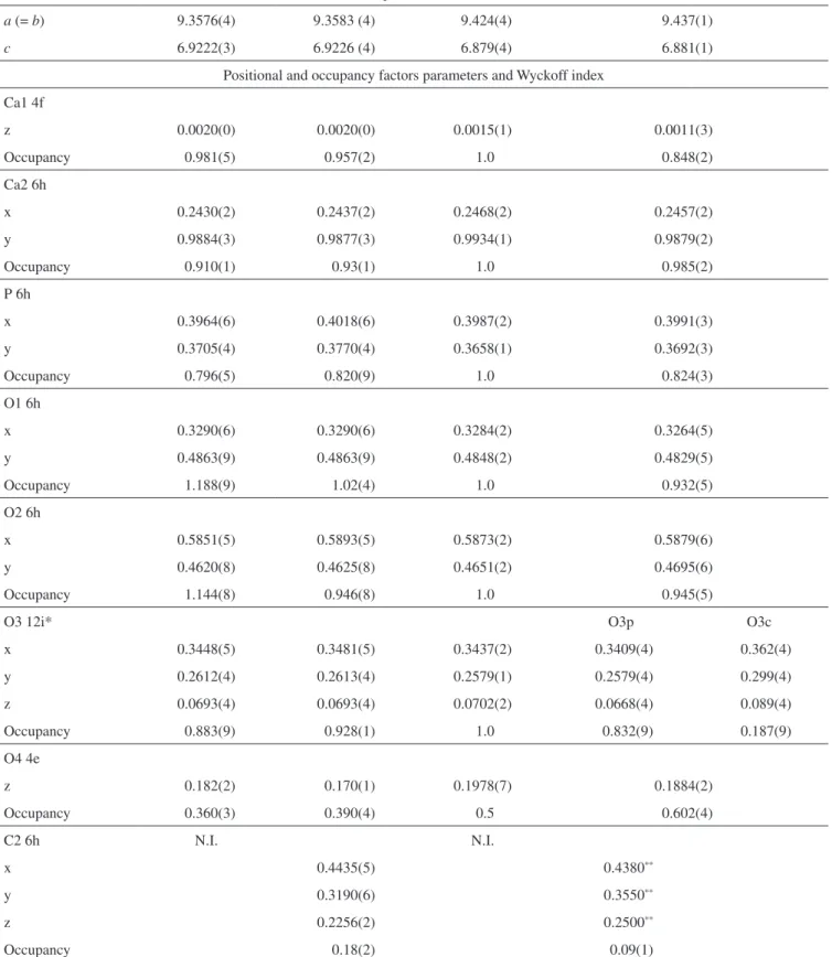
Table 1. Cell, positional and occupancy parameters of the crystal structure.
Method of Refinement
CO3-apatite sample
Model A Model B HA11 B-type CO
3-apatite2
Rietveld Rietveld Refinement with
neutrons
Rietveld
Cell parameters (Å)
a (= b) 9.3576(4) 9.3583 (4) 9.424(4) 9.437(1)
c 6.9222(3) 6.9226 (4) 6.879(4) 6.881(1)
Positional and occupancy factors parameters and Wyckoff index Ca1 4f
z 0.0020(0) 0.0020(0) 0.0015(1) 0.0011(3)
Occupancy 0.981(5) 0.957(2) 1.0 0.848(2)
Ca2 6h
x 0.2430(2) 0.2437(2) 0.2468(2) 0.2457(2)
y 0.9884(3) 0.9877(3) 0.9934(1) 0.9879(2)
Occupancy 0.910(1) 0.93(1) 1.0 0.985(2)
P 6h
x 0.3964(6) 0.4018(6) 0.3987(2) 0.3991(3)
y 0.3705(4) 0.3770(4) 0.3658(1) 0.3692(3)
Occupancy 0.796(5) 0.820(9) 1.0 0.824(3)
O1 6h
x 0.3290(6) 0.3290(6) 0.3284(2) 0.3264(5)
y 0.4863(9) 0.4863(9) 0.4848(2) 0.4829(5)
Occupancy 1.188(9) 1.02(4) 1.0 0.932(5)
O2 6h
x 0.5851(5) 0.5893(5) 0.5873(2) 0.5879(6)
y 0.4620(8) 0.4625(8) 0.4651(2) 0.4695(6)
Occupancy 1.144(8) 0.946(8) 1.0 0.945(5)
O3 12i* O3p O3c
x 0.3448(5) 0.3481(5) 0.3437(2) 0.3409(4) 0.362(4)
y 0.2612(4) 0.2613(4) 0.2579(1) 0.2579(4) 0.299(4)
z 0.0693(4) 0.0693(4) 0.0702(2) 0.0668(4) 0.089(4)
Occupancy 0.883(9) 0.928(1) 1.0 0.832(9) 0.187(9)
O4 4e
z 0.182(2) 0.170(1) 0.1978(7) 0.1884(2)
Occupancy 0.360(3) 0.390(4) 0.5 0.602(4)
C2 6h N.I. N.I.
x 0.4435(5) 0.4380**
y 0.3190(6) 0.3550**
z 0.2256(2) 0.2500**
Occupancy 0.18(2) 0.09(1)
N.I. = not included in the refinement. *The O3 atom was splitted into two O3p and O3c by D-Fourier maps. **Not refined by Ivanova et al. (2001).
A New Possible Atomic Arrangement for the Carbon Atom in the B-Sites of Ab-Type Carbonate Apatite
applied in the CO3 ion geometry to maintain the C carbon atom in the center of the equilateral triangle of oxygen atoms.
4. Conclusions
The Rietveld refinement of XRD data of the carbonated apatite crystal structure with explicit inclusion of CO3 ions considering B model showed that the C atom is weakly bounded with the only O2, O3 and O3´oxygen of the replaced PO4 ions and 0.18 Å distant of this plane face. The refinement in occupancies for the CO3 and PO4 ions resulted in ~9.5 wt. (%) of carbonate in this structure. The results seem to indicate a new possible crystal structure of carbonate group in phosphate site with CO3 ion occupying a small tetrahedral volume (0.164 Å3) when compared with the PO
4 tetrahedral (1.70 Å3).
Acknowledgements
The authors would like to thank Faperj, CNPq and CAPES for financial support.
References
1. Costa NMF, Melo BR, Brito RT, Fernandes GVO, Bernardo VG, Fonseca EC et al. Quality and Intensity of the Tissue Response to Two Synthetic Granular Hydroxyapatite Implanted in Critical Defects of Rat Calvaria. Materials Research, 2009; 12(2):245-51. http://dx.doi.org/10.1590/ S1516-14392009000200022
2. Ivanova TI, Frank-Kamenetskaya OV, Kol´tsov AB and Ugolkov VL. Crystal Structure of Calcium-Deficient Carbonated Hydroxyapatite.
Thermal Decomposition. Journal of Solid State Chemistry. 2001;
160(2):340-49. http://dx.doi.org/10.1006/jssc.2000.9238
3. Fleet, ME and Liu X. Location of type B carbonate ion in type
A-B carbonte apatite synthesized at high pressure. Journal of Solid
State Chemistry. 2004; 177:3174-82. http://dx.doi.org/10.1016/j. jssc.2004.04.002
4. Suetsugu Y, Takahashi Y, Okamura FP and Tanaka J. Structure Analysis of A-Type Carbonate Apatite by a Single-Crystal X-Ray Diffraction
Method. Journal of Solid State Chemistry. 2000; 155:292-97.
http://dx.doi.org/10.1006/jssc.2000.8887
5. Moreira EL, Araujo JC, Moraes VCA and Moreira APD. Análise por Difração de Raio-X de uma Hidroxiapatita Carbonatada Usando o Método
de Rietveld. Revista Matéria. 2007; 11(3):494-502.
6. Elliott JC. Structure and Chemistry of Apatites and Other Calcium
Orthophosphates. Amsterdam: Elsevier Science B.V.; 1994. Studies in Inorganic Chemistry 18.
7. Wilson RM, Stephanie EP and Elliot JC. Rietveld refinement and spectroscopic structural studies of a Na-free carbonate apatite made by
hydrolysis of monetite. Biomaterials. 2006; 27:4682-92. http://dx.doi.
org/10.1016/j.biomaterials.2006.04.033
8. Rietveld HM. Line Profiles of Neutron Powder-Diffraction Peaks for
Structure Refinement. Acta Crystallographica. 1967; 22(1):151-52.
http://dx.doi.org/10.1107/S0365110X67000234
9. Moreira APD. Síntese e Caracterização de Carbonato Apatitas
Nanoestruturadas. [Dissertação]. Rio de Janeiro: Universidade Federal do Rio de Janeiro, Instituto de Química; 2006.
10. Rodriguez-Carvajal J. FullProf 2000. Available from: <http://www-llb. cea.fr/fullweb>. Access in: 05/2010.
11. Sudarsanan K and Young RA. Significant Precision in Crystal Structural
Details: Holly Springs Hydroxyapatite. Acta Crystallographica. 1969;
B25:1534-43.
12. Wilson RM, Elliot JC, Stephanie EPD and Smith RI. Rietveld structure refinement of precipitated carbonate apatite using neutron diffraction
data. Biomaterials. 2003; 25:2205-13. http://dx.doi.org/10.1016/j.
biomaterials.2003.08.057
13. Bigi A, Ripamonti A, Brückner S, Gazzano M, Roveri N and Thomas AS. Structure Refinements of Lead-Substituted Calcium Hydroxyapatite
by X-Ray Powder Fitting. Acta Crystallographica. 1989; B45:247-51.
14. Astala R and Stott MJ. First Principles Investigation of Mineral
Component of Bone: CO3 Substitution in Hydroxyapatite. Chemistry
of Materials. 2005; 17:4125-33. http://dx.doi.org/10.1021/cm050523b