Linking immunity and hematopoiesis
by bone marrow T cell activity
1Divisão de Medicina Experimental, Coordenação de Pesquisa,
Instituto Nacional de Câncer, Rio de Janeiro, RJ, Brasil
2Programa de Formação em Pesquisa Médica, Faculdade de Medicina, 3Instituto de Microbiologia Prof. Paulo de Góes,
Universidade Federal do Rio de Janeiro, Rio de Janeiro, RJ, Brasil J.P. Monteiro1,2
and A. Bonomo1,3
Abstract
Two different levels of control for bone marrow hematopoiesis are believed to exist. On the one hand, normal blood cell distribution is believed to be maintained in healthy subjects by an “innate” hemato-poietic activity, i.e., a basal intrinsic bone marrow activity. On the other hand, an “adaptive” hematopoietic state develops in response to stress-induced stimulation. This adaptive hematopoiesis targets spe-cific lineage amplification depending on the nature of the stimuli. Unexpectedly, recent data have shown that what we call “normal hematopoiesis” is a stress-induced state maintained by activated bone marrow CD4+ T cells. This T cell population includes a large number
of recently stimulated cells in normal mice whose priming requires the presence of the cognate antigens. In the absence of CD4+ T cells or
their cognate antigens, hematopoiesis is maintained at low levels. In this review, we summarize current knowledge on T cell biology, which could explain how CD4+ T cells can help hematopoiesis, how
they are primed in mice that were not intentionally immunized, and what maintains them activated in the bone marrow.
Correspondence
A. Bonomo
Coordenação de Pesquisa Instituto Nacional de Câncer Rua André Cavalcanti, 37 20231-050 Rio de Janeiro, RJ Brasil
E-mail: [email protected]
Presented at SIMEC 2004 (International Symposium on Extracellular Matrix), Angra dos Reis, RJ, Brazil, September 27-30, 2004.
Publication supported by CNPq and FAF/INCA.
Received February 16, 2005 Accepted May 25, 2005
Key words
•T cell
•Hematopoiesis •Innate immunity •Adaptive immunity •Bone marrow
•Immunological memory
Introduction
Hematopoiesis control, i.e., the signals that govern lineage commitment and he-matopoietic stem cell maintenance in adult bone marrow, has been extensively studied in the last decades. A large part of this con-trol depends on the bone marrow microenvi-ronment, where a complex network includ-ing multiple cell types such as fibroblasts, osteocytes, endothelial cells, and all bone marrow-derived cells regulates hematopoie-sis through cytokine secretion and cellular interactions. In addition, hematopoiesis can
be “endocrinally” regulated through cyto-kines produced outside the bone marrow. While bone marrow-derived stimuli have been viewed as important to maintain basal blood cell production, peripheral stimuli have been related to hematopoiesis amplification during stress situations such as infection, inflammation, irradiation, or hypoxia. Thus, modulation of hematopoiesis has been viewed as a systemically controlled phenomenon (1-4).
by showing that blood cell counts in normal mice are not the result of basal “innate” hematopoietic activity (5). On the contrary, they reflect the antigenic stimulation of bone marrow CD4+ T cell. In other words,
“nor-mal hematopoiesis” is not an innate state, but an adaptive state, maintained by antigen stimulation. In view of these novel findings and considering the potential role of CD4+ T
cells as a source of hematopoietins, we pro-posed these cells to be key regulators of what we currently understand as “normal” hematopoiesis (5).
Here, we review the data that indirectly relate T cell and hematopoiesis and discuss the recent findings which redefine hemato-poiesis as a T cell-dependent phenomenon. We also summarize the current knowledge on T cell biology to explain how T cells can help hematopoiesis, how they are primed in untreated (not intentionally immunized) mice, and how they are maintained in an activated-state in the bone marrow.
T cells and hematopoiesis - the old history
The role of thymus-derived cells in he-matopoiesis was first suggested 30 years ago. Using the neonatal thymectomy model, several investigators described that 1-day thymectomized mice were anemic, showed arrested erythroid maturation and reduction in the number of spleen colony-forming units in the bone marrow and spleen (6,7). In addition, intravenous injection of live thy-mocytes accelerated hematopoiesis recon-stitution in sublethally irradiated mice (8). All of these studies claimed the occurrence of cooperation between hematopoietic cells and T cells for the establishment of optimal hematopoiesis in mice. Supporting these data, production of interleukin-3 (IL-3) and granu-locyte-macrophage colony-stimulating fac-tors (GM-CSF) by T cells was described in the early 80’s (9,10).
The increasing interest regarding the
re-lationship between T cells and hematopoie-sis was influenced by the description of fail-ure of bone marrow transplantation (BMT) in humans receiving T cell-depleted graft (11,12). At that time, BMT was already seen as an important component of the treatment of hematologic malignancies. However, the major problem regarding BMT is graft-ver-sus-host disease (GVHD) (13), a severe dis-ease mediated by T cells with high mortality and morbidity rates. Therefore, dissociation of T cell GVHD activity and bone marrow engraftment activity was essential for the advance in BMT therapy. In addition, it was shown that T cells also contribute to elimi-nating residual neoplastic cells in BMT re-cipient, introducing a new variable in this equation (12).
Thus, it is not surprising that almost all the literature on T cell/hematopoiesis in the last 25 years has been devoted to the role of T cells in hematopoiesis restoration after BMT. In fact, using bone marrow-radiation chimeras, several investigators reported the requirement for T cells in the establishment of hematopoiesis after BMT (14,15). Ildstad et al. (16) used mixed allogeneic chimeras (B10 + B10.D2 → B10) to show that selec-tive depletion of T cells in the bone marrow inoculum conditioned the pattern of hemato-poietic reconstitution: if B10 bone marrow cells were T cell-depleted, B10.D2-derived cells were the predominant hematopoietic cells in reconstituted mice, and vice-versa. These data showed that histocompatibility between hematopoietic and T cells is neces-sary for their cooperation despite previous interpretations that an allogeneic effect was necessary for engraftment.
pathogen-esis of GVHD seems to be very minor, if any (17). Only recently have these T cells been studied. The total bone marrow T cell popu-lation comprises 2-3% of all bone marrow cells. About one third of them are CD3+CD4
-CD8- or αßTCR+NK1.1+ cells (18). The role
of these unusual T cells in the bone marrow, specifically regarding hematopoiesis, has not been evaluated. The remaining two thirds are conventional αßTCR+ T cells enriched
in activated and memory CD4+ and CD8+
T cells (5,17,19,20). The potential of these cells as hematopoietin providers or their cy-tokine profile and the stimuli promoting their activation have never been investigated.
Besides conventional T cells, an unusual type of “T cells”, the “facilitating cells”, was described in bone marrow by Ildstad’s group (21). These cells comprise 0.4% of total bone marrow cells and are CD8+CD3+CD45
RB-Thy1+class IIinterm cells. Curiously, these
“T cells” do not express any known form of T cell antigen receptor (TCR). They are devoid of allospecific activity and can help hematopoiesis. Therefore, although func-tional data on the activity of these cells are abundant (21-23), their existence remains extremely controversial.
In addition to the BMT model, the role of T cells in hematopoiesis was also investi-gated in infectious disease models (24-26). It appears that hematopoiesis amplification, required to clear pathogens, is deficient in athymic mice. This is true for the diverse blood cell types, although it is more impor-tant within the granulocytic compartment. In these situations, T cells contribute secret-ing cytokines, includsecret-ing GM-CSF, 3, IL-4, IL-5, IL-6, IL-13, and oncostatin M, which all contribute to amplifying granulocyte gen-eration in bone marrow.
Introducing the recent findings
T cells have been shown to be involved in hematopoiesis in different stress situa-tions such as reconstitution after sublethal
irradiation, bone marrow engraftment after BMT, and amplification of hematopoiesis during infection, as cited above. However, it is not clear if T cells contribute to hemato-poiesis in the absence of such stresses. In other words, do T cells play any major role in the maintenance of normal hematopoie-sis? Can T cells contribute to normal he-matopoietic activity upon interaction with syngeneic bone marrow cells? And if so, what is the stimulus?
The closest replies to these questions were obtained in the experiments reported by Lord and Schofield (8) and by Bonomo et al. (27). The former showed that injection of syngeneic thymocytes accelerates bone mar-row reconstitution after sublethal irradia-tion. The latter, using an in vitro model, showed that syngeneic T cells stimulate the growth of hematopoietic progenitors.
With the above findings in mind, we devoted our efforts to the study of normal hematopoiesis in athymic nude mice since these animals lack all conventional T cells (5). Our findings were surprising: nude mice have a severe reduction in the number of granulocytes in peripheral blood, despite the high frequency of granulo-monocytic pro-genitors in the bone marrow. Nude mice reconstitution with fetal thymus or purified CD4+ T cells not only restores the normal
granulocyte counts in peripheral blood, but also reduces the frequency of progenitors in bone marrow. In contrast, purified CD8+
T cells are completely inefficient in restor-ing normal hematopoiesis. The progenitors that accumulate in the nude bone marrow are SCA1+CD11b+ cells, representing
commit-ted myeloid progenitors. These cells are not intrinsically unable to differentiate, since exogenous stimulation with growth factors promotes their differentiation in vitro.
promote terminal differentiation of these committed progenitors. These findings are not exclusive for athymic mice, since they have been confirmed in other T cell-deficient mouse lineages such as SCID and RAG-/-.
We also found a strong correlation be-tween the number of CD4+ T cells in bone
marrow and restoration of granulocyte counts in the peripheral blood of T cell-reconsti-tuted mice. The same was true for reduction in the number of bone marrow GM precur-sors (5). These data strongly suggest that bone marrow CD4+ T cells were responsible
for the effect on hematopoiesis. Thus, since to help hematopoiesis T cells must be acti-vated, we looked at the activation status of these cells in the bone marrow of normal mice. In agreement with previous descrip-tions (17,19) we found an increased propor-tion of activated CD4+ T cells in mouse bone
marrow - most of them showing an early activated phenotype (5).
The large number of activated T cells in the bone marrow of normal mice creates another problem: since these mice were not
intentionally immunized, how did their bone marrow T cells become primed? Possibly, T cells were being primed by their specific antigens (the cognate antigen) following the conventional pathway of T cell activation. This priming could be in the bone marrow or at the periphery, from where the antigen-primed cells would migrate to the bone mar-row. However, since our data showed that bone marrow T cells were activated while lymph node cells were not, it seemed pos-sible that bone marrow T cells had their own rules of activation. In this case, T cells would be primed by endogenous weak ligands that could work in synergy with other bone mar-row-specific stimuli for T cell activation.
In order to answer this question, we de-signed an experiment that dissociated these two hypotheses. We used RAG-/- TCR
trans-genic mice whose T cells all express TCR specific for an exogenous antigen, ovalbu-min (OVA), presented by the I-Ab major
histocompatibility complex (MHC) mole-cule (DO11.10) (28). If bone marrow T cells were activated in DO11.10 mice and he-matopoiesis was normal, this would mean that bone marrow T cells are primed by endogenous ligands in a distinct fashion. However, if bone marrow T cells were not activated, and hematopoiesis was abnormal, this would mean that bone marrow T cells must recognize the cognate antigen to be-come primed and help hematopoiesis. Through hematopoiesis and bone marrow T cell analyses, we found that the second hy-pothesis proved to be correct. In fact, DO11.10 RAG-/- mice had the same
he-matopoietic profile as nude mice, showing that bone marrow T cells must recognize their specific antigens in order to help he-matopoiesis (5).
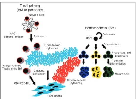
The conclusion was that, since hemato-poiesis in normal mice is maintained by antigenic stimulation of bone marrow T cells, it does not represent a basal state! On the contrary, it is already an induced state (Fig-ure 1). However, current literat(Fig-ure defines
Figure 1. Model of “adaptive” T cell-instructed hematopoiesis. T cells are primed by the cognate antigen and reach the bone marrow, where they can help hematopoiesis by i) direct cytokine production; ii) inducing cytokine production by the bone marrow stromal cells (through cytokines or cellular interaction). These cytokines act by promoting terminal differentiation of myeloid progenitors. BM = bone marrow; APC = antigen-presenting cell; HSC = hematopoietic stem cell.
Terminal differentiation
Progenitors and precursors
Antigen-primed T cells in the BM
APC +
cognate antigen Activation
T cell-derived cytokines
Cytokine stimulation
Stroma-derived cytokines
BM stroma
HSC
Self-renew
CD40/CD40L Naive T cells
T cell priming (BM or periphery)
Hematopoiesis (BM)
Commitment
normal hematopoiesis as “a basal state that maintains normal blood cell counts in the absence of hematopoietic stresses” (1-4). Can we consider antigenic stimulation a stress? We believe that the answer is yes. The source of antigen for bone marrow T cell activation is unknown. It may be a harm-less environmental antigen, a pathogen-de-rived antigen, or even a self antigen. We will discuss this question in more detail in the next sections. The fact is that normal he-matopoiesis does not represent a basal state, and our finding shows that the old view of a two layer-controlled hematopoiesis (normal x stress-induced) must be reviewed. Our suggestion would be to revisit the concept of “normal”, and replace it with innate hemato-poiesis, as the basal thymic-independent bone marrow activity based only on the constitu-ents of innate immunity.
The first problem: how T cells can help hematopoiesis
It seems obvious to think that CD4+ T
cells regulate hematopoiesis through cy-tokine secretion. Indeed, CD4+ T cells are
wonderful cytokine producers, not only quan-titatively, but also qualitatively, including the major T cell cytokines that control the immune responses such as IFN-γ, 4, IL-5, IL-13, IL-10 and TGF-ß, and the “minor” cytokines, whose effects on adaptive im-mune responses are less prominent (29). This group includes GM-CSF, IL-3, IL-6, IL-17, and oncostatin M - all involved in the regulation of hematopoiesis (30-37). The major problem is that most of these cyto-kines are not T cell exclusive. Therefore, it is very difficult to determine if the lack of T cells significantly affects the role of these cytokines.
In addition to having a direct activity as a source of hematopoietins, T cells can also stimulate cytokine production by bone mar-row stromal cells (Figure 1). It has been shown that oncostatin M and IL-17
pro-duced by T cells can stimulate IL-6 produc-tion by endothelial cells and osteocytes (32,36). IL-6 has an important effect on neutrophil production. IL-6-/- mice are
neu-tropenic. Also, in the absence of IL-6, re-sidual neutrophils found in G-CSFR-/- mice
are eliminated (34).
Besides cytokines, cell-cell interaction seems to be important for the contribution of T cells to hematopoiesis. T cells can interact with bone marrow stroma through the CD40-CD40L (CD154) pathway (Figure 1). In fact, hematologic recovery after syngeneic BMT is accelerated by treatment with soluble CD40L in mice (38). This protocol increases colony-forming units-GM frequency in mouse bone marrow and spleen, and stimu-lates platelet and leukocyte recovery in pe-ripheral blood. CD40L appears to directly stimulate the production of Flt3 by several cell types and of thrombopoietin by bone marrow stromal cells (39). This shows that, although T cells are not indispensable for megakaryopoiesis in normal mice, they can contribute to platelet generation under dam-age-induced conditions, i.e., after irradia-tion.
Further evidence for cellular interaction in T cell contribution to hematopoiesis came from mixed chimera experiments. Since MHC compatibility between T cells and he-matopoietic progenitors is required for bone marrow engraftment, it is clear that interac-tion between the two cell types, in an MHC-restricted way, is obligatory (16). How T cells are activated and what activates them is discussed in the next sections.
The second problem: how bone marrow T cells become antigen-primed cells
We and others have found an increased number of activated T cells in mouse bone marrow (5,17,19,20). This is true for both CD4+ and CD8+ T cells. However, normal
can they have such a high number of acti-vated T cells in bone marrow? We have found that only the cognate antigen can acti-vate bone marrow T cells (5). Which are the cognate antigens in “normal” mice? We en-vision two possible explanations: the stimu-lation by environmental antigens or by cog-nate self antigens. In fact, mice - like humans - are exposed to antigens present in the envi-ronment. These environmental antigens prob-ably prime some T cell clones, although their study has not attracted much interest lately.
The second explanation suggests that bone marrow T cells are primed by specific autoantigens present only in bone marrow. At present there are no data supporting or excluding this hypothesis, except for the fact that the CD4 T cell population of bone mar-row is composed of lymphocytes preferen-tially expressing Vß families, especially Vß3 and Vß7, suggesting that bone marrow T cells have some preferences in terms of anti-gen recognition (19). It is very difficult to choose between these two hypotheses. Our experimental model uses the DO11.10
RAG-/- mice in which all T cell clones
re-spond to OVA, excluding the possibility of studying both T cell priming by environ-mental antigens and cognate self-antigens (5). Experiments with DO11.10 in animals with a non-RAG-/- background have revealed
that their bone marrow T cells are activated, showing that T cells expressing endogenously rearranged TCR α-chains can be primed by other antigens. However, we could not dis-tinguish if these TCRs recognize self or environmental antigens.
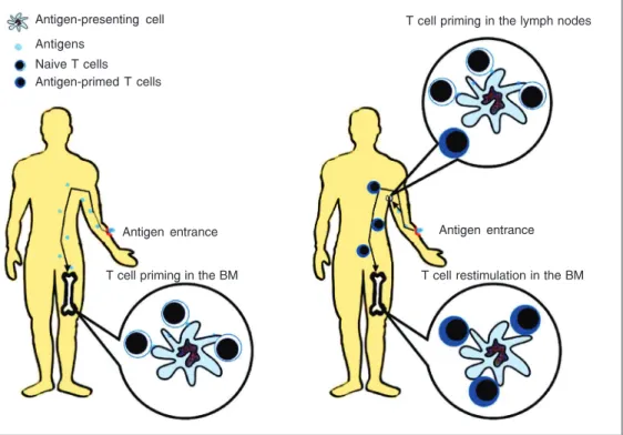
Another important issue is the priming site for bone marrow T cells (Figure 2). Although we know that T cells must be present in bone marrow to help hematopoie-sis, we do not know if they were primed in bone marrow or in the periphery. Activation of virgin T cells must include TCR recogni-tion of cognate antigens. Then, T cells can only be primed in the bone marrow if the antigens reach it. Feuerer et al. (40) showed that this is possible for blood-borne anti-gens. They transferred transgenic OT-I CD8+
T cells (which recognize pOVA + H-2Kb) to
normal B6 mice, and immunized these mice
Figure 2. Dynamics of bone mar-row T cell priming. The antigen could use the intravenous route to reach the bone marrow and prime specific T cells directly in the bone marrow (left). The anti-gens also could enter the body by a non-systemic route, i.e., subcutaneously, orally, or by the mucosal route. In this situation, T cells would be primed in the peripheral lymphoid organs, then migrate to the bone mar-row, where they would be re-stimulated to help hematopoie-sis. BM = bone marrow.
T cell restimulation in the BM Antigen entrance
T cell priming in the lymph nodes
Antigen entrance
T cell priming in the BM Antigen-presenting cell
with OVA intravenously. They found that 50% of bone marrow CD8+ T cells displayed
the early-activation marker CD69 6 h after immunization, while in the spleen and lymph nodes the frequency of activated cells was 25 and 5%, respectively. The authors con-cluded that priming of bone marrow T cells predominantly occurred in bone marrow. However, they did not show the total num-ber of CD8+ T cells in each organ. Probably
spleen and lymph nodes have a much higher number of activated CD8+ T cells than bone
marrow. In addition, Di Rosa and Santoni (41) showed that antigen-primed T cells have an increased capacity to migrate to bone marrow when compared to naive cells. Thus, it is possible that CD69+ T cells in the bone
marrow represent peripherally primed T cells that migrate to bone marrow after antigenic stimulation (Figure 2).
Although it is clear that bone marrow dendritic cells can present antigens in an immunogenic manner, this is restricted to antigens that reach the bone marrow. Prob-ably, this will not be the case for environ-mental antigens, which enter the body pre-dominantly through mucosal and subcuta-neous routes. If environmental antigens are the responsible for the activation of the ma-jority of bone marrow T cells in normal mice, probably these cells were primed at the periphery and then migrated to the bone marrow. As described above, antigen-primed T cells, both CD4+ and CD8+, have a high
capacity to migrate to the bone marrow (41). Lack of ICAM1 and CD18 molecules - which are important to direct T cells to lymph nodes - does not affect T cell migration to the bone marrow. Interestingly enough, adoptive trans-fer of activated T cells to 1-year-old mice thymectomized at 4 weeks of age precludes the access of activated cells to the bone marrow due to the presence of a pre-existing memory population in bone marrow (41).
The existence of a memory T cell pool in bone marrow was described by several groups (17,19,20). The CD8+ compartment
of memory T cells in bone marrow is par-ticularly interesting. Long-lived memory CD8+ T cells can be found in mouse bone
marrow 4-7 months after immunization (20). However, it is not clear if the bone marrow memory compartment is permanentor con-stantly renewed. Parabiosis experiments fa-vor the second hypothesis since, following parabiosis, memory CD8+ T cells rapidly
equilibrate into the lymphoid organs of each parabiont, including bone marrow (42). Re-cently, Di Rosa’s group showed that bone marrow is an important location for T cell memory maintenance (Di Rosa F, personal communication). They showed that memory bone marrow CD8+ T cells are in a highly
proliferative state and contribute to a large fraction of total CD8+ memory T cells in
normal mice. Regarding bone marrow CD4+
T cells, there is no definitive report showing that they are memory cells. However, if this possibility proves to be correct, it is possible that maintenance of hematopoiesis at an op-timal level in normal mice (and humans) is regulated by memory T cells.
The third problem: how the bone marrow maintains T cells activated
These findings show that these T cells are, in fact, antigen-primed, but also suggest that they are being restimulated in the bone marrow. The remaining question concerns the restimulation of these cells (Figure 3). Is it a cognate antigen/TCR stimulus? Is there a role for self-ligands and cytokines? Although the T cell population of bone marrow is composed of clones bearing a restricted Vß repertoire, the population is polyclonal (19). Thus, it is unlikely that all the antigens re-quired to activate a polyclonal T cell popula-tion will reach the bone marrow (Figure 3A). In other words, if bone marrow T cells are a resident population that is constantly restimu-lated, probably the source of this stimulation is not the cognate antigen, or at least not only the cognate antigen.
Self-recognition is a double-edge sword. If T cells recognize the peripheral self with too much affinity and avidity they initiate a deleterious autoimmune response. However, if T cells are unable to recognize the periph-eral self, they cannot survive or their activity is impaired. Then, self-recognition is a quan-titative problem, which is necessary for nor-mal T cell activity, but it must be controlled to avoid autoimmunity. The role of self-ligands varies according to the activation status of the T cell. While naive T cells cannot persist in the absence of the thymic positively selecting self-ligands, memory T cells are maintained for long periods of time in the absence of the selecting MHC (43,44). Moreover, self-recognition is required for a normal response of naive T cells to foreign antigens. Dendritic cells can signal T cells in the absence of exogenous antigens, induc-ing weak proliferation and some signs of activation, i.e., down-regulation of TCR and expression of CD25 and CD69 (45,46). En-dogenous peptide/MHC (pMHC) complexes appear to accumulate in the immunological synapse during antigen recognition, and al-though only agonist pMHC can initiate T cell activation, self pMHC seem to be re-quired for a maximal response (47). Li et al.
BM cognate ligands
A
B
C
BM non-cognate ligands
BM-derived cytokines
BM stromal cells T cells
T cells
T cells
BM stromal cells BM stromal cells
Cytokines APC APC Figure 3. The bone marrow
mi-croenvironment can provide several stimuli for antigen-primed T cell restimulation. Bone marrow antigen-experienced T cells can recognize high avidity cognate antigens, if they reach the BM (A). They also could be activated by non-cognate low avidity antigens, which normally could not activate naive cells (B). In addition to T cell antigen re-ceptor-mediated signals, BM T cells are under scrutiny of BM-derived cytokines such as IL-7, IL-15, IL-18, and IL-21. These cytokines act independently or synergistically with peptide/ma-jor histocompatibility complex recognition (C). BM = bone mar-row; APC = antigen-presenting cell.
(48) suggested that this cooperation between self and agonist ligands is required for opti-mal CD4-mediated Lck accumulation in the immunological synapse. Finally, Ron Germain’s group (49) showed that self-rec-ognition promotes the foreign antigen sensi-tivity of naive T cells. These cells deprived of contact with self-ligands show an acute loss of ζ-chain phosphorylation and respond poorly to subsequent cognate antigen stimu-lation. Likewise, blood naive T cells - which are naturally deprived of contact with self-ligands - also show decreased sensitivity to foreign antigens, confirming the physiologi-cal relevance of these findings.
The role of self-recognition in the behav-ior of already activated or memory T cells is less understood. Kassiotis et al. (50) showed that memory CD4+ T cells maintained in the
absence of any endogenous ligands are less responsive to subsequent antigenic stimula-tion. In addition, Stefanova et al. (51) sug-gested that 24-h pre-activated T cells (with the agonist ligand) become hypersensitive and can respond to different pMHC com-plexes, including some peptides which were antagonists for unprimed T cells. All of these findings strongly suggest that i) self-ligands have a function in T cell activation that is not uniquely related to survival signals, but is also related to maintenance of optimal TCR proximal signaling; ii) activated T cells are less demanding regarding antigen specific-ity, and can be restimulated by altered ligands which cannot activate the same naive T cells. Therefore, it seems appropriate that self-ligands should contribute to maintaining the activated status of bone marrow T cells (Fig-ure 3B). Further studies are required to con-firm this hypothesis.
In addition to self-recognition, some cy-tokines have remarkable effects on antigen-experienced T cell activation (Figure 3C). While naive T cells must find a cognate antigen to become activated, memory T cells can be activated through cytokines in deter-mined situations. Berg et al. (52) showed
that memory CD8+ T cells can secrete IFN-γ
when stimulated with IL-12 and IL-18 in the absence of a cognate antigen. Moreover, several investigators have shown that memory CD8+ T cell maintenance - that depends on
homeostatic proliferation - is regulated by IL-15 and, to a lesser extent, IL-7 (53,54). In fact, memory CD8+ T cells do not persist in
mice lacking IL-15 or IL-15Rα (55,56). In the CD4 compartment, IL-7 is necessary to maintain the memory, but appears to work in association with TCR-derived signals (57). Despite these findings, the existence and extension of cytokine-inducted activation of memory in normal mice have not been ad-dressed. However, considering that bone marrow T cells seem to include a large num-ber of memory cells, and that the bone mar-row microenvironment is an excellent source of cytokines - especially IL-7 and IL-15 - we cannot exclude a role for cytokines in the maintenance of the activated status of bone marrow T cells.
The possibility of cognate-antigen T cell activation in the bone marrow creates an-other problem: how to control T cells to avoid autoimmune responses against the bone marrow microenvironment? We believe that the answer is in this microenvironment it-self. Several investigators have reported that bone marrow stromal cells have a potent suppressive effect on T cell proliferation (58,59). This suppression has been shown for polyclonal and antigen-specific T cell activation, requires cell contact and appears to preserve cytokine production by the small number of quiescent T cells left after the suppressive interaction. Likewise, expres-sion of activation markers is not altered, suggesting that bone marrow stroma does not block T cell activation (60). The mech-anism for such effect has not been deter-mined.
cognate antigen) and negative signals which control the extent of this activation (the sup-pressive activity of bone marrow stroma). The tuning of these signals enables T cells to contribute to hematopoiesis in the absence of bone marrow damage.
Perspectives
The presence of activated T cells in the bone marrow is required for normal hemato-poiesis maintenance. Understanding the mechanisms underlying T cell activity in the bone marrow, can provide new clues not only about immune regulation and immuno-logical memory, but also about clinical prob-lems such as the physiopathology of GVHD, graft failure after BMT and hematologic manifestations in T cell-deficient patients. Finally, T cell activity in the bone marrow shows that immunity and hematopoiesis are intimately connected and that this connec-tion is required to establish the best defense
against foreign aggressors, keeping us in a healthy condition. Conceptually, it is impor-tant to mention that what we understand as normal hematopoiesis is the response of the basal or “innate” hematopoiesis to the adap-tive immune system.
Acknowledgments
We thank Dr. Marcello Barcinski for in-tellectual support and encouragement and Drs. Elaine Sobral, Flávio Paraguassú-Braga, Luciana Boffoni, and Aline Benjamin for criticisms and helpful suggestions. We are particularly indebted to Dr. Francesca Di Rosa for sharing data before publication, for comments on the manuscript and for intro-ducing us to her different view of T cell activity in the bone marrow. This is a publi-cation of the Millenium Institute for Tissue Bioengineering, a program of the Brazilian “Ministério de Ciência e Tecnologia” and CNPq.
References
1. Zhu J & Emerson SG (2002). Hematopoietic cytokines, transcription factors and lineage commitment. Oncogene, 21: 3295-3313. 2. Torok-Storb B (1988). Cellular interactions. Blood, 72: 373-385. 3. Payne KJ & Crooks GM (2002). Human hematopoietic lineage
com-mitment. Immunological Reviews, 187: 48-64.
4. Barreda DR, Hanington PC & Belosevic M (2004). Regulation of myeloid development and function by colony stimulating factors.
Developmental and Comparative Immunology, 28: 509-554. 5. Monteiro JP, Benjamin A, Costa ES et al. (2004). Normal
hemato-poiesis is maintained by activated bone marrow CD4+ T cells.
Blood, Oct 28 [Epub ahead of print]. DOI 10.1182/blood-2004-07-2856.
6. Trainin N & Resnitzky P (1969). Influence of neonatal thymectomy on cloning capacity of bone marrow cells in mice. Nature, 221: 1154-1155.
7. Resnitzky P, Zipori D & Trainin N (1971). Effect of neonatal thymec-tomy on hemopoietic tissue in mice. Blood, 37: 634-646.
8. Lord BI & Schofield R (1973). The influence of thymus cells in hemopoiesis: stimulation of hemopoietic stem cells in a syngeneic,
in vivo, situation. Blood, 42: 395-404.
9. Burgess AW & Metcalf D (1980). The nature and action of granulo-cyte-macrophage colony stimulating factors. Blood, 56: 947-958. 10. Ihle JN, Pepersack L & Rebar L (1981). Regulation of T cell
differen-tiation: in vitro induction of 20 alpha-hydroxysteroid dehydrogenase
in splenic lymphocytes from athymic mice by a unique lymphokine.
Journal of Immunology, 126: 2184-2189.
11. Martin PJ, Hansen JA, Buckner CD et al. (1985). Effects of in vitro
depletion of T cells in HLA-identical allogeneic marrow grafts. Blood, 66: 664-672.
12. Ho VT & Soiffer RJ (2001). The history and future of T-cell depletion as graft-versus-host disease prophylaxis for allogeneic hematopoi-etic stem cell transplantation. Blood, 98: 3192-3204.
13. Couriel D, Caldera H, Champlin R et al. (2004). Acute graft-versus-host disease: pathophysiology, clinical manifestations, and man-agement. Cancer, 101: 1936-1946.
14. Greinix HT, Ladiges WC, Graham TC et al. (1991). Late failure of autologous marrow grafts in lethally irradiated dogs given anti-class II monoclonal antibody. Blood, 78: 2131-2138.
15. Huss R, Beckham C, Storb R et al. (1994). Major histocompatibility complex class II expression is required for posttransplant immuno-logical but not hemopoietic reconstitution in mice. Transplantation, 58: 1366-1371.
16. Ildstad ST, Wren SM, Bluestone JA et al. (1986). Effect of selective T cell depletion of host and/or donor bone marrow on lymphopoietic repopulation, tolerance, and graft-versus-host disease in mixed allo-geneic chimeras (B10 + B10.D2→B10). Journal of Immunology, 136: 28-33.
receptors, cytokine secretion, and immune functions distinguish T cells in the bone marrow from those in the periphery: impact on allogeneic bone marrow transplantation. Blood, 99: 1449-1457. 18. Sykes M (1990). Unusual T cell populations in adult murine bone
marrow. Prevalence of CD3+CD4-CD8- and alpha beta TCR+ NK1.1+ cells. Journal of Immunology, 145: 3209-3215.
19. Price PW & Cerny J (1999). Characterization of CD4+ T cells in mouse bone marrow. I. Increased activated/memory phenotype and altered TCR Vbeta repertoire. European Journal of Immunology, 29: 1051-1056.
20. Di-Rosa F & Santoni A (2002). Bone marrow CD8 T cells are in a different activation state than those in lymphoid periphery. European Journal of Immunology, 32: 1873-1880.
21. Kaufman CL, Colson YL, Wren SM et al. (1994). Phenotypic charac-terization of a novel bone marrow-derived cell that facilitates en-graftment of allogeneic bone marrow stem cells. Blood, 84: 2436-2446.
22. Jacquet EG, Schanie CL, Fugier-Vivier I et al. (2003). Facilitating cells as a venue to establish mixed chimerism and tolerance. Pedi-atric Transplantation, 7: 348-357.
23. Neipp M, Zorina T, Domenick MA et al. (1998). Effect of FLT3 ligand and granulocyte colony-stimulating factor on expansion and mobili-zation of facilitating cells and hematopoietic stem cells in mice: kinetics and repopulating potential. Blood, 92: 3177-3188. 24. Ieki R, Furuta T, Asano S et al. (1989). Effect of recombinant human
granulocyte colony-stimulating factor on Pneumocystis carinii infec-tion in nude mice. Japanese Journal of Experimental Medicine, 59: 51-58.
25. Hamood M, Chatelain C, Fondu P et al. (1990). In vivo elaboration of CSF in acute inflammation: proportionality to the intensity of the inflammatory stimulus and requirement of T lymphocytes. European Journal of Haematology, 45: 244-249.
26. Magee DM, Williams DM, Wing EJ et al. (1991). Production of colony-stimulating factors during pneumonia caused by Chlamydia trachomatis. Infection and Immunity, 59: 2370-2375.
27. Bonomo AC, El-Cheikh MC, Borojevic R et al. (1990). Comparative analysis of splenic cell proliferation induced by interleukin 3 and by syngeneic accessory cells (syngeneic mixed leukocyte reaction): evidence that autoreactive T-cell functioning instructs hematopoietic phenomena. Cellular Immunology, 125: 210-224.
28. Murphy KM, Heimberger AB & Loh DY (1990). Induction by antigen of intrathymic apoptosis of CD4+CD8+TCRlo thymocytes in vivo.
Science, 250: 1720-1723.
29. Kelso A, Troutt AB, Maraskovsky E et al. (1991). Heterogeneity in lymphokine profiles of CD4+ and CD8+ T cells and clones activated
in vivo and in vitro. Immunological Reviews, 123: 85-114. 30. Greenberger JS, Krensky AM, Messner H et al. (1984). Production
of colony-stimulating factor(s) for granulocyte-macrophage and mul-tipotential (granulocyte/erythroid/megakaryocyte/macrophage) he-matopoietic progenitor cells (CFU-GEMM) by clonal lines of human IL-2-dependent T-lymphocytes. Experimental Hematology, 12: 720-727.
31. Migliaccio G, Migliaccio AR & Visser JW (1988). Synergism be-tween erythropoietin and interleukin-3 in the induction of hematopoi-etic stem cell proliferation and erythroid burst colony formation.
Blood, 72: 944-951.
32. Fossiez F, Djossou O, Chomarat P et al. (1996). T cell interleukin-17 induces stromal cells to produce proinflammatory and hematopoi-etic cytokines. Journal of Experimental Medicine, 183: 2593-2603. 33. Lai YH, Heslan JM, Poppema S et al. (1996). Continuous
adminis-tration of IL-13 to mice induces extramedullary hemopoiesis and
monocytosis. Journal of Immunology, 156: 3166-3173.
34. Liu F, Poursine-Laurent J, Wu HY et al. (1997). Interleukin-6 and the granulocyte colony-stimulating factor receptor are major independ-ent regulators of granulopoiesis in vivo but are not required for lineage commitment or terminal differentiation. Blood, 90: 2583-2590.
35. Roboz GJ & Rafii S (1999). Interleukin-5 and the regulation of eosinophil production. Current Opinion in Hematology, 6: 164-168. 36. Jovci……… G, Bugarski D, Petakov M et al. (2001). Effect of IL-17 on in
vitro hematopoietic progenitor cells growth and cytokine release in normal and post-irradiated murine bone marrow. Growth Factors, 19: 61-71.
37. Broxmeyer HE, Bruns HA, Zhang S et al. (2002). Th1 cells regulate hematopoietic progenitor cell homeostasis by production of onco-statin M. Immunity, 16: 815-825.
38. Funakoshi S, Taub DD, Anver MR et al. (1997). Immunologic and hematopoietic effects of CD40 stimulation after syngeneic bone marrow transplantation in mice. Journal of Clinical Investigation, 99: 484-491.
39. Solanilla A, Dechanet J, El Andaloussi A et al. (2000). CD40-ligand stimulates myelopoiesis by regulating flt3-ligand and thrombopoietin production in bone marrow stromal cells. Blood, 95: 3758-3764. 40. Feuerer M, Beckhove P, Garbi N et al. (2003). Bone marrow as a
priming site for T-cell responses to blood-borne antigen. Nature Medicine, 9: 1151-1157.
41. Di-Rosa F & Santoni A (2003). Memory T-cell competition for bone marrow seeding. Immunology, 108: 296-304.
42. Klonowski KD, Williams KJ, Marzo AL et al. (2004). Dynamics of blood-borne CD8 memory T cell migration in vivo. Immunity, 20: 551-562.
43. Tanchot C, Lemonnier FA, Perarnau B et al. (1997). Differential requirements for survival and proliferation of CD8 naive or memory T cells. Science, 276: 2057-2062.
44. Dorfman JR & Germain RN (2002). MHC-dependent survival of naive T cells? A complicated answer to a simple question. Microbes and Infection, 4: 547-554.
45. Kondo T, Cortese I, Markovic-Plese S et al. (2001). Dendritic cells signal T cells in the absence of exogenous antigen. Nature Immu-nology, 2: 932-938.
46. Bonomo A, Kehn PJ, Payer E et al. (1995). Pathogenesis of post-thymectomy autoimmunity. Role of syngeneic MLR-reactive T cells.
Journal of Immunology, 154: 6602-6611.
47. Wulfing C, Sumen C, Sjaastad MD et al. (2002). Costimulation and endogenous MHC ligands contribute to T cell recognition. Nature Immunology, 3: 42-47.
48. Li QJ, Dinner AR, Qi S et al. (2004). CD4 enhances T cell sensitivity to antigen by coordinating Lck accumulation at the immunological synapse. Nature Immunology, 5: 791-799.
49. Stefanova I, Dorfman JR & Germain RN (2002). Self-recognition promotes the foreign antigen sensitivity of naive T lymphocytes.
Nature, 420: 429-434.
50. Kassiotis G, Garcia S, Simpson E et al. (2002). Impairment of immunological memory in the absence of MHC despite survival of memory T cells. Nature Immunology, 3: 244-250.
51. Stefanova I, Dorfman JR, Tsukamoto M et al. (2003). On the role of self-recognition in T cell responses to foreign antigen. Immunologi-cal Reviews, 191: 97-106.
52. Berg RE, Crossley E, Murray S et al. (2003). Memory CD8+ T cells provide innate immune protection against Listeria monocytogenes
53. Schluns KS, Kieper WC, Jameson SC et al. (2000). Interleukin-7 mediates the homeostasis of naive and memory CD8 T cells in vivo.
Nature Immunology, 1: 426-432.
54. Tan JT, Ernst B, Kieper WC et al. (2002). Interleukin (IL)-15 and IL-7 jointly regulate homeostatic proliferation of memory phenotype CD8+ cells but are not required for memory phenotype CD4+ cells. Journal of Experimental Medicine, 195: 1523-1532.
55. Lodolce JP, Boone DL, Chai S et al. (1998). IL-15 receptor main-tains lymphoid homeostasis by supporting lymphocyte homing and proliferation. Immunity, 9: 669-676.
56. Kennedy MK, Glaccum M, Brown SN et al. (2000). Reversible defects in natural killer and memory CD8 T cell lineages in interleu-kin 15-deficient mice. Journal of Experimental Medicine, 191: 771-780.
57. Seddon B, Tomlinson P & Zamoyska R (2003). Interleukin 7 and T cell receptor signals regulate homeostasis of CD4 memory cells.
Nature Immunology, 4: 680-686.
58. Di Nicola M, Carlo-Stella C, Magni M et al. (2002). Human bone marrow stromal cells suppress T-lymphocyte proliferation induced by cellular or nonspecific mitogenic stimuli. Blood, 99: 3838-3843. 59. Krampera M, Glennie S, Dyson J et al. (2003). Bone marrow
mesen-chymal stem cells inhibit the response of naive and memory anti-gen-specific T cells to their cognate peptide. Blood, 101: 3722-3729. 60. Glennie S, Soeiro I, Dyson PJ et al. (2004). Bone marrow mesen-chymal stem cells induce division arrest anergy of activated T cells.