Dipeptidyl peptidase IV (CD26) activity
in the hematopoietic system: differences
between the membrane-anchored and
the released enzyme activity
1Divisão de Farmacologia, Coordenação de Pesquisa, Instituto Nacional do Câncer,
Rio de Janeiro, RJ, Brasil
2Departamento de Histologia e Embriologia, Instituto de Ciências Biomédicas,
Universidade Federal do Rio de Janeiro, Rio de Janeiro, RJ, Brasil
3Departamento de Biologia Celular, Instituto de Biologia, Universidade Estadual
de Campinas, Campinas, SP, Brasil
4Programa Avançado de Biologia Celular Aplicada à Medicina,
Hospital Universitário Clementino Fraga Filho,
Universidade Federal do Rio de Janeiro, Rio de Janeiro, RJ, Brasil D.A. Pereira1,2,
L. Gomes2,3,
M.C. El-Cheikh2
and R. Borojevic2,4
Abstract
Dipeptidyl peptidase IV (DPP-IV; CD26) (EC 3.4.14.5) is a mem-brane-anchored ectoenzyme with N-terminal exopeptidase activity that preferentially cleaves X-Pro-dipeptides. It can also be spontane-ously released to act in the extracellular environment or associated with the extracellular matrix. Many hematopoietic cytokines and chemokines contain DPP-IV-susceptible N-terminal sequences. We monitored DPP-IV expression and activity in murine bone marrow and liver stroma cells which sustain hematopoiesis, myeloid precur-sors, skin fibroblasts, and myoblasts. RT-PCR analysis showed that all these cells produced mRNA for DPP-IV. Partially purified protein reacted with a commercial antibody to CD26. The KM values for
Gly-Pro-p-nitroanilide ranged from 0.43 to 0.98 mM for the membrane-associated enzyme of connective tissue stromas, and from 6.76 to 8.86 mM for the enzyme released from the membrane, corresponding to a ten-fold difference, but only a two-fold difference in KM was found in
myoblasts. KM of the released soluble enzyme decreased in the
pres-ence of glycosaminoglycans, nonsulfated polysaccharide polymers (0.8-10 µg/ml) or simple sugars (320-350 µg/ml). Purified membrane lipid rafts contained nearly 3/4 of the total cell enzyme activity, whose KM was three-fold decreased as compared to the total cell membrane
pool, indicating that, in the hematopoietic environment, DPP-IV activity is essentially located in the lipid rafts. This is compatible with membrane-associated events and direct cell-cell interactions, whilst the long-range activity depending upon soluble enzyme is less prob-able in view of the low affinity of this form.
Correspondence
R. Borojevic Caixa Postal 68021 21941-970 Rio de Janeiro, RJ Brasil
Fax: +55-21-2562-6483 E-mail: [email protected]
Publication of the Millennium Institute of Tissue Bioengineering. Research supported by the National Cancer Institute, Rio de Janeiro (PROFIP-INCA), PRONEX, FINEP, CNPq and FAPERJ.
Publication supported by FAPESP.
Received June 25, 2002 Accepted December 2, 2002
Key words
·Hematopoiesis
·Stroma ·Exopeptidase
·Ectoenzyme
Introduction
Dipeptidyl peptidase IV (DPP-IV) (EC 3.4.14.5) is a member of the serine peptidase family, which cleaves N-terminal X-Pro-dipeptides from peptides and proteins. It is expressed in several tissues, with the highest levels being in kidney and small intestine, and lower ones in lungs, liver and spleen (1). It is an ectoenzyme frequently present on the apical surface of epithelial cells (2), and it is also present in connective tissue cells, in soluble form and/or associated with the sur-rounding extracellular matrix (3).
DPP-IV was originally described and characterized in liver and kidney (4). Later, the CD26 glycoprotein, characterized as a T cell differentiation or activation marker, was shown to be identical to DPP-IV (5). The mechanism of CD26-mediated lymphocyte stimulation has been ascribed in part to its association with CD45 and an increase of tyrosine phosphorylation in signal transduc-tion pathways, including activatransduc-tion of mito-gen-activated protein kinase (6,7). CD26 was later recognized to be identical to another relevant molecule in the immune system, i.e., the adenosine deaminase complexing protein. Binding to adenosine deaminase does not require the DPP-IV enzymatic activity and is involved in immunoregulatory mechan-isms through the control of adenosine-medi-ated inhibition of lymphocyte interleukin-2 (IL-2) production and proliferation (8).
Parallel studies of the lymphohemato-poietic system have shown that, in addition to the control of activation and proliferation of T cells, CD26 has a co-stimulating activ-ity for proliferation of granulocytes and mac-rophages (9). Inhibition of the DPP-IV enzy-matic activity was reported to increase granu-locyte-macrophage colony formation as well as immature thymocyte proliferation (10). These data contrast with the reported in-crease of IL-2 production associated with increased DPP-IV activity in mitogen-stimu-lated T cells (11), and the decrease of IL-2
production and antigen-stimulated prolifera-tion of peripheral T cells caused by DPP-IV inhibitors (12).
Biological activities that have been pro-posed for DPP-IV include degradation of denatured collagen, intestinal and renal han-dling of proline-containing peptides, as well as metabolism of neuropeptides and gluca-gon-like peptides (13,14). Many cytokines involved in hematopoiesis, such as IL-1ß, IL-2, IL-3, IL-5, IL-6, IL-8, IL-10, IL-13, granulocyte-macrophage colony-stimulating factor, granulocyte colony-stimu-lating factor and erythropoietin contain the DPP-IV-susceptible N-terminal amino acid sequence with proline in the second posi-tion. However, the direct demonstration of the biological significance of DPP-IV-medi-ated cleavage of hematopoietic cytokines has been elusive. However, recent studies have shown that the biological activities of chemokines can be regulated by the DPP-IV-mediated cleavage of their N-terminal region (15-17).
Hematopoiesis is dependent upon the tis-sue microenvironment, composed of stroma cells, extracellular matrix and cytokines, which can be produced locally or transported to the tissue by circulating biological fluids. The bioavailability of cytokines for the he-matopoietic precursors depends upon their input, stability, distribution, and association with the extracellular matrix. Consequently, the presence and catalytic activity of various peptidases can determine the availability of cytokines at a particular site in the hemato-poietic environment.
view of the fact that CD26 can both poten-tially act as a membrane ectoenzyme and be released in a soluble form into the intercellu-lar biological fluids, we have addressed the question of the relative importance and ac-tivity of the two forms of DPP-IV in the hematopoietic environment. We have also monitored its activity in stroma cells and in their supernatants. We have found that all the cells studied expressed DPP-IV, whose enzymatic activity was modulated by the cell membrane background and the insertion of the enzyme into membrane lipid rafts.
Material and Methods
Cell cultures
All cell lines were obtained from the Rio de Janeiro Cell Bank (PABCAM, Fed-eral University of Rio de Janeiro, Rio de Janeiro, RJ, Brazil). We used the following permanent murine cell lines: a) growth fac-tor-dependent myeloid precursor cell line FDC-P1, b) murine hematopoiesis-support-ive bone marrow stroma cell line S17, and c) myoblast cell line C2C12. The S17 cell line was used with the authorization of K. Dorshkind (20). The following primary mu-rine cell lines were used: a) normal fetal liver connective tissue cells FF18, b) connective tissue cells isolated from fibrogranulomatous reactions elicited in mouse liver by schisto-somal infection, named GR cells, and c) newborn skin fibroblasts (SF). The former two cell types sustain myelopoiesis whilst the last one does not. These cells were iso-lated, maintained, and characterized as pre-viously described (21-23). Cells were main-tained routinely in Dulbeccos minimum es-sential medium (DMEM; Sigma, St. Louis, MO, USA) supplemented with 10% fetal bovine serum (FBS; Cultilab, Campinas, SP, Brazil). FDC-P1 cells were maintained in the medium supplemented with the superna-tant of the WeHi3B cell line as a source of IL-3.
DPP-IV gene expression
Total RNA was obtained from cells lysed with Trizol reagent (Gibco-BRL, Gaithers-burg, MD, USA), and purified according to the manufacturers protocol. RNA was scribed into cDNA using a reverse tran-scriptase (Gibco-BRL) following the stan-dard protocol. It was amplified by PCR in 40 cycles (92ºC for 1 min, 40ºC for 1 min, 72ºC for 1 min) with primers specific for murine CD26 (5'-ATG GAA TAA CTG ACT GGG TTT ATG A-3' and 5'-TGT ACA GTC TTT CTT ATC TTT CGG G-3') and ß-actin (5'-GTG GGC CGC TCT AGG CAC CA-3and 5'-CTC TTT GAT GTC ACG CAC GAT TTC-3'). These CD26 primers amplify the region between the 9th and the 16th exons, between bp 686 and 1284, and provide a product of 599 bp. An RT-PCR, in which the reverse transcriptase was omitted in the preparation of the cDNA, was always used as a control in order to monitor possible DNA contamination and amplification of the genomic DNA sequences.
Enzyme kinetics and validation of the experimental model
The cells were plated onto 24-well cul-ture plates (1 x 10⁵ cells/well), and incubated
spectrophotometrically at 405 nm. Both the reactions and the control assays were carried out in triplicate. Porcine kidney DPP-IV (Sigma) was used as a positive control.
Cell viability during the enzyme kinetic study was monitored by the Trypan blue exclusion assay. S17 cells were plated onto 24-well tissue culture plates (Nunc, Roskilde, Denmark) and maintained until reaching con-fluence. The cultures were washed and incu-bated for 1 h at 37ºC in a) 0.1 M Tris-HCl buffer with the highest concentration of the substrate (5 mM Gly-Pro-p-nitroanilide used in the kinetic study, b) the same buffer with-out the substrate, c) the standard phosphate-buffered calcium-magnesium-free saline so-lution, d) DMEM, and e) DMEM supple-mented with 10% FBS (each one in tripli-cate). Cell cultures were washed, trypsinized and incubated with Trypan blue and cell viability was monitored under the micro-scope. Viability ranged from 88 to 93% for the first three solutions, and from 91 to 97% for the last two culture media. The differ-ences between these controls were not sig-nificant (Mann-Whitney U-test).
Since the cells were maintained in the presence of FBS prior to the enzyme activity assays, we determined whether the DPP-IV present in the serum could bind to culture dishes or to the cell surface and be respon-sible for the DPP-IV activity observed. Con-fluent S17 cell cultures were prepared in 24-well tissue culture plates (105 cells/well) and
maintained for 24 h at 37ºC in DMEM supple-mented with 10% FBS. The cultures were placed on ice and washed twice for 1 min with Tris-HCl buffer, followed by 30-s incu-bation with iced 2 M NaCl in order to dis-lodge the noncovalently associated mol-ecules. Subsequently, cells were washed with buffer and the activity of the enzyme was monitored as described. The KM of DPP-IV was 0.98 ± 0.093 mM in assays without the 2 M NaCl treatment (with a confidence inter-val of 0.77-1.19 at P = 0.05), and 1.13 ± 0.088 mM after the 2 M NaCl treatment
(with a confidence interval of 0.93-1.33 at P = 0.05). The difference was not significant.
Culture flasks (175 cm2) containing cells
in confluence were incubated at 37ºC under 5% CO2 for 1 h in DMEM without phenol red, and supplemented with 0.5% bovine serum albumin. The cell supernatants were filtered, and aliquots of 150 µl were used for the enzyme kinetic studies for 2 h, as de-scribed above. Fitting of the Michaelis-Menten equation to the data obtained in the kinetic studies with the aid of a nonlinear regression computer program (24) provided estimates of KM. The confidence intervals were calculated using the Student t-test (con-sidered to be significant at P<0.05) (25).
Partial purification of DPP-IV and inhibition of enzyme activity by diprotin-A
To determine the sensitivity of DPP-IV obtained from different cells to inhibition by diprotin-A (Ile-Pro-Ile; Bachem), we carried out a partial purification of the enzyme. S17 cells were maintained in six culture flasks (175 cm2 each) until reaching confluence,
and washed with 0.01 M Tris-HCl buffer, pH 7.5. Cells were harvested with a rubber policeman and transferred to the centrifuge tube, and the final volume was adjusted to 5 ml with the same buffer. Cells were submit-ted to nine freeze and thaw cycles in liquid nitrogen, and centrifuged for 10 min at 5000
g. The floating membrane-containing frac-tion, the aqueous fracfrac-tion, and the pellet were harvested and stored for further analy-sis.
above, and the fractions with the highest total activity were pooled and used for fur-ther study. For the study of diprotin-A inhi-bition of the enzyme activity, the samples were incubated with 1.5 mM Gly-Pro-p-ni-troanilide and diprotin-A at concentrations ranging from 0 to 10 mM, and the enzyme activity was monitored as described above.
Immunoblots
DPP-IV purified from S17 cells as de-scribed above and porcine kidney DPP-IV (Sigma) were blotted onto Hybond-P hydro-phobic polyvinylidene difluoride membranes, using a Minifold II blotter (SRC 072/0, Schleicher and Schuell Inc., Keene, NH, USA), 10 or 20 µl per slot. The membrane was saturated with Tris-HCl buffer contain-ing 0.15 M NaCl, 0.2% Tween-20 (Sigma), and 5% skim milk for 1 h at room tempera-ture in a rotary shaker. The polyclonal goat anti-CD26 antibody (SC-7042, Santa Cruz Biotechnology, Santa Cruz, CA, USA) was diluted 1:200 in the same buffer lacking skim milk, and incubated for 1 h at room temperature. The membranes were washed 3 x 5 min with the same buffer, followed by incubation with the polyclonal anti-goat im-munoglobulin coupled with horseradish per-oxidase (Santa Cruz, SC-2020), diluted 1:20,000 for 45 min. After washing, the membranes were incubated for 5 min with the ECL-Plus Western Blotting Detection System (RPN 2132, Amersham-Pharmacia Biotech, Bucks, UK) following the manu-facturers protocols, and exposed to X-OMAT 500 RA X-ray film (BRAF, Manaus, AM, Brazil).
Isolation of cell membrane lipid rafts
Rafts were isolated from S17 cell mem-branes by the method of Kabouridis et al. (26). Briefly, 5 x 10⁷ cells were washed and
treated on ice with a 1% Triton X-100 solu-tion containing protease inhibitors (4 µg/ml
leupeptin, 10 µg/ml soybean trypsin inhibi-tor, 100 µg/ml phenylmethylsulfonyl fluo-ride, 5 µg/ml aprotinin). The suspension was centrifuged on a discontinuous 5, 30 and 40% sucrose gradient at 200,000 g at 4ºC for 15 h. The fraction located at the 5-30% sucrose gradient interphase, containing low density compounds including membrane rafts, was harvested and compared to the pellet (below the 40% sucrose solution) that contained the high density compounds. Pro-teins from both fractions were precipitated with iced acetone for quantification and Western blots. For enzyme activity assays, cells were processed without protease in-hibitors. They were centrifuged at 25,000 g
at 4ºC for 20 min, and the supernatant was harvested and centrifuged at 200,000 g at 4ºC for 90 min. The precipitate was har-vested and resuspended in 300 µl 20 mM Tris-HCl buffer, pH 7.4. The enzyme activ-ity was monitored in 15-µl samples as de-scribed above.
Results
Expression of DPP-IV
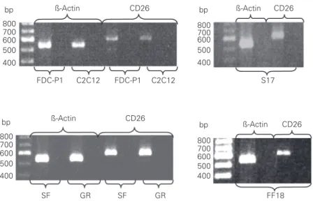
The expression of the DPP-IV (or CD26) gene was studied by RT-PCR in connective stroma cells that sustain hematopoiesis or myelopoiesis, i.e., S17 (bone marrow), FF18 (fetal liver) and GR (liver cells isolated from fibrogranulomatous inflammatory reactions), those that do not sustain hematopoiesis, i.e., SF (newborn skin fibroblast), and an unre-lated cell line C2C12 (myoblasts), as well as in the established myeloid precursor cell lineage FDC-P1. The DPP-IV gene was ex-pressed equally in all the cells tested. They produced the single mRNA band, with no alternative splicing in the region between exons 9 and 16 that was amplified by the primers used (Figure 1).
purified enzyme from S17 cells showed a immunoreactivity similar to that observed for the DPP-IV obtained from porcine kid-ney (Figure 2).
Inhibition of DPP-IV activity by diprotin-A
In order to test the specificity of the proteolytic activity of DPP-IV, we moni-tored its inhibition by diprotin-A, which is a competitive inhibitor of the enzyme. The maximal inhibition of the DPP-IV activity, obtained with 10 mM inhibitor and 1.5 mM substrate, was 23% for the total S17 cell extract, 46% for the S17 cell supernatant, 53% for the crude S17 membrane fraction, 80% for the protein partially purified on a DEAE-Sephacel column, and 100% for the purified porcine kidney DPP-IV. The partial inhibition in the nonpurified extracts was probably due to the presence of other pro-teases, which may hydrolyze the inhibitor, or of other molecules that may bind to the inhibitor. The presence of other enzymes that specifically cleave the substrate at the Pro-anilide bond is not probable, in view of the absence of this type of activity in DEAE-Sephacel-purified fractions other than the one containing DPP-IV (data not shown).
DPP-IV activity depends on the cell membrane environment
The saturation curve of DPP-IV (Figure 3) was analyzed and showed differences between the membrane-associated and re-leased enzymes. The observed apparent KM
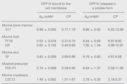
and the confidence intervals showed also that the KM were not the same for all the assayed cell lines (Table 1). S17 cells and all the primary cell lines obtained from connec-tive tissue stromas showed similar apparent
KM values, with no clear difference between those that sustain hemato- or myelopoiesis (bone marrow and liver) or do not (skin). The apparent KM of myeloid progenitors was within the same magnitude range. The ap-parent KM of myoblasts (C2C12 cells) was significantly different from those observed for the connective tissue stromas, indicating that the membrane environment relevant for the enzyme affinity is tissue specific.
bp
800 700 600 500 400
bp
800 700 600 500 400
ß-Actin CD26 bp ß-Actin CD26
800 700 600 500 400
bp
800 700 600 500 400
FDC-P1 C2C12 FDC-P1 C2C12 S17
ß-Actin CD26
ß-Actin CD26
FF18
SF GR SF GR
Figure 1. RT-PCR detection of DPP-IV (CD26) expression in the myeloid progenitor cell line FDC-P1, myoblast cell line C2C12, murine bone marrow stroma cell line S17, primary murine liver cell lines FF18 and GR, and primary murine newborn skin fibroblasts (SF). ß-Actin expression is used as an internal control.
v
(nmol/h)
40
30
20
10
0
0 1 2 3 4 5
Glycine-proline-p-nitroanilide (mM) Figure 3. Analysis of DPP-IV
en-zyme kinetics using glycine-pro-line-p-nitroanilide as substrate.
Circles, Enzyme activity of the cell membrane-associated en-zyme. Squares, Enzyme activity in the cell culture supernatant. A typical assay is shown, with the curves that represent the best adjustment values for enzyme ki-netics calculated from fittings of the Michaelis-Menten equation to the experimental data.
A B C D E
Figure 2. Immunoblot analysis of DPP-IV (CD26) using commercial polyclonal goat DPP-IV anti-body (Santa Cruz). A and B, DPP-IV purified from porcine kidney
Upon the spontaneous release of DPP-IV into the cell supernatant, the kinetics of its hydrolysis of Gly-Pro-p-nitroanilide was con-siderably modified, with the KM being larger roughly by one order of magnitude. These differences were statistically significant, as shown by analysis of the intervals of confi-dence (Table 1). The enzyme released by myoblasts was again significantly different from those released from connective tissue stromas and myeloid cells, with the KM of myoblast DPP-IV being only slightly differ-ent under such conditions.
Dependence of DPP-IV activity on the environment
Our previous studies, as well as studies from other laboratories, have shown that the ability of a stroma to sustain myelopoiesis depends upon the stroma-associated glyco-conjugates, and glycosaminoglycans in par-ticular (23,27-29). In order to determine whether the differences in the KM values for membrane-bound versus spontaneously re-leased DPP-IV were a consequence of the loss of interactions of the enzyme with the cell membrane-associated molecules such as proteoglycans, we first assayed the effect of sulfated glycosaminoglycans on the stroma-derived enzyme KM. Heparin had no effect on the activity of DPP-IV anchored to the hematopoietic stroma cell membrane, or on the activity of DPP-IV purified from por-cine kidney (Figure 4A,B). Conversely, both heparin and heparan sulfate reduced the ap-parent KM of stroma-derived enzymes in the soluble form apparently in a concentration-dependent manner (Figure 4C,D). An in-verse linear relationship was obtained be-tween the molecular mass and the glycosami-noglycan concentration in the solution that elicited a maximal modification of the en-zyme affinity (Figure 5). In view of the fact that DPP-IV is a highly glycosylated mem-brane-anchored molecule, we further ques-tioned whether the molecular structure and
the presence of negative charges on the as-sayed glycoconjugates was determinant for their modulation of DPP-IV activity. This was apparently not the case, since dextran sulfate and nonsulfated dextran had similar effects in increasing the KM of the enzyme, as also did smaller sugars such as sucrose and trehalose that are also known to organize the water molecules (30) (Table 2). These results were supported in assays using the treatment of S17 cells with chlorate that inhibits glycosaminoglycan sulfation, or treat-ment with heparitinase prior to monitoring the enzyme activity. In both cases there was no effect on the apparent DPP-IV KM under such experimental conditions (data not shown). Taken together, these experiments suggested that DPP-IV affinity was depend-ent upon the environmdepend-ent, which provided an optimal relationship between the enzyme and the substrates.
Since being attached to the cell mem-brane provided conditions for a low appar-ent KM of DPP-IV, we questioned whether
Table 1. Apparent KM for dipeptidyl peptidase IV (DPP-IV) activity in the cell layer and released in the soluble form into the supernatants of cell cultures.
DPP-IV bound to the DPP-IV released in
cell membrane a soluble form
KM (mM)a CIb KM (mM)a CIb
Murine bone marrow
S17 0.98 ± 0.093 0.77-1.19 8.86 ± 0.93 5.02-12.69
Murine liver
FF18 0.53 ± 0.074 0.37-0.70 8.44 ± 0.66 6.97-9.92
GR 0.63 ± 0.102 0.40-0.85 7.95 ± 1.34 4.99-10.91
Murine skin
SF 0.82 ± 0.059 0.69-0.96 6.76 ± 0.80 4.97-8.56
Myeloid precursor
FDC-P1 0.74 ± 0.069 0.59-0.90 8.64 ± 1.37 5.58-11.69
Murine myoblasts
C2C12 1.49 ± 0.082 1.31-1.67 2.78 ± 0.26 2.18-3.37
Data are reported as means ± SEM.
aDetermined from fitting the Michaelis-Menten equation to data obtained from kinetic
studies.
in the cell extract prepared on ice, rich in lipid rafts, where the enzyme velocity at-tained 18.6 ± 1.6 nmol/h, while in extracts prepared at room temperature in which lipid rafts were dissolved it was 5.1 ± 0.2 nmol/h. Moreover, the apparent KM of DPP-IV in the raft-enriched extract was very low, roughly three times lower than that observed for intact cell membranes, and more than one order of magnitude lower than the one ob-served in the cell supernatant (Table 3).
Taken together, these data indicate that DPP-IV is located in specialized microdomains on membranes of the connective tissue stromas, the lipid membrane rafts, which provide the molecular environment required and sufficient for optimal enzyme activity.
Discussion
We demonstrated the peptidolytic activity
KM
(mM)
1.5
1.0
0.5
0.0
0 0.1 1 10
Heparin (µg/ml)
A
KM
(mM)
0.60
0.40
0.20
0.00
Heparin (µg/ml)
0 5 10 16
B
Heparan sulfate (µg/ml)
0 5 10
KM
(mM)
14
10
6
0 2 4 8 12
D
KM
(mM)
14
10
6
0
0.1 1 10 100
Heparin (µg/ml) 0
2 4 8 12
C
Figure 4. Determination of KM for DPP-IV (CD26) in the pres-ence of heparin or heparan sul-fate. A, DPP-IV associated with S17 cell membranes in the pres-ence of heparin (0 to 10 µg/ml).
B, DPP-IV purified from porcine kidney (Sigma) in the presence of heparin (0 to 16 µg/ml). C, DPP-IV in the supernatant of S17 cell cultures in the presence of heparin (0 to 100 µg/ml). D, DPP-IV in the supernatant of S17 cell cultures in the presence of hep-aran sulfate (0 to 10 µg/ml).
Figure 5. Linear inverse ratio be-tween the concentration of the assayed glycosaminoglycans (GAG) that gave the maximal modification of the DPP-IV KM when assayed in the superna-tant of S17 cells and their molec-ular mass.
Molecular mass (kDa)
500
400
300
200
0
0 1 2 3 4
1/[GAG] 100
y = 124.56x
R2 = 0.9998
of DPP-IV in all the cell cultures studied, including the connective tissue stromas of bone marrow and liver that sustain hematopoiesis in vitro, skin fibroblasts that do not, a myeloid progenitor cell line, and unrelated cells such as myoblasts. Furthermore, we showed that DPP-IV mRNA was expressed in various tissues. The mouse DPP-IV gene is approximately 90 kb long, comprises 26 exons, and an alterna-tive splicing of DPP-IV has not been reported (31,32). Accordingly, our data show that the observed peptidase activity corresponded to expression of the DPP-IV gene in all the mu-rine cell lines studied. A single mRNA band was observed by RT-PCR analysis, showing that in all the cells studied the enzyme is a product of a single gene with no alternative splicing under the conditions used, and within the limits of the RT-PCR-amplified fragment. The immunoreactivity of the protein purified from the S17 cell extract was compared with that of the porcine kidney DPP-IV, and both proteins were recognized by the commercially available antibody for CD26.
In contrast to the homogeneity of the enzyme expression, we show that the DPP-IV apparent KM depended upon whether the enzyme was membrane bound or released from the membrane. The apparent KM was significantly different and dependent upon the tissue origin of the cells studied. When DPP-IV was released from the cell mem-brane environment, its apparent KM increased by one order of magnitude in murine con-nective tissue stromas and myeloid cells, but only doubled in myoblasts. The apparent KM
does not depend upon the total quantity of the assayed enzyme and indirectly reflects the relative affinity of the enzyme for a given substrate. Although the assayed substrate is an artificial one and does not necessarily re-flect the affinity of the enzyme for potential natural substrates, the observed differences in the apparent KM for diverse molecular back-grounds indicated that the enzyme affinity was modulated by the associated molecules.
DPP-IV is a highly sialylated
glycopro-Table 2. Effect of glycosaminoglycans and sugars on the KM of dipeptidyl peptidase IV (DPP-IV) in S17 cells.
Conditions KM (mM)a CIb
DPP-IV bound to the 0.98 ± 0.093 0.77-1.19
membrane of S17 cells
DPP-IV released from S17 cells 8.86 ± 0.93 5.02-12.69
in the soluble form
DPP-IV released from S17 cells in 3.06 ± 0.34 2.32-3.81
the presence of heparin (10 µg/ml)
DPP-IV released from S17 cells in 3.34 ± 0.47 2.30-4.39
the presence of heparan sulfate (5 µg/ml)
DPP-IV released from S17 cells in 4.62 ± 0.53 3.43-5.81
the presence of dextran sulfate (0.25 µg/ml)
DPP-IV released from S17 cells in 3.89 ± 0.37 3.07-4.70
the presence of chondroitin sulfate (2 µg/ml)
DPP-IV released from S17 cells in 4.81 ± 0.39 3.95-5.68
the presence of dextran (0.8 µg/ml)
DPP-IV released from S17 cells in 5.00 ± 0.71 3.11-6.59
the presence of trehalose (320 µg/ml)
DPP-IV released from S17 cells in 5.15 ± 0.42 4.20-6.08
the presence of sucrose (350 µg/ml)
The concentrations of saccharide that gave the maximum effect on KM are reported. Data are reported as means ± SEM.
aDetermined from fitting the Michaelis-Menten equation to data obtained from kinetic
studies.
bCI, confidence interval calculated by the Student t-test (P = 0.05).
tein, and inactivation of a single N-glycosy-lation site resulted in changes of enzymatic activity, subcellular localization, and bio-logical stability of the protein (33,34). The T cell membrane-bound DPP-IV is a homo-dimer, with two chains of 110 kDa molecu-lar mass. The soluble form is produced by proteolytic cleavage, and several molecular forms have been reported to be present in the human serum (35,36). The activity of
DPP-Table 3. The apparent KM for dipeptidyl peptidase IV (DPP-IV) from hematopoietic stroma cells.
Stroma-derived DPP-IV KM (mM)a CIb
Bound to cell membrane 0.98 ± 0.093 0.77-1.19
Released in a soluble form 8.86 ± 0.93 5.02-12.69
Cell membrane preparations enriched with 0.29 ± 0.027 0.23-0.35
lipid rafts
Data are reported as means ± SEM.
aDetermined from fitting the Michaelis-Menten equation to data obtained in kinetic
studies.
IV purified from serum has a KM value of 0.22 mM for the Gly-Pro-p-nitroanilide sub-strate, similar to that of the membrane-bound DPP-IV purified from T cells (37), suggest-ing that the glycosylation or release from the cell membrane did not modify the enzyme
KM in this cell type. In contrast, our studies showed a considerable modification in ap-parent KM when DPP-IV was released spon-taneously from the pericellular environment of connective tissue stromas into the super-natant. Addition of hydrophilic molecules could partially reverse the loss of affinity, in a well-defined range of their ratio with DPP-IV. This suggested the existence of ordered supramolecular membrane structures in which DPP-IV acts on the pericellular envi-ronment. Indeed, extraction of cell mem-branes by a method that preserved the organ-ization of membrane lipid rafts was suffi-cient to fully preserve the affinity of DPP-IV observed on intact cells. DPP-IV was re-ported to be integrated into lipid rafts on lymphocytes, but not on epithelial cells (38,39). Targeting of DPP-IV to rafts is re-quired for stimulation of T cells, albeit its enzyme activity is not (40), with the possibil-ity that the enzymatic activpossibil-ity released from lymphocytes that fully preserves its substrate affinity is an independent enzyme pool, not related to lipid rafts. In our study, the en-zyme fraction inserted into lipid rafts had a lower KM than the total enzyme in intact membranes. We understand that this may also indicate the presence of two membrane pools, one in lipid rafts with the optimal substrate affinity, and the other dispersed in the membrane with a lower affinity. Alterna-tively, the presence of membrane-associated molecules not participating in rafts, which are absent in raft-enriched fractions, may interfere with the optimal enzyme affinity.
Our study suggests that, in the hematopoi-etic environment, DPP-IV activity is essen-tially associated with short-range controls which are dependent upon the stroma cell membrane organization and direct cell-cell
interactions, while long-range controls depend-ing upon soluble enzyme forms are less prob-able in view of the low catalytic activity of the enzyme. Simultaneously, the released enzyme can act by interaction with other molecular systems that do no require its catalytic activity, such as adenosine deaminase binding. This is in keeping with the concept of micromental niches in the bone marrow environ-ment with rigorously controlled cell-cell inter-actions, which can retain early progenitors in a low-cycling state, whilst the more mature ones follow commitment and intense production of specific cell lineages in a highly ordered and programmed pattern.
It is generally accepted that DPP-IV is a product of a single gene. The highly divergent enzymatic behavior of DPP-IV is understand-able in the context of the membrane back-ground, since supramolecular complexes in lipid rafts are known to be required for optimal molecular activity in transduction pathways and cell activation. However, the spontane-ously released DPP-IV molecule is cleaved at a conserved site close to the membrane, fully preserving the structure of its catalytic site. It is thus surprising that in hematopoietic and con-nective tissues the enzyme release is associ-ated with such a great loss of affinity, not observed in myoblasts, lymphocytes or kidney extract. Post-translational modifications of DPP-IV may be determinant for the wide tis-sue-specific differences in its membrane loca-tion and enzyme kinetics, such as observed in the present study, and they are the subject of ongoing studies.
Acknowledgments
References
1. Hong WJ, Petell JK, Swank D, Sanford J, Hixson DC & Doyle D (1989). Expression of dipeptidyl peptidase IV in rat tissues is mainly regulated at the mRNA levels. Experimental Cell Research, 182: 256-266.
2. Tsugiki M, Kobayashi Y, Kawasaki T & Yoshimi T (1998). Identifica-tion of bile canalicular cell surface antigen HAM.4 as dipeptidyl peptidase IV (DPPIV) and characterization of its role in hepatic regen-eration after partial hepatectomy in rats. Digestive Diseases and Sciences, 43: 2591-2600.
3. Levy MT, McCoughan GW, Abbott CA, Park JE, Cunningham AM, Muller E, Rettig WJ & Gorrell MD (1999). Fibroblast activation pro-tein: a cell surface dipeptidyl peptidase and gelatinase expressed by stellate cells at the tissue remodelling interface in human cirrhosis.
Hepatology, 29: 1768-1778.
4. Hopsu-Havu VK & Glaner GG (1966). A new dipeptide naphthylami-dase hydrolyzing glycyl-prolyl-ß-naphthylamide. Histochimie, 7: 197-201.
5. Hegen M, Niedobitek G, Klein CE, Stein H & Fleischer B (1990). The T cell triggering molecule Tp103 is associated with dipeptidyl ami-nopeptidase IV activity. Journal of Immunology, 144: 2908-2914. 6. Torimoto Y, Dang NH, Vivier E, Tanaka T, Schlossman SF & Morimoto
C (1991). Coassociation of CD26 (dipeptidyl peptidase IV) with CD45 on the surface of human T lymphocytes. Journal of Immunology, 147: 2514-2517.
7. Hegen M, Kameoka J, Dong RP, Schlossman SF & Morimoto C (1997). Cross-linking of CD26 by antibody induces tyrosine phos-phorylation and activation of mitogen-activated protein kinase. Im-munology, 90: 257-264.
8. Dong RP, Kameoka J, Hegen M, Tanaka T, Xu Y, Schlossman SF & Morimoto C (1996). Characterization of adenosine deaminase bind-ing to human CD26 on T cells and its biologic role in immune response. Journal of Immunology, 156: 1349-1355.
9. Bristol LA, Sakaguchi K, Appella E, Doyle D & Takács L (1992). Thymocyte costimulating antigen is CD26 (dipeptidyl-peptidase IV): Co-stimulation of granulocyte, macrophage, and T lineage cell prolif-eration via CD26. Journal of Immunology, 149: 367-372.
10. Bristol LA, Bachvshin W & Takács L (1995). Inhibition of CD26 enzyme activity with Pro-boropro stimulates rat granulocyte/macro-phage colony formation and thymocyte proliferation in vitro. Blood, 85: 3602-3609.
11. Scholz W, Mentlein R, Heymann E, Feller AC, Ulmer AJ & Flad H (1985). Interleukin 2 production by human T lymphocytes identified by antibodies to dipeptidyl peptidase IV. Cellular Immunology, 93: 199-211.
12. Schön E & Ansorge S (1990). Dipeptidyl peptidase IV in the immune system. Biological Chemistry Hoppe-Seyler, 371: 699-705. 13. Holst JJ & Deacon CF (1998). Inhibition of the activity of
dipeptidyl-peptidase IV as a treatment for type 2 diabetes. Diabetes, 47: 1663-1670.
14. Augustyns K, Bal G, Thonus G, Belyaev A, Zhang XM, Bollaert W, Lambier AM, Durinx C, Goossens F & Haemers A (1999). The unique properties of dipeptidyl-peptidase IV (DPP IV/CD26) and the thera-peutic potential of DPP IV inhibitors. Current Medicinal Chemistry, 6: 311-327.
15. Oravecz T, Pall M, Roderiquez G, Gorrell MD, Ditto M, Nguyen NY, Boykins R, Unsworth E & Norcross MA (1997). Regulation of the receptor specificity and of the function of the chemokine RANTES by dipeptidyl peptidase IV (CD-26)-mediated cleavage. Journal of Experimental Medicine, 186: 1865-1872.
16. Van Coillie E, Proost P, Van Aelst I, Struyf S, Polfliet M, De Meester I, Harvey DJ, Van Damme J & Opendakker G (1998). Functional comparison of two human monocyte chemotatic protein-2 isoforms, roles of the amino-terminal pyroglutamic acid and processing by CD26/dipeptidyl peptidase IV. Biochemistry, 37: 12672-12680. 17. Lambier AM, Proost P, Durinx C, Bal G, Senten K, Augustyns K,
Schrape S, Van Damme J & De Meester I (2001). Kinetic investiga-tion of chemokine truncainvestiga-tion by CD26/dipeptidyl peptidase IV re-veals a striking selectivity within the chemokine family. Journal of Biological Chemistry, 276: 29839-29845.
18. Iwaki-Egawa S, Watanabe Y & Fujimoto Y (1998). Dipeptidyl pepti-dase IV from human serum: purification, characterization, and N-terminal amino acid sequence. Journal of Biochemistry, 124: 428-433.
19. Loster K, Zeilinger K, Schuppan D & Reuter W (1995). The cysteine-rich region of dipeptidyl peptidase IV (CD26) is the collagen-binding site. Biochemical and Biophysical Research Communications, 217: 341-348.
20. Collins LS & Dorshkind K (1987). A stromal cell line from myeloid long-term bone marrow culture can support myelopoiesis and B lymphopoiesis. Journal of Immunology, 138: 1082-1087.
21. Alvarez-Silva M & Borojevic R (1996). GM-CSF and IL-3 activities in schistosomal liver granulomas are controlled by stroma-associated heparan-sulfate proteoglycans. Journal of Leukocyte Biology, 59: 435-441.
22. Dutra HS, Rossi MID, Azevedo SP, El-Cheikh MC & Borojevic R (1997). Haematopoietic capacity of colony-forming cells mobilized into hepatic inflammatory reactions as compared to that of normal bone marrow cells. Research in Immunology, 148: 437-444. 23. Carvalho MA, Arcanjo K, Silva LCF & Borojevic R (2001). The
capac-ity of connective tissue stromas to sustain myelopoiesis depends both upon the growth factors and the local intercellular environ-ment. Biology of the Cell, 92: 605-614.
24. Paiva LMC, Pinto GF & Oestreicher EG (1991). Inhibition of horse liver alcohol dehydrogenase and rabbit muscle lactate dehydrogen-ase by phenylhydrazine. Biomedica Biochimica Acta, 50: 25-29. 25. Metzler CM (1981). Statistical properties of kinetics estimates. In:
Endrenyi L (Editor), Kinetic Data Analysis: Design and Analysis of Enzyme and Pharmacokinetic Experiments. Plenum Press, New York, NY, USA, 25-37.
26. Kabouridis PS, Magee AI & Ley SC (1997). S-acylation of LCK protein tyrosine kinase is essential for its signaling function in T lympho-cytes. EMBO Journal, 16: 4983-4998.
27. Alvarez-Silva M, Silva LC & Borojevic R (1993). Cell membrane-associated proteoglycans mediate extramedullar myeloid prolifera-tion in granulomatous inflammatory reacprolifera-tions to schistosome eggs.
Journal of Cell Science, 104: 477-484.
28. Alvarez-Silva M, Pinazo AC, El-Cheikh MC & Borojevic R (1994). Myelopoietic competence of stroma composed of hepatic granu-loma-derived connective tissue cells or skin fibroblasts. Brazilian Journal of Medical and Biological Research, 27: 2143-2152. 29. Gupta P, Oegema TR, Brazil JJ, Dudek AZ, Slungaard A & Verfaillie
CM (1998). Structurally specific heparan sulfates support primitive human hematopoiesis by formation of a multimolecular stem cell niche. Blood, 92: 4641-4651.
30. Panek AD (1995). Trehalose metabolism - new horizons in techno-logical applications. Brazilian Journal of Medical and Biological Re-search, 28: 169-181.
(1992). cDNA cloning for mouse thymocyte-activating molecule: A multifunctional ectodipeptidyl peptidase IV (CD26) included in a subgroup of serine proteases. Journal of Biological Chemistry, 267: 2200-2208.
32. Bernard AM, Mattei MG, Pierres M & Marguet D (1994). Structure of the mouse dipeptidyl peptidase IV (CD26) gene. Biochemistry, 33: 15204-15214.
33. Fan H, Meng W, Kilian C, Grams S & Reutter W (1997). Domain-specific N-glycosylation of the membrane glycoprotein dipeptidyl-peptidase IV (CD26) influences its subcellular trafficking, biological stability, enzyme activity and protein folding. European Journal of Biochemistry, 246: 243-251.
34. Smith RE, Talhouk JW, Brown EE & Edgar SE (1998). The signifi-cance of hypersialylation of dipeptidyl peptidase IV (CD26) in the inhibition of its activity by tat and other cationic peptides. CD26: A subverted adhesion molecule for HIV peptide binding. AIDS Re-search and Human Retroviruses, 14: 851-868.
35. Duke-Cohan JS, Morimoto C, Rocker JA & Schlossman SF (1995). A novel form of dipeptidylpeptidase in human serum. Isolation, charac-terization, and comparison with T lymphocyte membrane dipepti-dase IV (CD26). Journal of Biological Chemistry, 270: 14107-14114. 36. Kähne T, Kröning H, Thiel U, Ulmer AJ, Flad HD & Ansorge S (1996).
Alteration in structure and cellular localization of molecular forms of DPP IV/CD26 during cell activation. Cellular Immunology, 170: 63-70.
37. Morimoto C & Schlossman SF (1998). The structure and function of CD26 in the T-cell immune response. Immunological Reviews, 161: 55-70.
38. Ilangumaran S, Briol A & Hoessli DC (1997). Distinct interactions among GPI-anchored, transmembrane and membrane associated intracellular proteins and sphingolipids in lymphocyte and endotheli-al cell plasma membranes. Biochimica et Biophysica Acta, 1328: 227-236.
39. Ait Slimane T, Lenoir C, Bello V, Delaunay JL, Goding JW, Chwetzoff S, Maurice M, Fransen JA & Trungan G (2001). The cytoplasmic/ transmembrane domain of dipeptidyl peptidase IV, a type II glyco-protein, contains an apical targeting signal that does not specifically interact with lipid rafts. Experimental Cell Research, 270: 45-55. 40. Ishii T, Ohnuma K, Murakami A, Takasawa N, Kobayashi S, Dang NH,