Correlation between heart rate variability and
pulmonary function adjusted by confounding
factors in healthy adults
M.S. Bianchim
1, E.F. Sperandio
1, G.S. Martinhão
1, A.C. Matheus
1, V.T. Lauria
1, R.P. da Silva
1,
R.C. Spadari
2, A.R.T. Gagliardi
3, R.L. Arantes
3, M. Romiti
3and V.Z. Dourado
1 1Laboratório de Epidemiologia e Movimento Humano, Departamento de Ciências do Movimento Humano, Universidade Federal de São Paulo, Santos, SP, Brasil 2Departamento de Biociências, Universidade Federal de São Paulo, Santos, SP, Brasil 3AngioCorpore Instituto de Medicina Cardiovascular, Santos, SP, Brasil
Abstract
The autonomic nervous system maintains homeostasis, which is the state of balance in the body. That balance can be determined simply and noninvasively by evaluating heart rate variability (HRV). However, independently of autonomic control of the heart, HRV can be influenced by other factors, such as respiratory parameters. Little is known about the relationship between HRV and spirometric indices. In this study, our objective was to determine whether HRV correlates with spirometric indices in adults without cardiopulmonary disease, considering the main confounders (e.g., smoking and physical inactivity). In a sample of 119 asymptomatic adults (age 20–80 years), we evaluated forced vital capacity (FVC) and forced expiratory
volume in 1 s (FEV1). We evaluated resting HRV indices within a 5-min window in the middle of a 10-min recording period,
thereafter analyzing time and frequency domains. To evaluate daily physical activity, we instructed participants to use a triaxial accelerometer for 7 days. Physical inactivity was defined aso150 min/week of moderate to intense physical activity. We found
that FVC and FEV1, respectively, correlated significantly with the following aspects of the RR interval: standard deviation of the
RR intervals (r=0.31 and 0.35), low-frequency component (r=0.38 and 0.40), and Poincaré plot SD2 (r=0.34 and 0.36). Multivariate regression analysis, adjusted for age, sex, smoking, physical inactivity, and cardiovascular risk, identified the SD2 and dyslipidemia as independent predictors of FVC and FEV1(R2=0.125 and 0.180, respectively, for both). We conclude that
pulmonary function is influenced by autonomic control of cardiovascular function, independently of the main confounders. Key words: Autonomic nervous system; Spirometry; Smoking
Introduction
The autonomic nervous system maintains visceral functions through the activity of its sympathetic and parasympathetic branches. At times, the two branches operate in an antagonistic manner, generating a dynamic balance known as autonomic control. Analysis of heart rate variability (HRV) is a noninvasive and simple method for assessing autonomic control of the heart. The oscillation in the interval between consecutive heartbeats is an indicator of the integrity of the cardiovascular system and its ability to adapt to environmental changes (1).
Decreased HRV is associated with increased risk of morbidity and mortality after acute myocardial infarction (2). In addition, autonomic imbalance has been linked to the development of a wide range of diseases, including arterio-sclerosis, congestive heart failure, diabetic neuropathy, obesity, depression, and stress (3–5).
The respiratory cycle also affects autonomic control. The heart rate increases during inspiration and decreases during expiration, causing fluctuations in HRV (6). This physiological phenomenon is known as respiratory sinus arrhythmia (RSA). There are several ways to measure RSA, the most common being through analysis of the high-frequency (HF) component of HRV (7). The spectral variable HF is also referred to as RSA or respiratory rate because it has the same range as typical healthy adult respiration (7). This indicates that there is functional syn-chrony between the heart and lungs. However, the relation-ship between HRV and pulmonary function is unclear.
Many factors influence pulmonary and cardiac func-tion. Pulmonary function is influenced by lifestyle and cardiovascular risk factors such as obesity, high blood pressure, high cholesterol, metabolic syndrome, physical
Correspondence: M.S. Bianchim:<[email protected]>
inactivity, and smoking (8–14). Dyslipidemia and elevated heart rate are independent risk factors for pulmonary func-tion impairment (15). In addifunc-tion, pulmonary funcfunc-tion is inversely associated with levels of inflammation-sensitive plasma proteins (16). The role of lipids in inflammatory processes is well known and might explain the role of dyslipidemia in the development of pulmonary diseases (16). Physical activity also plays an important role in regulating cardiac and pulmonary function. Furthermore, regular physical activity increases HRV by increasing parasympathetic activity at rest. Moderate to vigorous physical activity can reduce the decline in pulmonary function among smokers, preventing the development of chronic obstructive pulmonary disease (COPD) as well as reducing mortality (17). Nevertheless, longtime smokers present lung tissue remodeling and a pro-nounced decline in pulmonary function with aging. Moreover, HRV is compromised in smokers (18). Those effects are attributable to increased release and reduced catabolism of catecholamines, together with decreased vagal tone (18). Increased activation of the sympathetic nervous system in smokers has an important clinical role, as has been widely reported (18–20).
Although the correlation between HRV and pulmonary function has been investigated in respiratory diseases such as COPD and asthma (21,22), few studies have evaluated that correlation in healthy adults (23). We hypothesized that lower HRV indices are associated with impaired pulmonary function, independently of factors such as smoking, level of physical activity in daily life, and cardiovascular risk. Therefore, the objective of the present study was to determine whether HRV correlates with the main spirometric indices in asymptomatic adults, as well as whether those correlations remain significant after being adjusted for the main confounders.
Material and Methods
The Epidemiological Study of Human Movement and Hypokinetic Diseases is a longitudinal, population-based cohort study investigating whether sedentary behavior and physical inactivity are associated with the occurrence of hypokinetic diseases, especially cardiorespiratory dis-eases. From those participating in this ongoing study, we recruited 119 participants (50 men and 69 women) who were asymptomatic and free of cardiorespiratory disease. We excluded individuals with Chagas disease, acute myocardial infarction, coronary heart disease, COPD, uncontrolled hypertension, diabetes, evidence of osteoar-ticular problems, or a recent history of respiratory infection, as well as those with a high risk of cardiac disease and those using cardiovascular drugs (e.g., beta-adrenoceptor antago-nists). The study was approved by the Ethics Committee for Research in Humans, Universidade Federal de São Paulo (Protocol #186.796), and all participants provided their written informed consent.
Health evaluation
During clinical evaluation of participants, we collected data related to level of education and medication use. In addition, we collected detailed information on the following cardiovascular risk factors: age, obesity, family history of cardiovascular disease, hypertension, dyslipidemia, seden-tary lifestyle, angina (stable or unstable), dizziness, and syncope. Dyslipidemia was defined as total cholesterol or triglyceride levels higher than 240 mg/dL. Body mass index (BMI) was calculated after measuring weight and height on a scale equipped with a stadiometer (2124; Toledo, Brazil). Participants with BMI X30 kg/m2 were considered obese (24). Cardiovascular risk was classified as mild or moderate according to the number of risk factors (o2 orX2, respectively). Participants who reported current smoking and having smoked at least 100 cigarettes in their lifetime were classified as smokers (25,26). Smoking history was calculated in pack-years. Participants were asked about their history of COPD and asthma, as well as about expo-sure to dusty environments and chemicals within the last year.
Pulmonary function
We performed pulmonary function tests with a spirometer (Quark PFT; Cosmed, Italy) using the forced vital capacity (FVC) maneuver. The maneuver could be repeated up to seven times until three results were reproducible. The turbines were calibrated before each test. Forced expiratory volume in 1 s (FEV1), FVC, and the
FEV1/FVC ratio were determined. Spirometric indices are
expressed as absolute values and as percentages of the predicted values (27).
HRV
frequency domain, we obtained the following linear indices: the HF component, the low-frequency (LF) component, and the LF/HF ratio. The geometric indices assessed were short-term variability (SD1) and long-short-term variability (SD2) of the Poincaré plot (4). We also analyzed the nonlinear indices
a1 anda2 (28).
HRV can be influenced by various conditions including blood pressure, anxiety, left ventricular ejection fraction, lung volume, breathing pattern, respiratory frequency, and medication use. To minimize such interference, all analyses were performed at the same time of day, with participants at rest in the supine position. Participants were instructed to avoid drinking coffee, tea, soft drinks, and alcoholic bever-ages, as well as to avoid engaging in physical activities and avoid smoking before the HRV test. All participants remained at rest for 5 min before the test. They were instructed to breathe normally and avoid speaking during the test.
Physical activity in daily life
The level of physical activity in daily life was measured with a triaxial accelerometer (GT3X+; Actigraph, USA). Each instrument was programmed according to the char-acteristics of the participant (sex, age, dominant side, height, and body mass). Participants were instructed to wear the accelerometer at the waist above the domi-nant hip for 7 days. They were instructed to remove the accelerometer during sleeping and water activities, including bathing. Only days with at least 12 h of continuous moni-toring were considered valid. Energy expenditure was measured and physical activity was classified as mild, moderate, vigorous, or very vigorous (29). Participants who were unable to engage in moderate to vigorous physical activity for at least 150 min/week were consid-ered physically inactive. Therefore, physical inactivity was analyzed as a categorical variable.
Statistical analysis
Sample size was calculated based on the number of predictors in the multiple regression models, as follows: age, sex, BMI, HRV, smoking, physical inactivity, and cardiovas-cular risk factors (family history of cardiovascardiovas-cular disease, hypertension, dyslipidemia, angina, and syncope). Consid-ering a correction coefficient (r) of 0.80 and a coefficient of determination (R2) of 0.64, with 11 predictors, the minimum sample size required for this study would be 110 participants. In multivariate linear regressions, spirometric indices were analyzed as outcomes.
Statistical analysis was performed using the IBM SPSS Statistics, Version 23.0 (IBM Corp., USA). Data were analyzed using descriptive statistics. We used the Kolmogorov-Smirnov to assess data normality. Continu-ous variables are presented as mean±standard deviation
or as median (interquartile range), depending on the distri-bution of the data (symmetrical or asymmetrical). Categorical variables are presented as frequencies. The Pearson or
Spearman correlation coefficient was used to evaluate the correlations, also depending on the distribution of the data. Stepwise multiple linear regressions were used to identify correlations between HRV indices as predictors and spirometric indices as outcomes. The HRV indices were divided into time, frequency, and nonlinear domains. Those HRV indices that presented the strongest correla-tions with FEV1and FVC in each category were selected
as predictors for inclusion in the regression models. The multiple regression models were adjusted for the main confounders, such as smoking and cardiovascular risk including physical inactivity assessed directly by triaxial accelerometry. We also adjusted the models for the use of medications other than cardiovascular drugs. The prob-ability of a type I error was set at 5%.
Results
Participants were, on average, middle-aged adults, ranging in age from 20 to 80 years (Table 1). The mean BMI was 27±5 kg/m2(within the range of overweight),
and 31 (26.1%) of the 119 participants were obese. According to the spirometric indices, the participants were free of respiratory disturbances. However, nine partici-pants (7.6%) had arterial hypertension, seven (5.9%) had diabetes, and 24 (20.2%) had dyslipidemia. In addition, 19 (16%) were smokers and 14 (11.9%) were physically inactive.
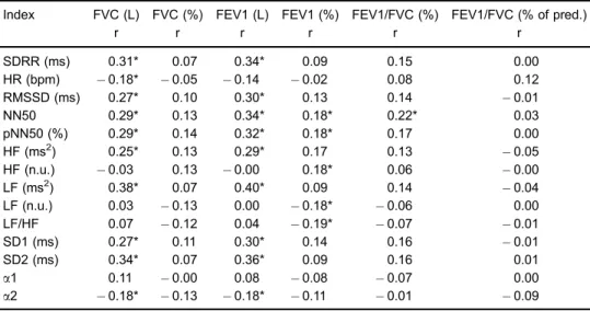
We found that HRV correlated moderately, but signifi -cantly, with the spirometric indices (Table 2). The indices representing parasympathetic and overall autonomic control (standard deviation of the RR intervals, root mean square of successive differences between adjacent normal RR intervals, number of adjacent normal RR intervals differing by 450 ms, proportion of adjacent normal RR intervals differing by450 ms, HF component, SD1, and SD2) presented positive correlations with spirometric indices. Those representing sympathetic modulation (the LF component and
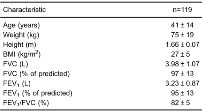
Table 1.General characteristics and pulmonary function in the study sample.
Characteristic n=119
Age (years) 41±14
Weight (kg) 75±19
Height (m) 1.66±0.07
BMI (kg/m2) 27±5
FVC (L) 3.98±1.07
FVC (% of predicted) 97±13
FEV1(L) 3.23±0.87
FEV1(% of predicted) 95±13
FEV1/FVC (%) 82±5
LF/HF ratio) presented negative correlations with those indices. The nonlinear index a2 correlated significantly with FVC. After multiple regression analysis, adjusted for the main confounders, only SD2 of the Poincaré plot and dyslipidemia remained as determinants of the spirometric indices (Table 3).
Discussion
Here, we have demonstrated that pulmonary func-tion correlated significantly with several HRV indices in asymptomatic adults. Although moderate, those correlations
remained significant regardless of cardiovascular risk. Among cardiovascular risk factors, dyslipidemia was found to be the most important predictor of pulmonary function.
After adjusting for the main confounders, we found that the SD2 of the Poincaré plot remained as a determinant of FVC and FEV1. SD2 has been associated with overall
HRV (1–3). Therefore, higher SD2 values indicate the predominance of the parasympathetic nervous system in autonomic balance. Although there have been few studies evaluating the Poincaré plot in healthy participants at rest, the evidence suggests that an increase in SD1 indicates increased parasympathetic activity, whereas an increase in SD2 indicates decreased sympathetic activity (1–3). Given that FEV1has been shown to be an important risk
factor for mortality and respiratory diseases (30) and that FVC is considered a measure of pulmonary capacity (30), our finding that autonomic control presented significant positive correlations with spirometric indices suggests that there is synchronicity between the activities of the lung and those of the heart. The predominance of the para-sympathetic nervous system might be related to respira-tory efficiency. Thatfinding is in agreement with those of Hayano et al. (31), who showed that the HF component is a measure of parasympathetic nervous system activity. In the cardiopulmonary system, the HF component is an intrinsic resting function aimed at optimizing pulmonary function. It has also been suggested that the HF com-ponent creates functional synchrony between the heart and lungs by matching the timing of alveolar ventilation to
Table 3.Results of multiple linear regression analysis for FEV1(L) and FVC (L) models.
Coefficients (b) Standard error P
FEV1
Intercept 2.874 0.156 0.000
SD2 (ms) 0.007 0.002 0.001
Dyslipidemia 0.508 0.192 0.009
FVC
Intercept 3.632 0.220 0.000
SD2 (ms) 0.007 0.003 0.012
Dyslipidemia 0.541 0.245 0.029
Adjusted FEV1 R 2
=0.180; adjusted FVC R2=0.125. FEV1: forced expiratory volume in 1 s; FVC: forced vital capacity; SD2: long-term variability of Poincaré plot. Adjusted for the main confounders (i.e., age, sex, smoking, cardiovascular risk, physical inactivity).
Table 2.Simple correlations between indices of heart rate variability (HRV) and spirometric indices.
Index FVC (L) FVC (%) FEV1 (L) FEV1 (%) FEV1/FVC (%) FEV1/FVC (% of pred.)
r r r r r r
SDRR (ms) 0.31* 0.07 0.34* 0.09 0.15 0.00
HR (bpm) 0.18* 0.05 0.14 0.02 0.08 0.12
RMSSD (ms) 0.27* 0.10 0.30* 0.13 0.14 0.01
NN50 0.29* 0.13 0.34* 0.18* 0.22* 0.03
pNN50 (%) 0.29* 0.14 0.32* 0.18* 0.17 0.00
HF (ms2) 0.25* 0.13 0.29* 0.17 0.13 0.05
HF (n.u.) 0.03 0.13 0.00 0.18* 0.06 0.00
LF (ms2) 0.38* 0.07 0.40* 0.09 0.14 0.04
LF (n.u.) 0.03 0.13 0.00 0.18* 0.06 0.00
LF/HF 0.07 0.12 0.04 0.19* 0.07 0.01
SD1 (ms) 0.27* 0.11 0.30* 0.14 0.16 0.01
SD2 (ms) 0.34* 0.07 0.36* 0.09 0.16 0.01
a1 0.11 0.00 0.08 0.08 0.07 0.00
a2 0.18* 0.13 0.18* 0.11 0.01 0.09
capillary perfusion during the respiratory cycle, increasing the rate of gas exchange (32). However, even though the HF component is considered a valid index of parasympa-thetic nervous system input, the respiratory rate and tidal volume might be confounders of the association between the HF component and cardiac vagal tone (7). At rest (when the cardiac vagal tone is constant), decreased tidal volume and increased respiratory rate can attenuate the HF component (8). Our results show that, in addition to the HF component, SD2 is also related to synchronization of cardiac and pulmonary function, being representative of the autonomic balance. Rapid breathing is known to greatly attenuate the HF component (32), which could explain why we found that the HF component did not correlate with the spirometric indices when we used the FVC maneuver. Despite the modestR2values, our results
suggest that autonomic control has an independent effect on pulmonary function in adults.
To date, there have been few studies investigating the correlations evaluated in the present study. To our knowl-edge, only one study has addressed such correlations in healthy adults; Behera et al. (23) also found that pulmonary function presented positive bivariate correlations with the parasympathetic and overall HRV indices, the HF compo-nent and FEV1, and negative correlations with the
sympa-thetic HRV indices, the LF component and peak expiratory
flow in healthy adults. After linear regression analysis, the authors observed a significant correlation between the HF component and FEV1/FVC ratio, indicating that HRV is a
determinant of pulmonary function. However, only the frequency domain of HRV was used and the regression analysis was not adjusted for the main confounders, such as physical inactivity and other cardiovascular risk factors. In contrast, after adjusting for cardiovascular risk factors, we found that the frequency domain of HRV was not an important determinant of pulmonary function.
In the present study, multiple regression analysis revealed that dyslipidemia was also a determinant of pulmonary function (FEV1and FVC). The important role of
cholesterol as an inflammatory regulator might partially explain the relationship between pulmonary function and dyslipidemia. In addition, there is a correlation between total cholesterol and mortality from respiratory disease (15). Furthermore, dyslipidemia has been shown to corre-late negatively with FEV1 (33). Our results underscore
previous data indicating an inverse relationship between dyslipidemia and pulmonary function. That relationship might also be explained by the role of low density lipoprotein as an optimizer of inflammation, which, in conjunction with oxida-tive stress, increases the severity of pulmonary diseases (34). Age also plays an important role in the association between dyslipidemia and pulmonary function because aging individuals tend to show declines in FEV1and FVC,
as well as increased dyslipidemia (35). However, in the present study, we found that dyslipidemia was predictive of pulmonary function, even after adjusting for age. Therefore,
it is reasonable to assert that dyslipidemia could have dele-terious effects on lung tissue, affecting spirometric indices independently of other factors.
The association between pulmonary function and HRV was also independent of smoking, physical inactivity, and other cardiovascular risk factors. The influence of those factors on pulmonary function has previously been described (16,18,19). Among such factors, special atten-tion should be given to physical activity in daily life, measured directly as in the present study. In a recent prospective study, an increase in physical activity level was found to prevent a decline in FVC among adolescents and young adults (36). In a 5-year cohort study (37), FEV1
was shown to increase by 50 mL in participants who remained active, whereas it declined by 40 mL in those who remained inactive. In an epidemiological study (38), a relatively large proportion of never smokers were found to have COPD (38). Certainly, there are other modifiable genetic or environmental risk factors that determine individual susceptibility (39). Although a low level of physical activity in daily life has been described as a consequence of COPD, recent studies raise the possibility that inactivity is actually a risk factor for the development and progression of the disease. It is plausible to suggest that a low level of physical activity in daily life has negative repercussions for pulmonary function because it increases oxidative stress and inflammation, which are commonly observed in sedentary individuals (40). In the present study, we observed an influence of HRV on FEV1 and
FVC, independent of the well-established association between daily physical activity and pulmonary function. Therefore, our results suggest a complex interaction among cardiovascular risk factors, autonomic balance, and pulmonary function. Future studies should investigate these relationships in a longitudinal manner.
The present study has certain limitations. Because this was a cross-sectional study, we cannot know whether improvement of the HRV indices would prevent a decline in pulmonary function over time. In addition, we assessed cardiovascular risk factors through interviews, which could have led us to underestimate the influence of factors such as hypertension and diabetes on pulmonary func-tion. However, the previously described interaction among inflammation, autonomic control, smoking, and physical inactivity supports our results. Furthermore, the correla-tions we observed between pulmonary function and auto-nomic control in adults free of cardiorespiratory disease have clinical relevance and should be considered when assessing the risk of respiratory diseases.
related to decreased pulmonary function. Therefore, strate-gies for improving autonomic control and reducing the impact of dyslipidemia in asymptomatic adults should be investigated in cohort studies, which might help prevent a decline in pulmonary function over time. Our results highlight the importance of the integrity of autonomic control to pulmonary function in asymptomatic adults.
Acknowledgments
This study was fully sponsored by the Fundac¸ão de
Amparo à Pesquisa do Estado de São Paulo (FAPESP, 2011/07282-6). The Angiocorpore Institute of Cardiovas-cular Medicine provided the necessary infrastructure for the cardiopulmonary exercise tests.
References
1. Malik M, Bigger JT, Camm AJ, Kleiger RE, Malliani A, Moss AJ, et al. Heart rate variability standards of measurement, physiological interpretation, and clinical use. Eur Heart J 1996; 17: 354–381, doi: 10.1093/oxfordjournals.eurheartj. a014868.
2. Kleiger RE, Miller JP, Bigger JT Jr, Moss AJ. Decreased heart rate variability and its association with increased mortality after acute myocardial infarction. Am J Cardiol 1987; 59: 256–262, doi: 10.1016/0002-9149(87)90795-8. 3. Vanderlei LC, Pastre CM, Hoshi RA, Carvalho TD, Godoy
MF. Basic notions of heart rate variability and its clinical applicability.Rev Bras Cir Cardiovasc2009; 24: 205–217, doi: 10.1590/S0102-76382009000200018.
4. Windham BG, Fumagalli S, Ble A, Sollers JJ, Thayer JF, Najjar SS, et al. The relationship between heart rate variability and adiposity differs for central and overall adiposity.J Obes2012; 2012: 149516, doi: 10.1155/2012/149516.
5. Hayano J, Yamada M, Sakakibara Y, Fujinami T, Yokoyama K, Watanabe Y, et al. Short- and long-term effects of cigarette smoking on heart rate variability. Am J Cardiol 1990; 65: 84–88, doi: 10.1016/0002-9149(90)90030-5. 6. Grossman P, Wilhelm FH, Spoerle M. Respiratory sinus
arrhythmia, cardiac vagal control, and daily activity.
Am J Physiol Heart Circ Physiol2004; 287: H728–H734,
doi: 10.1152/ajpheart.00825.2003.
7. Grossman P, Taylor EW. Toward understanding respiratory sinus arrhythmia: relations to cardiac vagal tone, evolution and biobehavioral functions.Biol Psychol 2007; 74: 263– 285, doi: 10.1016/j.biopsycho.2005.11.014.
8. Laurendi G, Donfrancesco C, Palmieri L, Vanuzzo D, Scalera G, Giampaoli S. Association of lifestyle and cardiovascular risk factors with lung function in a sample of the adult Italian population: a cross-sectional survey. Respiration 2015; 89: 33–40, doi: 10.1159/000369035.
9. Li AM, Chan D, Wong E, Yin J, Nelson EA, Fok TF. The effects of obesity on pulmonary function. Arch Dis Child 2003; 88: 361–363, doi: 10.1136/adc.88.4.361.
10. Schnabel E, Karrasch S, Schulz H, Glaser S, Meisinger C, Heier M, et al. High blood pressure, antihypertensive medi-cation and lung function in a general adult population.Respir Res2011; 12: 50, doi: 10.1186/1465-9921-12-50.
11. Melo S, Melo VA, Menezes Filho RS, Santos F. Effects of progressive increase in body weight on lung function in six groups of body mass index.Rev Assoc Med Bras2011; 57: 509–515.
12. Gopal DM, Santhanakrishnan R, Wang YC, Ayalon N, Donohue C, Rahban Y, et al. Impaired right ventricular hemodynamics indicate preclinical pulmonary hypertension in patients with metabolic syndrome. J Am Heart Assoc 2015; 4: e001597, doi: 10.1161/JAHA.114.001597.
13. Cheng ST, Wu YK, Yang MC, Huang CY, Huang HC, Chu WH, et al. Pulmonary rehabilitation improves heart rate variability at peak exercise, exercise capacity and health-related quality of life in chronic obstructive pulmonary disease.Heart Lung2014; 43: 249–255, doi: 10.1016/j.hrtlng.2014.03.002.
14. Paulose-Ram R, Tilert T, Dillon CF, Brody DJ. Cigarette smoking and lung obstruction among adults aged 40–79: United States, 2007–2012.NCHS Data Brief2015; 1–8. 15. Naveed B, Weiden MD, Kwon S, Gracely EJ, Comfort AL,
Ferrier N, et al. Metabolic syndrome biomarkers predict lung function impairment: a nested case-control study. Am J
Respir Crit Care Med 2012; 185: 392–399, doi: 10.1164/
rccm.201109-1672OC.
16. Engstrom G, Lind P, Hedblad B, Wollmer P, Stavenow L, Janzon L, et al. Lung function and cardiovascular risk: relationship with inflammation-sensitive plasma proteins.
Circulation 2002; 106: 2555–2560, doi: 10.1161/01.CIR.
0000037220.00065.0D.
17. Pelkonen M, Notkola IL, Lakka T, Tukiainen HO, Kivinen P, Nissinen A. Delaying decline in pulmonary function with physical activity: a 25-year follow-up.Am J Respir Crit Care Med2003; 168: 494–499, doi: 10.1164/rccm.200208-954OC. 18. Grassi G, Seravalle G, Calhoun DA, Bolla GB, Giannattasio C,
Marabini M, et al. Mechanisms responsible for sympathetic activation by cigarette smoking in humans.Circulation1994; 90: 248–253, doi: 10.1161/01.CIR.90.1.248.
19. Flouris AD, Dinas PC, Tzatzarakis MN, Metsios GS, Kostikas K, Jamurtas AZ, et al. Exposure to secondhand smoke promotes sympathetic activity and cardiac muscle cachexia.Int J Environ Health Res2014; 24: 189–194, doi: 10.1080/09603123.2013.800966.
20. Yotsukura M, Koide Y, Fujii K, Tomono Y, Katayama A, Ando H, et al. Heart rate variability during the first month of smoking cessation.Am Heart J1998; 135: 1004–1009, doi: 10.1016/S0002-8703(98)70065-1.
21. Pantoni CB, Reis MS, Martins LE, Catai AM, Costa D, Borghi-Silva A. Study on autonomic heart rate modulation at rest among elderly patients with chronic obstructive pulmonary disease.Braz J Phys Ther2007; 11: 35–41. 22. Lewis MJ, Short AL, Lewis KE. Autonomic nervous system
control of the cardiovascular and respiratory systems in asthma. Resp Med 2006; 100: 1688–1705, doi: 10.1016/ j.rmed.2006.01.019.
23. Behera JK, Sood S, Kumar N, Sharma K, Mishra R, Roy PS. Heart rate variability and its correlation with pulmonary function test of smokers.Heart Views2013; 14: 22–25, doi: 10.4103/1995-705X.107116.
24. World Health Organization.Obesity: preventing and
mana-ging the global epidemic (No. 894). city: World Health
25. Santos JD, Silveira DV, Oliveira DF, Caiaffa WT. [Instruments used to evaluate smoking habits: a systematic review].
Cien Saude Colet2011; 16: 4707–4720, doi:
10.1590/S1413-81232011001300020.
26. US Department of Health and Human Services.Reducing the health consequences of smoking: 25 years of progress.
A Report of the US Surgeon GeneralPublic Health Service,
Centers for Disease Control, Center for Health Promotion and Education, Office on Smoking and Health. DHHS pub. No.(CDC), 89–8411; 1989.
27. Pereira CA, Sato T, Rodrigues SC. New reference values for forced spirometry in white adults in Brazil. J Bras Pneumol 2007; 33: 397–406, doi: 10.1590/S1806-37132007000400008. 28. Wagner CD, Persson PB. Chaos in the cardiovascular system: an update. Cardiovasc Res 1998; 40: 257–264, doi: 10.1016/S0008-6363(98)00251-X.
29. Santos-Lozano A, Santin-Medeiros F, Cardon G, Torres-Luque G, Bailon R, Bergmeir C, et al. Actigraph GT3X: validation and determination of physical activity intensity cut points.Int J Sports Med2013; 34: 975–982, doi: 10.1055/s-0033-1337945. 30. Drummond M. Bradley, et al Spirometric predictors of lung
function decline and mortality in early chronic obstructive pulmonary disease. American journal of respiratory and
critical care medicine. 2012; 12 : 1301–1306, doi: 10.1164/
rccm.201202-0223OC.
31. Hayano J, Yasuma F. Hypothesis: respiratory sinus arrhythmia is an intrinsic resting function of cardiopulmonary system.
Cardio-vasc Res2003; 58: 1–9, doi: 10.1016/S0008-6363(02)00851-9.
32. Censi F, Calcagnini G, Cerutti S. Coupling patterns between spontaneous rhythms and respiration in cardiovascular variability signals. Comput Methods Programs Biomed 2002; 68: 37–47, doi: 10.1016/S0169-2607(01)00158-4.
33. Jacobs DR Jr, Iribarren C. Invited commentary: low cholesterol and nonartherosclerotic disease risk: a persistently perplexing question.Am J Epidemiol2000; 151: 748–751, doi: 10.1093/ oxfordjournals.aje.a010273.
34. Cirillo DJ, Agrawal Y, Cassano PA. Lipids and pulmonary function in the Third National Health and Nutrition Examina-tion Survey. Am J Epidemiol 2002; 155: 842–848, doi: 10.1093/aje/155.9.842.
35. Hering D, Somers VK, Kara T, Kucharska W, Jurak P, Bieniaszewski L, et al. Sympathetic neural responses to smoking are age dependent.J Hypertens 2006; 24: 691– 695, doi: 10.1097/01.hjh.0000217851.95583.57.
36. Twisk JW, Staal BJ, Brinkman MN, Kemper HC, van Mechelen W. Tracking of lung function parameters and the longitudinal relationship with lifestyle.Eur Respir J1998; 12: 627–634, doi: 10.1183/09031936.98.12030627.
37. Cheng YJ, Macera CA, Addy CL, Sy FS, Wieland D, Blair SN. Effects of physical activity on exercise tests and respiratory function.Br J Sports Med2003; 37: 521–528, doi: 10.1136/bjsm.37.6.521.
38. Celli BR, Halbert RJ, Nordyke RJ, Schau B. Airway obstruction in never smokers: results from the Third National Health and Nutrition Examination Survey.Am J Med2005; 118: 1364–1372, doi: 10.1016/j.amjmed.2005.06.041. 39. Lamprecht B, Schirnhofer L, Kaiser B, Buist S, Studnicka M.
Non-reversible airway obstruction in never smokers: results from the Austrian BOLD study. Respir Med 2008; 102: 1833–1838, doi: 10.1016/j.rmed.2008.07.007.