Propranolol inhibits the proliferation, migration and
tube formation of hemangioma cells through HIF-1
a
dependent mechanisms
Y.Z. Chen
1,2, N. Bai
2, J.H. Bi
1, X.W. Liu
1, G.Q. Xu
1, L.F. Zhang
1, X.Q. Li
1and R. Huo
1 1Department of Aesthetic, Plastic, and Burn Surgery, Shandong Provincial Hospital Affiliated to Shandong University, Jinan, Shandong, China 2
Department of Aesthetic, Plastic, and Burn Surgery, Linyi People’s Hospital, Linyi, Shandong, China
Abstract
The aim of this study was to investigate the mechanism of propranolol on the regression of hemangiomas. Propranolol-treated hemangioma tissues were collected and the expression of hypoxia inducible factor-1a(HIF-1a) was examined. We also established HIF-1a overexpression and knockdown hemangioma cells, and determined the effects of HIF-1a on the hemangioma cells proliferation, apoptosis, migration and tube formation. Significantly increased HIF-1alevel was found in the hemangioma tissues compared to that in normal vascular tissues, whereas propranolol treatment decreased the HIF-1alevel in hemangioma tissues in a time- and dose-dependent manner. Moreover, propranolol treatment significantly decreased cell proliferation, migration and tube formation as well as promoted cell apoptosis in HIF-1a overexpression and knockdown hemangioma cells. Propranolol suppressed the cells proliferation, migration and tube formation of hemangioma cells through HIF-1adependent mechanisms. HIF-1acould serve as a novel target in the treatment of hemangiomas.
Key words: Propranolol; Hemangiomas; Hypoxia inducible factor-1a; Knockdown; Regression
Introduction
Infantile hemangioma (IH), one of the most common vascular tumors in infants, is composed of disorganized blood vessels and immature endothelial cells (ECs) (1,2). The prevalence of IH is about 5–10% and it occurs more frequently in female, premature, and low-birth-weight new-borns (3,4). Clinicopathologically, IH is a unique category of benign tumors, characterized by abnormal ECs pro-liferation without a regular vascular architecture during the first year of life, and spontaneous regression of the neovasculature in early childhood. Most IH have a simple course and a general watchful waiting policy is often recommended (5). However, 10% of IHs may grow dra-matically, destroy tissue, and impair function, resulting in long-term cutaneous residua, permanent disfigurement, and even danger to life (6). Hence, it is necessary to clarify the relevant mechanism for IH and develop rational ther-apeutic strategies.
In clinical practice, corticosteroids have been consid-ered thefirst-line medical management for aggressive IH with local or systemic complications (7). However, the recent advent of propranolol for treatment of severe cases (8) following the unexpected discovery of its effects on
hemangioma regression has led to its subsequent popu-larization of this drug asfirst-line treatment (9,10). Although some possible explanations for propranolol’s regression have been proposed, there is still no evidence to support this conclusion in hemangiomas. Despite the remarkable effects of propranolol on hemangiomas regression, little is known about its mechanism.
In this study, we collected tissue samples from IH patients and isolated the hemangioma cells for investigat-ing the mechanism of propranolol on the regression of hemangiomas.
Material and Methods
Sample collection
From March 2015 to February 2016, 10 IH patients in the present study were recruited for this study and were orally treated with propranolol (Tianjin Lisheng Pharma-ceutical Co., Ltd., China). Patients had received no surgery or drug treatments prior to the propranolol treatment. Before and after the propranolol treatment, hemangioma tissues were collected from the patients. The vascular tissues from
Correspondence: R. Huo:<huorr10@tom.com>
surgical operations were used as normal control. The study protocol was approved by the Ethics Committee of Shan-dong Provincial Hospital Affiliated to Shandong University and the written informed consents were provided by family members.
Isolation of hemangioma cells
According to a previous description, hemangioma cells were isolated from patient tissues (11). Cells were cultured under standard conditions in a-MEM supplemented with 10% FBS, 0.25% fungizone (Sigma-Aldrich, USA) and 1% gentamicin (Sigma-Aldrich). Cells were passaged up to a maximum offive times before use in experiments.
Reverse transcription-quantitative PCR (RT-qPCR) Total RNA was isolated using TRIzol Reagent (Invitro-gen, USA) according to the manufacturers’ instruction. cDNA was synthesized using the first-strand synthesis and qPCR kit (Qiagen, Germany) according to the manu-facturer’s instructions. RT-qPCR detection was performed using SYBR Green PCR Kit (Qiagen). RT-qPCR condi-tions were an initial 95°C for 15 min, followed by 40 cycles of 94°C for 15 s, 56°C for 30 s, and 70°C for 30 s. PCR products were subsequently analyzed by a melting curve analysis. Humanb-actin mRNA was used as the internal control to normalize the HIF-1a expression and the
DDCt method was used to analyze the data as described
previously (12).
Western blotting
Cells and tissue samples were lysed in RIPA buffer supplemented with protease inhibitors cocktail, followed by high-speed centrifugation at 16,000gat 4oC for 30 min and protein quantification using a bicinchoninic acid assay. The proteins were subjected to sodium dodecyl sulfate-polyacrylamide gel electrophoresis (SDS-PAGE) and the separated proteins were transferred to polyvinylidene difluoride (PVDF, Millipore, USA) membranes. After blocking, the membranes were immunostained with anti-HIF-1amonoclonal primary antibodies (Cell Signaling Tech-nology, USA) overnight, and then the membranes were incubated with appropriate horseradish peroxidase (HRP)-conjugated secondary antibody at room temperature. The protein bands were developed with SuperSignal Ultra Chemiluminescent Substrate (USA). GAPDH (Santa Cruz Biotechnology, USA) was used as the loading control.
Cell treatment
Hemangioma cells were treated with concentrations of propranolol (0, 5, 50, and 200mM) for 48 h or treated with 50mM propranolol for different time courses (24, 48, and 72 h). To overexpress or knockdown HIF-1aprotein, hemangioma cells were infected with lentivirus parti-cles overexpressing or with knockdown HIF-1aplasmids (Hanbio, China) for 48 h according to the manufacturer’s instructions.
Cell proliferation
Hemangioma cells (1104 cells) were seeded onto 96-well plates and cultured ina-MEM supplemented with 10% FBS until they reached 80% confluence. Thereafter, the cells were infected with lentivirus particles over-expressing or with knockdown HIF-1a plasmids with or without propranolol treatment. After 48 h, cellular pro-liferation was evaluated by CCK-8 kit (Dojindo, Japan) according to the manufacturer’s instructions using a microplate reader (Biotek ELX-800, USA) to measure the absorbance.
Cell apoptosis
Hemangioma cells (1105cells) were trypsinized and seeded into 6-well plates in 2 ml of culture medium for attachment. The cells infected with lentivirus particles for 48 h were trypsinized, collected, washed and then stained with annexin V-FITC and propidium iodide (PI; BD Bioscience, USA) for 10 min at 4°C in dark according to a standard protocol. Apoptotic cells were determined by flow cytometry analysis (Becton-Dickinson FACScan, BD Biosciences, USA).
Transwell migration assay
Hemangioma cells migration assay was determined by transwell system. Briefly, resuspended hemangioma cells (2105cells) were seeded onto the upper chamber of the insert (BD Biosciences, 8-mm pore size) coated with matrigel (BD Biosciences), and the lower chamber was filled with a-MEM supplemented with 10% FBS. After maintaining in a cell culture incubator for 12 h the mem-branes of the upper chamber were stained by crystal violet and 3 randomfields (200 magnification) of each mem-brane were picked for cell counting. All treatments were performed in triplicates.
Tube formation assay
In vitrotube formation assay was performed for assess-ing the neovascularization capacity. Thawed matrigel (BD Biosciences) was plated onto 96-well plates at 37°C for up to 1 h to form a reconstituted basement membrane. Trypsinized hemangioma cells were harvested and resuspended (1104 cells/100 mL medium), seeded into matrigel, then incubated at 37°C for 6 h. Tube struc-tures were inspected under an inverted light microscope (200 magnification). The images were quantified using the Image-Pro Plus 6.0 software from Media Cybernetics (USA), as previously described (13). The quantified values are reported as the fold of control.
Statistical analyses
Statistical analyses were performed using SPSS v18 (USA). Data are reported as means±SD. The Student’s
t-test or one-way analysis of variance was used to deter-mine differences among groups. A P value of less than 0.05 was considered to be significant.
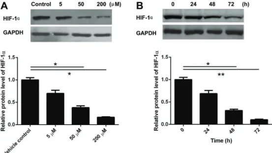
Figure 2.A, Hemangioma cells treated with different concentrations of propranolol for 48 h.B, Hemangioma cells treated with 50mM propranolol for 24, 48, and 72 h. The expression of hypoxia inducible factor-1a(HIF-1a) protein level was determined by western blotting. Data are reported as means±SD absorbance of HIF-1aprotein of three independent experiments. *Po0.05, **Po0.01 (ANOVA).
Results
Increased expression of HIF-1ain hemangioma tissues
Significantly increased levels of HIF-1a mRNA and protein were found in hemangioma tissues compared with normal vascular tissues. However, HIF-1amRNA and protein levels remarkably decreased in the propranolol-treated hemangiomas (Figure 1A and B).
Propranolol treatment decreased the expression of HIF-1aprotein in a dose- and time-dependent manner
The treatment with gradient concentrations of propra-nolol (0, 5, 50, and 200 mM) showed that it gradually decreased the expression of HIF-1a protein in a dose-dependent manner (Figure 2A). Next, 50mM propranolol used to treat the hemangioma cells for 24, 48, and 72 h showed that the propranolol gradually decreased the expression of HIF-1a level in a time-dependent manner (Figure 2B).
Overexpression of HIF-1aabolished the inhibitory effects of propranolol on hemangioma cells proliferation
To further validate the role of HIF-1ain the propranolol-treated cells, we established HIF-1a overexpressed and knockdown hemangioma cells. The results showed that cell proliferation was significantly inhibited by propranolol treatment of hemangiomas and significantly increased by HIF-1a overexpression, whereas propranolol treatment completely abolished HIF-1aoverexpression-induced heman-gioma cells proliferation (Figure 3A). Differently, HIF-1a knockdown remarkably inhibited hemangioma cells prolif-eration (Figure 3B).
Propranolol treatment promoted hemangioma cell apoptosis
As shown in Figure 4, the cell apoptotic percentage in propranolol treatment accounted for 17.9% (9.2+8.7%), almost 2-fold than control group (8.4%, 4.1+4.3%), suggesting that propranolol treatment induced heman-gioma cell apoptosis. On the other hand, the cell apoptotic percentage accounted for 9.1% (3.9+5.2%) in HIF-1a overexpression group and 16.7% (8.2+8.5%) in HIF-1a knockdown group, suggesting that HIF-1aoverexpression abrogated propranolol-induced hemangioma cells apoptosis.
Overexpression of HIF-1aabrogated the inhibitory effects of propranolol on hemangioma cells migration
The results showed that propranolol treatment decreased the number of migrated cells. Contrarily, HIF-1aoverexpression significantly increased the cell migra-tion and HIF-1a knockdown significantly decreased cell migration. Propranolol treatment completely abrogated the HIF-1a overexpression-increase in migrated cells (Figure 5A and B).
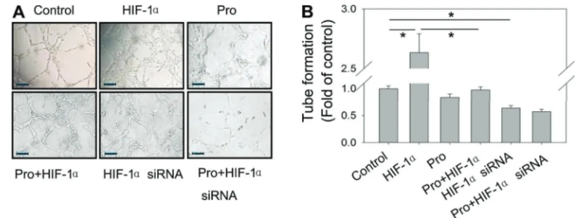
Overexpression of HIF-1aabrogated the inhibitory effects of propranolol on tube formation in hemangioma cells
The tube formation test results showed that proprano-lol treatment slightly decreased the level of tube formation. HIF-1a overexpression, on the other hand, significantly increased the level of tube formation and HIF-1a knock-down decreased the level of tube formation. Propranolol treatment completely abrogated the HIF-1a overexpres-sion-increase in tube formation (Figure 6A and B).
Discussion
In the present study, we demonstrated that signifi -cantly increased HIF-1alevels were found in hemangioma tissues compared to normal vascular tissues, and its levels were decreased by propranolol treatment. The in vitro study demonstrated that propranolol treatment decreased the expression of HIF-1aprotein in a dose- and time-dependent manner. Moreover, propranolol treatment
Figure 3. Overexpressed hypoxia inducible factor-1a(HIF-1a) hemangioma cells (A) and knockdown hemangioma cells (B) were treated or not with 50mM propranolol (Pro) for 48 h. Cell proliferation was measured by CCK-8 kit assay. Data are reported as means±SD of one representative experiment of at least three experiments with quintuplicates or sextuplicates. *Po0.05 (ANOVA).
significantly decreased hemangioma cell proliferation, migration and tube formation in HIF-1a-overexpressed and knockdown cells.
Traditionally, corticosteroids as well as other drugs such as interferonaand vincristine have been considered asfirst-line therapeutic approaches for the treatment of IH.
However, multiple and serious side-effects that are very painful accompany these drugs. Recent evidence shows that propranolol effectively regressed IH in a patient with obstructive hypertrophic cardiomyopathy. Further evidence has shown that the use of propranolol in the treatment of IH can achieve satisfactory outcomes and no serious
Figure 4.Overexpressed hypoxia inducible factor-1a(HIF-1a) hemangioma cells and knockdown hemangioma cells were treated or not with 50mM propranolol (Pro) for 48 h. Cell apoptosis was determined by annexin V-FITC. Representative images of cell apoptosis are shown for each group. PI: propidium iodide
side-effects. Furthermore, propranolol may be effective in IH patients who are allergic to corticosteroids and inter-feron or in severe IH. It has been suggested that pro-pranolol exerts its effects as a non-selectiveb-adrenergic receptor (AR) blocker that inhibits cell growth and induces cell apoptosis of the endothelial cells (14). Our results confirmed the cytotoxic effect of propranolol with an annexin V-FITC apoptosis assay, which demonstrated that high concentrations of propranolol resulted in increased apoptosis. However, the precise molecular mechanism of its action remains poorly understood.
Studies have shown that a causal relationship are observed between the expressions ofb-AR and HIF-1ain pancreatic, breast and prostate cancer cells (15,16). Accumulation of HIF-1a was found to correlate with b-adrenergic agonists treatment, whereas norepinephrine-induced expressions of HIF-1a and vascular endothelial growth factor (VEGF) are blocked by propranolol treat-ment. Besides HIF-1a, there are numbers of other VEGF regulators. Several other non-canonical pathways are not dependent on hypoxia and HIF-1a also regulate VEGF and angiogenesis (17,18). However, several studies have demonstrated that children with proliferative hemangio-mas showed upregulation and stabilization of HIF-1a
compared to normal controls (19–21), suggesting that these lesions may be intrinsically hypoxic, resulting in hemangioma endothelial cell proliferation through VEGF-mediated downstream signaling. This intrinsic difference in HIF-1a expression between hemangiomas and nor-mal tissues could result in predominance of the canoni-cal HIF-1a pathway in VEGF signaling. A reliance on HIF-1a signaling and dependence on VEGF (22) for survival could explain the specific response of heman-giomas rather than other tissues treated with pro-pranolol.
In conclusion, significantly increased HIF-1a levels were found in the hemangioma tissues. Propranolol treat-ment inhibited cell proliferation, migration, tube formation and promoted apoptosis in the hemangioma cells, sug-gesting that HIF-1acould serve as a therapeutic target in the treatment of IH.
Acknowledgments
This work was supported by the National Natural Science Foundation of China (No. 8167080540) and Shandong Province Natural Science Foundation of China (ZR2015HL012).
References
1. Bruckner AL, Frieden IJ. Hemangiomas of infancy.J Am Acad Dermatol 2003; 48: 477–493; quiz 494–496, doi: 10.1001/archdermatol.2011.122.
2. Drolet BA, Esterly NB, Frieden IJ. Hemangiomas in children. N Engl J Med 1999; 341: 173–181, doi: 10.1056/NEJM 199907153410307.
3. Zou HX, Jia J, Zhang WF, Sun ZJ, Zhao YF. Propranolol inhibits endothelial progenitor cell homing: a possible treat-ment mechanism of infantile hemangioma.Cardiovasc Pathol 2013; 22: 203–210, doi: 10.1016/j.carpath.2012.10.001.
4. Frieden IJ, Eichenfield LF, Esterly NB, Geronemus R, Mallory SB. Guidelines of care for hemangiomas of infancy. American Academy of Dermatology Guidelines/Outcomes Committee.J Am Acad Dermatol1997; 37: 631–637, doi: 10.1016/S0190-9622(97)70183-X.
5. Haggstrom AN, Drolet BA, Baselga E, Chamlin SL, Garzon MC, Horii KA, et al. Prospective study of infantile heman-giomas: clinical characteristics predicting complications and treatment. Pediatrics 2006; 118: 882–887, doi: 10.1542/ peds.2006-0413.
Figure 6.Overexpressed hypoxia inducible factor-1a(HIF-1a) hemangioma cells and knockdown hemangioma cells treated or not with 50mM propranolol (Pro) for 48 h.A, Representative images of tube formation (200 magnification).B, Relative quantified values of numbers of tubes in each group. Data are reported as means±SD of 3 randomfields. All treatments were performed in triplicate. *Po0.05 (ANOVA).
6. Lin RL, Schwartz RA. Hemangiomas of infancy - a clinical review.Acta Dermatovenerol Croat2006; 14: 109–116. 7. Gampper TJ, Morgan RF. Vascular anomalies: hemangiomas.
Plast Reconstr Surg2002; 110: 572–585; quiz 586; discus-sion 587–578.
8. Leaute-Labreze C, Dumas de la Roque E, Hubiche T, Boralevi F, Thambo JB, Taieb A. Propranolol for severe hemangiomas of infancy.N Engl J Med2008; 358: 2649– 2651, doi: 10.1056/NEJMc0708819.
9. Arneja JS, Pappas PN, Shwayder TA, Cullen ML, Becker CJ, Hamzavi FH, et al. Management of complicated facial hemangiomas with beta-blocker (propranolol) therapy.Plast Reconstr Surg 2010; 126: 889–895, doi: 10.1097/PRS. 0b013e3181e5f8b6.
10. Sans V, de la Roque ED, Berge J, Grenier N, Boralevi F, Mazereeuw-Hautier J, et al. Propranolol for severe infantile hemangiomas: follow-up report.Pediatrics2009; 124: e423– e431, doi: 10.1542/peds.2008-3458.
11. Li P, Xiao XE, Xu Q, Guo ZT. [Establishment of human infancy hemangioma-derived endothelial cell line XPTS-1 and animal model of human infancy hemangioma). Zhon-ghua Kou Qiang Yi Xue Za Zhi2011; 46: 129–133. 12. Livak KJ, Schmittgen TD. Analysis of relative gene
expres-sion data using real-time quantitative PCR and the 2(-Delta Delta C(T)) Method.Methods2001; 25: 402–408, doi: 10.1006/ meth.2001.1262.
13. Jing Z, Xu H, Chen X, Zhong Q, Huang J, Zhang Y, et al. The proton-sensing G-protein coupled receptor GPR4 promotes angiogenesis in head and neck cancer.PLoS One2016; 11: e0152789, doi: 10.1371/journal.pone.0152 789.
14. Storch CH, Hoeger PH. Propranolol for infantile haeman-giomas: insights into the molecular mechanisms of action. Br J Dermatol2010; 163: 269–274, doi: 10.1111/j.1365-2133. 2010.09848.x.
15. Hu HT, Ma QY, Zhang D, Shen SG, Han L, Ma YD, et al. HIF-1alpha links beta-adrenoceptor agonists and pancreatic cancer cells under normoxic condition.Acta Pharmacol Sin 2010; 31: 102–110, doi: 10.1038/aps.2009.181.
16. Park SY, Kang JH, Jeong KJ, Lee J, Han JW, Choi WS, et al. Norepinephrine induces VEGF expression and angiogen-esis by a hypoxia-inducible factor-1alpha protein-dependent mechanism.Int J Cancer2011; 128: 2306–2316, doi: 10.1002/ ijc.25589.
17. Hu YL, Tee MK, Goetzl EJ, Auersperg N, Mills GB, Ferrara N, et al. Lysophosphatidic acid induction of vascular endothelial growth factor expression in human ovarian cancer cells.J Natl Cancer Inst2001; 93: 762–768, doi: 10.1093/jnci/93.10.762. 18. Arany Z, Foo SY, Ma Y, Ruas JL, Bommi-Reddy A, Girnun
G, et al. HIF-independent regulation of VEGF and angiogen-esis by the transcriptional coactivator PGC-1alpha.Nature 2008; 451: 1008–1012, doi: 10.1038/nature06613. 19. Comati A, Beck H, Halliday W, Snipes GJ, Plate KH, Acker
T. Upregulation of hypoxia-inducible factor (HIF)-1alpha and HIF-2alpha in leptomeningeal vascular malformations of Sturge-Weber syndrome.J Neuropathol Exp Neurol 2007; 66: 86–97, doi: 10.1097/nen.0b013e31802d9011.
20. Chang EI, Thangarajah H, Hamou C, Gurtner GC. Hypoxia, hormones, and endothelial progenitor cells in hemangioma. Lymphat Res Biol 2007; 5: 237–243, doi: 10.1089/lrb. 2007.1014.
21. Kelly BD, Hackett SF, Hirota K, Oshima Y, Cai Z, Berg-Dixon S, et al. Cell type-specific regulation of angiogenic growth factor gene expression and induction of angiogenesis in nonischemic tissue by a constitutively active form of hypoxia-inducible factor 1.Circ Res2003; 93: 1074–1081, doi: 10.1161/01.RES.0000102937.50486.1B.