Pleiotropic effects of simvastatin in
physically trained ovariectomized rats
N. Bernardes
1,4, J.O. Brito
1, T.G. Fernandes
2, S.F. Llesuy
3, M.C. Irigoyen
4, A. Be´llo-Klein
2and
K. De Angelis
1 1Universidade Nove de Julho (UNINOVE), Sa˜o Paulo, SP, Brasil 2Laborato´rio de Fisiologia Cardiovascular, Departamento de Fisiologia, Instituto de Cieˆncias Ba´sicas da Sau´de,Universdade Federal do Rio Grande do Sul, Porto Alegre, RS, Brazil 3University of Buenos Aires, Buenos Aires, Argentina 4Unidade de Hipertensa˜o, Instituto do Corac¸a˜o, Faculdade de Medicina, Universidade de Sa˜o Paulo, Sa˜o Paulo, SP, Brazil
Abstract
This study tested the hypothesis that simvastatin treatment can improve cardiovascular and autonomic functions and membrane lipoperoxidation, with an increased effect when applied to physically trained ovariectomized rats. Ovariectomized rats were divided into sedentary, sedentary++simvastatin and trained++simvastatin groups (n = 8 each). Exercise training was performed on a treadmill for 8 weeks and simvastatin (5 mg/kg) was administered in the last 2 weeks. Blood pressure (BP) was recorded in conscious animals. Baroreflex sensitivity was evaluated by the tachycardic and bradycardic responses to BP changes. Cardiac vagal and sympathetic effects were determined using methylatropine and propranolol. Oxidative stress was evaluated based on heart and liver lipoperoxidation using the chemiluminescence method. The simvastatin-treated groups presented reduced body weight and mean BP (trained++simvastatin = 99 ± 2 and sedentary++simvastatin = 107 ± 2 mmHg) compared to the sedentary group (122 ± 1 mmHg). Furthermore, the trained group showed lower BP and heart rate compared to the other groups. Tachycardic and bradycardic responses were enhanced in both simvastatin-treated groups. The vagal effect was increased in the trained++simvastatin group and the sympathetic effect was decreased in the sedentary++simvastatin group. Hepatic lipoperoxidation was reduced in sedentary++simvastatin (<21%) and trained+ +sim-vastatin groups (<57%) compared to the sedentary group. Correlation analysis involving all animals demonstrated that cardiac lipoperoxidation was negatively related to the vagal effect (r = -0.7) and positively correlated to the sympathetic effect (r = 0.7). In conclusion, improvement in cardiovascular and autonomic functions associated with a reduction of lipoperoxidation with simvastatin treatment was increased in trained ovariectomized rats.
Key words: Menopause; Exercise training; Simvastatin; Autonomic function; Oxidative stress
Introduction
Cardiovascular diseases remain the leading cause of death among women in the most developed areas of the world (1). Studies have demonstrated that menopause can induce metabolic, endothelial and autonomic dys-functions and increase oxidative stress in women, thus increasing cardiovascular risk (2-5). Similarly, rats sub-jected to ovarian hormone deprivation presented increased blood pressure (BP) and sympathetic tonus and reduced baroreflex sensitivity (BRS) compared to control female rats (6).
On the other hand, simvastatin treatment reduces both mortality and coronary events in humans, and also prevents and controls atherosclerosis (7,8). The beneficial pleiotropic effects of simvastatin treatment may be due to
the endothelial protection, oxidative stress reduction, sympathetic decrease, and control of inflammatory reac-tions it seems to offer (9-12).
In addition, exercise training has been recognized as a non-pharmacological treatment for cardiovascular dis-ease. A recent systematic review of randomized, con-trolled trials reported benefits of regular exercise after menopause (13). Furthermore, previous studies per-formed in our laboratory have demonstrated that exercise training induced a reduction in body weight, BP and cardiac oxidative stress and an increase in BRS in an experimental model of menopause (6,14,15).
However, there are few data in the literature concerning the effects of either pharmacological or non-pharmacological
Correspondence: K. De Angelis, Laborato´rio de Fisiologia Translacional, Universidade Nove de Julho, Rua Vergueiro 235/249, 2osubsolo, Mestrado, 01504-001 Sa˜o Paulo, SP, Brasil. E-mail: prof.kangelis@uninove.br
approaches on hemodynamic, autonomic or oxidative stress profile changes induced by ovarian hormone deprivation. Also, there are no data on the effects of the association of simvastatin treatment with exercise training, two common approaches used in postmenopausal women in clinical practice.
Therefore, the purpose of the present study was to test the hypothesis that short-term simvastatin treatment (2 weeks) can induce cardiovascular and autonomic improvement and reduce oxidative stress when evaluated by membrane lipoperoxidation. Moreover, we hypothe-sized that simvastatin treatment can induce additional benefits in physically trained ovariectomized rats.
Material and Methods
Animals and groups
Experiments were performed on 24 female Wistar rats (215-230 g) from the animal house of the Universidade Sa˜o Judas Tadeu, Sa˜o Paulo, Brazil. The rats were given standard laboratory chow and waterad libitumand were housed in individual cages in a temperature-controlled room (226C) with a 12-h dark/light cycle. All rats were treated similarly in terms of daily manipulation. The experimental protocol was approved by the institutional Animal Care and Use Committee of Universidade Sa˜o Judas Tadeu and the investigation was conducted in accordance with the Guide for the Care and Use of Laboratory Animals published by the US National Institutes of Health (NIH Publication No. 85-23, revised 1985). Ovariectomized rats were randomly assigned to the following groups: sedentary (SO, n = 8), sedentary treated with simvastatin (SSO, n = 8), and exercise trained treated with simvastatin (STO, n = 8).
Ovariectomy
At 10-12 weeks of age, animals were anesthetized (80 mg/kg ketamine and 12 mg/kg xylazine), and a small abdominal incision was made. The ovaries were then located, and a silk thread was tightly tied around the oviduct, including the ovarian blood vessels. The oviduct was sectioned and the ovaries removed. The skin and muscle wall were then sutured with silk thread. After surgery, the animals received an injection of antibiotics (40,000 U/kg penicillin G procaine,im) (6,14,16).
Exercise training
Exercise training was performed on a motor treadmill at low-moderate intensity (<40 to 65% maximal running
speed) for 1 h a day, 5 days a week, for 8 weeks, with a gradual increase in speed from 0.3 to 1.2 km/h. All animals were adapted to the procedure (10 min/day, 0.3 km/h) for 1 week before beginning the exercise training protocol. This adaptation period began 24 h after ovariectomy (6,14). All groups were subjected to a maximal treadmill test, as described in detail in a previous publication (17,18). The
tests were performed three times: 1) at the beginning of the experiment, 2) in the fourth week, and 3) in the eighth week of the training protocol. The purpose was to determine maximal physical capacity and exercise training intensity.
Simvastatin treatment
Simvastatin treatment (5 mg kg-1?day-1) was performed daily by gavage in the last 2 weeks of sedentarism or training. The SO group was submitted to a daily gavage of water. The choice of a simvastatin dose of 5 mg kg-1
?day-1
was based on a pilot study in which we compared the dose of 20 mg kg-1
?day-1to a dose of 5 mg kg-1
?day-1in male
rats. The results of these experiments showed that the dose of 20 mg kg-1?day-1 induced further reduction in triglycerides and total cholesterol in treated male rats, which could induce changes in cardiac and autonomic functions, independently of the pleiotropic effects of simvastatin. Considering these results and the fact that the dose of 5 mg/kg did not induce changes in the metabolic parameters evaluated, for the present experi-ments we chose the simvastatin dose of 5 mg/kg.
Metabolic evaluations
Body weight was measured at the beginning and at the end of the protocol. Blood glucose and triglycerides were evaluated after 4 h fasting at the beginning and at the end of the protocol using the Roche GCT Accutrend (Roche, Brazil).
Cardiovascular assessments
After the last training session, 2 catheters filled with 0.06 mL saline were implanted into the carotid artery and jugular vein (PE-10) of anesthetized rats (80 mg/kg ketamine and 12 g/kg xylazine) for direct measurement of BP and for drug administration, respectively. The rats were conscious and allowed to move freely during the hemodynamic experiments. The arterial cannula was connected to a strain-gauge transducer (Blood Pressure XDCR, Kent Scientific, USA), and blood pressure signals were recorded over a 30-min period by a microcomputer equipped with an analog-to-digital converter board (CODAS, 2-kHz sampling frequency; Dataq Instruments, Inc., USA). The recorded data were analyzed on a beat-to-beat basis to quantify changes in mean BP (MBP) and heart rate (HR). Increasing doses of phenylephrine (0.25 to 32.0mg/kg, iv) and sodium nitroprusside (0.05 to
1.6mg/kg, iv) were given as sequential bolus injections
tachycardia. The mean index was expressed as bpm/ mmHg, as described elsewhere (14,15).
Vagal and sympathetic effects were studied by injecting methylatropine (3 mg/kg, iv) and propranolol (4 mg/kg, iv) at a dose of 0.1 mL/100 g body weight. Resting HR was recorded while the rats were in their cages in an unrestrained state, and methylatropine was injected immediately after the recording. Because the HR response to these drugs reaches its peak within 5 min, this time interval was allowed to elapse before the HR measurement was made. On the following day, the sequence of injection was inverted, with propranolol given first. The sympathetic effect was determined as the difference between the basal HR and the lowest HR after propranolol. The vagal effect was obtained by the difference between the maximum HR after methylatropine injection and the basal HR (19).
Evaluation of membrane lipoperoxidation
The animals were killed by decapitation and the heart (ventricles) and liver were immediately removed, rinsed in saline, and trimmed to remove fat tissue and visible connective tissue. These tissues were cut into small pieces, placed in ice-cold buffer, and homogenized in an ultra-Turrax blender with 1 g tissue per 5 mL 150 mM KCl and 20 nM sodium phosphate buffer, pH 7.4. The homogenates were centrifuged at 600g for 10 min at -26C. Tissue membrane lipoperoxidation was evaluated by chemilumi-nescence. The chemiluminescence assay was carried out with an LKB Rack Beta liquid scintillation spectrometer 1215 (LKB Producer AB, USA) in the out-of-coincidence mode at room temperature (256to 276C). The supernatants were diluted in 140 mM KCl and 20 mM sodium phosphate buffer, pH 7.4, and added to glass tubes, which were placed in scintillation vials; 3 mM tert-butylhydroperoxide was added, and chemiluminescence was determined up to the maximal level of emission (14,20,21).
Statistical analysis
Data are reported as means ± SE. Comparisons between the 3 groups were performed by one-way ANOVA followed by the Student-Newman-Keuls post hoc test. Pearson’s correlation was used to determine association among variables. The level of significance was established at P,0.05.
Results
There were no differences in body weight between groups at the beginning of the protocol (SO = 207 ± 2.5 g). At the end of the training period, SSO and STO animals (SSO = 306 ± 5 g; STO = 308 ± 8 g) had a smaller increase in body weight compared to SO animals (323 ± 4 g). No difference in blood metabolic parameters was observed between groups at the beginning of the protocol. After 4 h of fasting, blood glucose (SO = 89 ±
2 mg/dL) and triglycerides (SO = 97 ± 5 mg/dL) did not differ between groups at the end of the protocol.
Maximal physical performance was evaluated by the response to the maximal treadmill test. At the beginning of the experiment, the physical performance was similar for all groups (SO = 2 ± 0.08 km/h). However, the animals submitted to the exercise training protocol (STO = 2.4 ± 0.09 km/h) showed an increase in maximum running speed compared to the SO (2 ± 0.09 km/h) and SSO (1.9 ± 0.07 km/h) groups after 8 weeks of exercise training.
Simvastatin treatment associated or not with exercise training induced reduction in systolic BP (SBP; STO = 120 ± 3 and SSO = 123 ± 2vsSO = 140 ± 2 mmHg; P,0.001), diastolic BP (DBP; STO = 82 ± 2 and SSO = 92 ± 2vsSO = 104 ± 2 mmHg; P,0.001) and MBP (STO = 99 ± 2 and SSO = 107 ± 2vsSO = 122 ± 1 mmHg; P , 0.001). The exercise training associated with simvastatin treatment (STO) induced an additional reduction in MBP compared to the animals treated with simvastatin alone (SSO) (Figure 1A). HR was lower in trained rats (STO = 329 ± 6 bpm) when compared to sedentary rats treated or not with simvastatin (SSO = 366 ± 8 and SO = 374 ± 7 bpm) (Figure 1B).
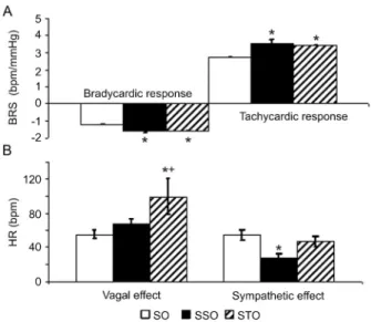
The tachycardic (SSO = 3.5 ± 0.2 and STO = 3.4 ± 0.2vsSO = 2.7 ± 0.2 bpm/mmHg) and the bradycardic (SSO = -1.6 ± 0.10 and STO = -1.6 ± 0.15vs SO = -1.2 ± 0.1 bpm/mmHg) responses evoked by the barore-flex during BP reductions and increases, respectively, were increased in rats treated with simvastatin, regardless of any association with exercise training, when compared to sedentary ovariectomized rats (SO; Figure 2A).
The vagal effect was increased in trained rats (STO = 99 ± 21 bpm) compared to sedentary rats treated or not
with simvastatin (SO = 55 ± 5 and SSO = 67 ± 6 bpm). However, the sympathetic effect was decreased in seden-tary rats treated with simvastatin (SSO = 28 ± 4 bpm) compared to sedentary rats (SO = 54 ± 6 bpm; Figure 2B). Oxidative stress evaluated by membrane lipoperoxida-tion showed a significant reduclipoperoxida-tion in liver homogenates from animals submitted to simvastatin treatment (<21%)
and from animals submitted to exercise training associated with simvastatin treatment (<57%) compared to the
sedentary group (SO). No difference in cardiac lipoperox-idation was observed between the two groups (Table 1).
Correlation analysis involving all animals studied demonstrated that cardiac lipoperoxidation was nega-tively related to the vagal effect (r = -0.7; r2= 0.49; P,
0.05) and positively correlated to the sympathetic effect (r = 0.7; r2= 0.49; P,0.05).
Discussion
Important insights were obtained in the present study. Firstly, although the simvastatin-treated ovariectomized rats, either trained or not, showed unchanged blood triglycerides and glucose levels (after 4 h of fasting), they indeed presented decreased body weight, SBP, DBP, and MBP compared to sedentary ovariectomized rats. Secondly, simvastatin treatment of sedentary or trained ovariectomized rats induced improved bradycardic and tachycardic baroreflex responses compared to sedentary untreated rats, a fact possibly related to a decrease in cardiac sympathetic effect (sedentary group treated with simvastatin) or with an increase in vagal effect on the heart (trained group). Thirdly, simvastatin treatment of ovariectomized animals (SSO and STO groups) induced a reduction in membrane lipoperoxidation of the liver, the target organ for the lipid-regulating effect of this drug. Fourthly and importantly, the BP and liver lipoperoxidation reductions in the simvastatin-treated groups were more pronounced in trained rats and were accompanied by resting bradycardia and improved physical capacity. Finally, the levels of cardiac lipoperoxidation were correlated with the autonomic control of HR, suggesting that improved autonomic cardiac control could be associated with reduced oxidative stress in the heart.
The incidence of cardiovascular disease in women increases sharply after menopause and probably involves changes in BP and its regulation associated with estrogen loss (2-3). In the present study, we showed elevated BP values in ovariectomized rats when compared to BP values previously found not only in intact female rats but also in male rats (6,16,21-23). In fact, ovariectomized rats showed an increase in BP values, reaching values similar to the ones observed in our experiments (6). Moreover, some previous studies have not shown a decrease in BP values after simvastatin treatment in male rabbits, rats or humans (9,24-26). In the present study, we showed a decrease in BP after simvastatin treatment, corroborating previous data that demonstrated BP reduction after long-term simvastatin treatment (27,28). Reduction in BP values after training was reported in normotensive postmenopausal women (13,29). Moreover, previous studies have shown that low- to moderate-intensity exercise training is an efficient nonpharmacological treatment for hypertension (30-32). This is the first study to demonstrate that simvastatin treatment in trained rats increases the BP reduction observed in rats only exposed to this pharmacological approach.
The mechanisms involved in BP reduction may be related to changes in cardiac output or peripheral resistance. It is well established that increased oxidative stress plays an important role in endothelial and cardiac dysfunctions, which are involved in the vasomotor and cardiac tonus. It is important to emphasize that an additional BP reduction was observed in STO animals.
Figure 2.Baroreflex sensitivy (BRS) evaluated by bradycardic and tachycardic responses to blood pressure changes (A) and autonomic control of heart rate (HR) reflected in vagal and sympathetic effects (B) in sedentary ovariectomized rats (SO), sedentary ovariectomized rats treated with simvastatin (SSO) and trained ovariectomized rats treated with simvastatin (STO). *P,0.05 vsSO group; +P
,0.05 vsSSO group (one-way ANOVA followed by the Student-Newmann-Keuls test).
Table 1.Oxidative stress evaluated by chemiluminescence (CL) in sedentary ovariectomized rats (SO), sedentary ovariectomized rats treated with simvastatin (SSO), and trained ovariectomized rats treated with simvastatin (STO).
SO SSO STO
CL (cps/mg protein)
Heart 6614 ± 392 5236 ± 339 5244 ± 230
Liver 2511 ± 428 1985 ± 538* 1068 ± 203*
This change may be associated with the resting brady-cardia observed in this group, which could induce a reduction in cardiac output. In fact, some investigators (33) have reported that exercise training causes a reduction in cardiac output in hypertensive subjects, whereas others (30) have observed a decrease in total peripheral resistance in humans. The resting bradycardia verified in the trained ovariectomized rats treated with simvastatin can be attributed to the increased cardiac vagal tonus obtained in this group as previously docu-mented in males and ovariectomized females (6,15,19).
BRS dysfunction has been documented in cardiovas-cular diseases as an important predictor of mortality and survival (34). Thus, the identification of the mechanism underlying a depressed BRS, as well as the study of therapies to improve this cardiovascular reflex, have important clinical implications. Davy et al. (35) have reported that physically active postmenopausal women present higher BRS when compared to age-matched less active women, providing insights into a possible cardioprotective mechanism in physically active postmenopausal women. Pliquett et al. (9) have also shown baroreflex improvement in rabbits with heart failure after statin treatment without changes in total plasma or high-density cholesterol. In the present study, we demonstrated that simvastatin treatment, associated or not with exercise training in an experimental model of ovarian hormone deprivation, induced an improve-ment in BRS for both the bradycardic and tachycardic responses evoked by BP rises and falls, respectively.
The improvement in this important cardiovascular reflex could be due to changes in the efferent pathways, since the vagal effect was increased in the trained group and the sympathetic effect was decreased in the sedentary simvas-tatin group. Moreover, the BP reduction may have con-tributed to improving the sensitivity of this reflex. However, additional changes in afferent pathways (36) or in the central component of this reflex should not be excluded.
Studies from our laboratory have shown an increase in cardiac vagal activity in trained mice and rats (6,15,19). Corroborating these data, in the present study we showed an increased vagal effect in the group subjected to both treatments (simvastatin++exercise). Other studies, how-ever, have reported that exercise training decreased or had no effect on sympathetic activity in spontaneously hyper-tensive rats and in healthy and ovariectomized female rats (6,15,23,31). Moreover, Gentlesk et al. (37) have demon-strated a reduction in cardiac death after simvastatin treatment, which was attributed to changes in sympathetic modulation. In the present study, we observed a decrease in sympathetic effect in simvastatin-treated rats.
Importantly, these beneficial changes in cardiac autonomic control observed in the simvastatin groups, trained or not, were related to reduced cardiac oxidative stress as evaluated by membrane lipoperoxidation. Exercise training (14,21,32) and simvastatin (38) have been reported as therapies, which improve the antioxidant
mechanism and reduce oxidative stress. Corroborating these data, we observed reduced hepatic lipoperoxidation in the simvastatin-treated groups (trained or not) com-pared to the untreated group, but no significant changes were obtained in cardiac lipoperoxidation. Two aspects deserve a special note: first, the trained group presented about 50% reduction in hepatic lipoperoxidation compared to the sedentary group treated with simvastatin alone. This suggests an additional effect obtained by associating the therapies. Second, despite the fact that cardiac lipoperoxidation was similar in all groups studied, the correlations observed between cardiac lipoperoxidation and sympathetic and parasympathetic effects suggest that the animals presenting a decreased sympathetic effect and/or increased vagal effect also presented lower cardiac membrane lipoperoxidation. In the present study, correlation analysis (r2) showed that cardiac lipoperoxida-tion variability explained 50-60% of cardiac sympathetic or vagal effect variability. This reinforces the role of oxidative stress in cardiovascular autonomic dysfunction as pre-viously reported by our group (14,32).
Importantly, the trained females treated with simvasta-tin showed a marked increase in estimated physical capacity as evaluated by their response to the maximal exercise test, indicating that the training protocol used was effective. Significant correlation between oxygen consump-tion and maximum running velocity was reported in untrained rats (17,18). Furthermore, the findings for resting bradycardia and reduced body weight are also good indications of the efficacy of the exercise training protocol in promoting overall fitness. In a recent review, Asikainen et al. (13) have shown that 9 studies on postmenopausal women have reported improvement in exercise capacity and body weight reduction after regular physical activity.
The improvement in cardiac autonomic control after short-term simvastatin treatment in an experimental model of ovarian hormone deprivation in the present study was independent of the classical plasma lipid effects of this drug, thus reinforcing the hypothesis that statin would reduce cardiovascular risk in females. These autonomic pleiotropic effects of statins may account for the patients’ outcome and should be further character-ized. In this respect, compelling evidence suggests that statins have beneficial effects on survival, even for normal lipid levels (8,39). However, additional studies are needed to confirm the pleiotropic effects of long-term statin treatment as well as the additional effects of associated physical training on the autonomic dysfunction and on the outcome of women in the climacterium.
Acknowledgments
References
1. Mosca L, Mochari H, Christian A, Berra K, Taubert K, Mills T, et al. National study of women’s awareness, preventive action, and barriers to cardiovascular health.Circulation2006; 113: 525-534, doi: 10.1161/CIRCULATIONAHA.105.588103. 2. Lima SM, Aldrighi JM, Consolim-Colombo FM, Mansur AP, Rubira MC, Krieger EM, et al. Acute administration of 17beta-estradiol improves endothelium-dependent vasodi-lation in postmenopausal women.Maturitas2005; 50: 266-274, doi: 10.1016/j.maturitas.2004.05.010.
3. Sowers MR, La Pietra MT. Menopause: its epidemiology and potential association with chronic diseases.Epidemiol Rev1995; 17: 287-302.
4. Farag NH, Bardwell WA, Nelesen RA, Dimsdale JE, Mills PJ. Autonomic responses to psychological stress: the influence of menopausal status.Ann Behav Med2003; 26: 134-138, doi: 10.1207/S15324796ABM2602_05.
5. Tchernof A, Calles-Escandon J, Sites CK, Poehlman ET. Menopause, central body fatness, and insulin resistance: effects of hormone-replacement therapy. Coron Artery Dis
1998; 9: 503-511, doi: 10.1097/00019501-199809080-00006. 6. Flues K, Paulini J, Brito S, Sanches IC, Consolim-Colombo F, Irigoyen MC, et al. Exercise training associated with estrogen therapy induced cardiovascular benefits after ovarian hormones deprivation. Maturitas 2010; 65: 267-271, doi: 10.1016/j.maturitas.2009.11.007.
7. Randomised trial of cholesterol lowering in 4444 patients with coronary heart disease: the Scandinavian Simvastatin Survival Study (4S).Lancet1994; 344: 1383-1389. 8. Heart Protection Study Collaborative Group. MRC/BHF
Heart Protection Study of antioxidant vitamin supplementa-tion in 20,536 high-risk individuals: a randomised placebo-controlled trial. Lancet 2002; 360: 23-33, doi: 10.1016/ S0140-6736(02)09328-5.
9. Pliquett RU, Cornish KG, Zucker IH. Statin therapy restores sympathovagal balance in experimental heart failure.J Appl Physiol2003; 95: 700-704.
10. Ridker PM, Rifai N, Lowenthal SP. Rapid reduction in C-reactive protein with cerivastatin among 785 patients with primary hypercholesterolemia.Circulation2001; 103: 1191-1193, doi: 10.1161/01.CIR.103.9.1191.
11. Bellosta S, Bernini F, Ferri N, Quarato P, Canavesi M, Arnaboldi L, et al. Direct vascular effects of HMG-CoA reductase inhibitors. Atherosclerosis 1998; 137 (Suppl): S101-S109, doi: 10.1016/S0021-9150(97)00319-5. 12. Scalia R, Appel JZ III, Lefer AM. Leukocyte-endothelium
interaction during the early stages of hypercholesterolemia in the rabbit: role of P-selectin, ICAM-1, and VCAM-1.
Arterioscler Thromb Vasc Biol 1998; 18: 1093-1100, doi: 10.1161/01.ATV.18.7.1093.
13. Asikainen TM, Kukkonen-Harjula K, Miilunpalo S. Exercise for health for early postmenopausal women: a systematic review of randomised controlled trials.Sports Med2004; 34: 753-778, doi: 10.2165/00007256-200434110-00004. 14. Irigoyen MC, Paulini J, Flores LJ, Flues K, Bertagnolli M,
Moreira ED, et al. Exercise training improves baroreflex sensitivity associated with oxidative stress reduction in ovariectomized rats.Hypertension2005; 46: 998-1003, doi: 10.1161/01.HYP.0000176238.90688.6b.
15. Souza SB, Flues K, Paulini J, Mostarda C, Rodrigues B,
Souza LE, et al. Role of exercise training in cardiovascular autonomic dysfunction and mortality in diabetic ovariecto-mized rats.Hypertension2007; 50: 786-791, doi: 10.1161/ HYPERTENSIONAHA.107.095000.
16. Hernandez I, Delgado JL, Diaz J, Quesada T, Teruel MJ, Llanos MC, et al. 17Beta-estradiol prevents oxidative stress and decreases blood pressure in ovariectomized rats.Am J Physiol Regul Integr Comp Physiol 2000; 279: R1599-R1605.
17. Brooks GA, White TP. Determination of metabolic and heart rate responses of rats to treadmill exercise.J Appl Physiol
1978; 45: 1009-1015.
18. Rodrigues B, Figueroa DM, Mostarda CT, Heeren MV, Irigoyen MC, De Angelis K. Maximal exercise test is a useful method for physical capacity and oxygen consumption determination in streptozotocin-diabetic rats. Cardiovasc Diabetol2007; 6: 38, doi: 10.1186/1475-2840-6-38. 19. De Angelis K, Wichi RB, Jesus WR, Moreira ED, Morris M,
Krieger EM, et al. Exercise training changes autonomic cardiovascular balance in mice.J Appl Physiol2004; 96: 2174-2178, doi: 10.1152/japplphysiol.00870.2003. 20. Gonzalez Flecha B, Llesuy S, Boveris A.
Hydroperoxide-initiated chemiluminescence: an assay for oxidative stress in biopsies of heart, liver, and muscle.Free Radic Biol Med
1991; 10: 93-100, doi: 10.1016/0891-5849(91)90002-K. 21. De Angelis KL, Oliveira AR, Werner A, Bock P, Bello-Klein A,
Fernandes TG, et al. Exercise training in aging: hemodynamic, metabolic, and oxidative stress evaluations. Hypertension
1997; 30: 767-771, doi: 10.1161/01.HYP.30.3.767.
22. Nickenig G, Baumer AT, Grohe C, Kahlert S, Strehlow K, Rosenkranz S, et al. Estrogen modulates AT1 receptor gene expressionin vitroandin vivo.Circulation1998; 97: 2197-2201, doi: 10.1161/01.CIR.97.22.2197.
23. Sanches IC, Sartori M, Jorge L, Irigoyen MC, De Angelis K. Tonic and reflex cardiovascular autonomic control in trained-female rats.Braz J Med Biol Res 2009; 42: 942-948, doi: 10.1590/S0100-879X2009001000011.
24. de Sotomayor MA, Perez-Guerrero C, Herrrera MD, Jimenez L, Marin R, Marhuenda E, et al. Improvement of age-related endothelial dysfunction by simvastatin: effect on NO and COX pathways.Br J Pharmacol2005; 146: 1130-1138, doi: 10.1038/sj.bjp.0706420.
25. Ledingham JM, Laverty R. Fluvastatin remodels resistance arteries in genetically hypertensive rats, even in the absence of any effect on blood pressure. Clin Exp Pharmacol Physiol 2002; 29: 931-934, doi: 10.1046/ j.1440-1681.2002.03752.x.
26. Ichihara A, Hayashi M, Koura Y, Tada Y, Kaneshiro Y, Saruta T. Long-term effects of statins on arterial pressure and stiffness of hypertensives.J Hum Hypertens2005; 19: 103-109, doi: 10.1038/sj.jhh.1001786.
27. Sicard P, Lauzier B, Oudot A, Busseuil D, Collin B, Duvillard L, et al. [A treatment with rosuvastatin induced a reduction of arterial pressure and a decrease of oxidative stress in spontaneously hypertensive rats]. Arch Mal Coeur Vaiss
2005; 98: 804-808.
Cardiovasc Pharmacol 2000; 35: 549-555, doi: 10.1097/ 00005344-200004000-00006.
29. Asikainen TM, Miilunpalo S, Kukkonen-Harjula K, Nenonen A, Pasanen M, Rinne M, et al. Walking trials in postmenopausal women: effect of low doses of exercise and exercise fractioniza-tion on coronary risk factors.Scand J Med Sci Sports2003; 13: 284-292, doi: 10.1034/j.1600-0838.2003.00331.x. 30. Jennings GL, Dart A, Meredith I, Korner P, Laufer E, Dewar
E. Effects of exercise and other nonpharmacological measures on blood pressure and cardiac hypertrophy. J Cardiovasc Pharmacol 1991; 17: S70-S74, doi: 10.1097/ 00005344-199117002-00015.
31. Gava NS, Veras-Silva AS, Negra˜o CE, Krieger EM. Low-intensity exercise training attenuates cardiac beta-adrener-gic tone during exercise in spontaneously hypertensive rats.
Hypertension 1995; 26: 1129-1133, doi: 10.1161/ 01.HYP.26.6.1129.
32. Bertagnolli M, Campos C, Schenkel PC, de Oliveira V, De Angelis K, Bello-Klein A, et al. Baroreflex sensitivity improvement is associated with decreased oxidative stress in trained spontaneously hypertensive rat.J Hypertens2006; 24: 2437-2443, doi: 10.1097/01.hjh.0000251905.08547.17. 33. Pagani M, Somers V, Furlan R, Dell’Orto S, Conway J,
Baselli G, et al. Changes in autonomic regulation induced by physical training in mild hypertension.Hypertension1988; 12: 600-610, doi: 10.1161/01.HYP.12.6.600.
34. La Rovere MT, Bigger JT Jr, Marcus FI, Mortara A,
Schwartz PJ. Baroreflex sensitivity and heart-rate variability in prediction of total cardiac mortality after myocardial infarction. ATRAMI (Autonomic Tone and Reflexes After Myocardial Infarction) Investigators.Lancet1998; 351: 478-484, doi: 10.1016/S0140-6736(97)11144-8.
35. Davy KP, Miniclier NL, Taylor JA, Stevenson ET, Seals DR. Elevated heart rate variability in physically active postme-nopausal women: a cardioprotective effect?Am J Physiol
1996; 271: H455-H460.
36. Brum PC, Da Silva GJ, Moreira ED, Ida F, Negra˜o CE, Krieger EM. Exercise training increases baroreceptor gain sensitivity in normal and hypertensive rats. Hypertension
2000; 36: 1018-1022, doi: 10.1161/01.HYP.36.6.1018. 37. Gentlesk PJ, Wiley T, Taylor AJ. A prospective evaluation of
the effect of simvastatin on heart rate variability in non-ischemic cardiomyopathy.Am Heart J2005; 150: 478-483, doi: 10.1016/j.ahj.2004.10.031.
38. Rugale C, Delbosc S, Mimran A, Jover B. Simvastatin reverses target organ damage and oxidative stress in angiotensin II hypertension: comparison with apocynin, tempol, and hydralazine. J Cardiovasc Pharmacol 2007; 50: 293-298, doi: 10.1097/FJC.0b013e3180a72606. 39. Pedersen TR, Wilhelmsen L, Faergeman O, Strandberg TE,
Thorgeirsson G, Troedsson L, et al. Follow-up study of patients randomized in the Scandinavian Simvastatin Survival Study (4S) of cholesterol lowering.Am J Cardiol