Effe ct o f 1 7 ß-e stradio l o r ale ndro nate
o n the bo ne de nsito m e try, bo ne
histo m o rpho m e try and bo ne
m e tabo lism o f o varie cto m ize d rats
Departamentos de 1Reumatologia and 2Nefrologia,
Faculdade de Medicina, Universidade de São Paulo, São Paulo, SP, Brasil
L.H.B.C. da Paz1,
V. de Falco1, N.C. Teng2,
L.M. dos Reis2, R.M.R. Pereira1
and V. Jorgetti2
Abstract
The objective of the present study was to evaluate the effect of 17ß-estradiol or alendronate in preventing bone loss in 3-month-old ovari-ectomized Wistar rats. One group underwent sham ovariectomy (con-trol, N = 10), and the remaining three underwent double ovariectomy. One ovariectomized group did not receive any treatment (OVX, N = 12). A second received subcutaneous 17ß-estradiol at a dose of 30 µg/ kg for 6 weeks (OVX-E, N = 11) and a third, subcutaneous alendronate at a dose of 0.1 mg/kg for 6 weeks (OVX-A, N = 8). Histomorphom-etry, densitomHistomorphom-etry, osteocalcin and deoxypyridinoline measurements were applied to all groups. After 6 weeks there was a significant decrease in bone mineral density (BMD) at the trabecular site (distal femur) in OVX rats. Both alendronate and 17ß-estradiol increased the BMD of ovariectomized rats, with the BMD of the OVX-A group being higher than that of the OVX-E group. Histomorphometry of the distal femur showed a decrease in trabecular volume in the untreated group (OVX), and an increase in the two treated groups, principally in the alendronate group. In OVX-A there was a greater increase in trabecular number. An increase in trabecular thickness, however, was seen only in the OVX-E group. There was also a decrease in bone turnover in both OVX-E and OVX-A. The osteocalcin and deoxy-pyridinoline levels were decreased in both treated groups, mainly in OVX-A. Although both drugs were effective in inhibiting bone loss, alendronate proved to be more effective than estradiol at the doses used in increasing bone mass.
Co rre spo nde nce
L.H.B.C. da Paz
Departamento de Reumatologia Faculdade de Medicina, USP Av. Dr. Arnaldo, 455, 3º andar 01246-930 São Paulo, SP Brasil
Fax: + 55-11-3088-7626 E-mail: vandajor@ usp.br or lhemrbcp@ usp.br
Research supported in part by FAPESP (No. 96/07001-5).
Received January 4, 2001 Accepted May 17, 2001
Ke y wo rds
·Rat
·17ß-Estradiol
·Alendronate
·Densitometry
·Histomorphometry
Intro ductio n
Estrogen deficiency as a consequence of menopause causes osteopenia in about two-thirds of women. Thus, estrogen replacement is commonly used as a prophylactic and thera-peutic measure. Despite the beneficial effects of estrogen on bone mass, many patients
different animal models (13-16) and meth-ods (17) applied in studies inducing os-teoporosis, the model most frequently used is the ovariectomized rat (18-21). This mo-del allows us to study the bone loss and to evaluate the effects of drugs commonly used to treat osteoporosis.
Although there have been many clinical and experimental studies on the effects of estrogen or bisphosphonate on osteoporosis, the present study is one of the first to analyze the effects of both drugs at the same time by means of invasive and noninvasive methods. The aim of the present study was to compare the effects of 17ß-estradiol and the bisphos-phonate alendronate in preventing bone loss in ovariectomized rats. To determine the ef-fects of the drug, we measured bone mineral density (BMD), performed histomorphomet-ric analysis of the distal femur and measured biochemical markers of bone turnover.
Mate rial and Me tho ds
Anim als
Three-month-old female Wistar rats were maintained under constant conditions of tem-perature (20 ± 1oC) and light (12-h light-dark
cycle) with ad libitum access to food and
water.
Surgical pro ce dure s
The rats were sham operated or under-went bilateral ovariectomy after being anes-thetized with ketamine (Ketalar, Parke-Davis, Buenos Aires, Argentina) and Xylazine (Rompum, Bayer, São Paulo, SP, Brazil). In the ovariectomized rats a ventral incision was made to expose the ovaries which were removed after ligation of the uterine horn.
Pro to co l
The following groups were formed: sham-operated control rats (N = 10),
ovariecto-mized rats receiving saline only (OVX, N = 12), ovariectomized rats receiving 17ß-es-tradiol (Sigma Chemical Co., St. Louis, MO, USA) dissolved in small amounts of ethanol with the volume adjusted with olive oil to give a concentration of 30 µg/kg body weight and administered daily subcutane-ously for 6 weeks (OVX-E, N = 11), ovariec-tomized rats receiving alendronate (Merck Sharp and Dohme, Ranway, NJ, USA) dis-solved in saline and administered daily sub-cutaneously for 6 weeks at a dose of 0.1 mg/ kg body weight (OVX-A, N = 8). All rats were sacrificed after 6 weeks. On the 2nd, 3rd, 28th, and 29th days prior to sacrifice, they received oxytetracycline (Terramycin, Pfizer, Guarulhos, São Paulo, Brazil) admin-istered intramuscularly at a dose of 20 mg/kg for bone labeling. Femora were then ob-tained for mineralized bone histology and histomorphometry.
Bo ne m ine ral m e asure m e nts
BMD was measured by dual-energy X-ray absorptiometry (DXA; Hologic QDR-2000, Bedford, MA, USA) adapted to the measurement of BMD in small animals. A distal femur scan was performed.
In vivo reproducibility was evaluated by
measuring the coefficient of variation (CV = 100 x SD/mean) of five BMD measurements in one rat weighing 220 g, each time reposi-tioning the rat at the two different sites. The variation was 1.4% in distal femur. All pa-rameters were measured twice, i.e., at base-line and after 6 weeks.
Histo m o rpho m e try
0.1% toluidine blue, pH 6.4, and at least two nonconsecutive sections were examined for each sample. Static and structural param-eters of bone formation and resorption were measured at a standardized site below the growth plate in the secondary spongiosa us-ing a semi-automatic method (Osteometrics, Inc., Atlanta, GA, USA). Kinetic data were obtained by means of a Zeiss integrating eyepiece II or a calibrated eyepiece. Kinetic bone parameters were obtained from un-stained 10-µm sections examined by fluo-rescent light microscopy (Nikon, Tokyo, Ja-pan). All histomorphometric indices were reported according to the standardized no-menclature recommended by the American Society of Bone and Mineral Research (22). All animal data were obtained by blind meas-urements.
Blo o d and urine co lle ctio n and assays
Urine was collected in metabolic cages. Urinary deoxypyridinoline (Dpyr)was meas-ured by ELISA (Metra Biosystems, Palo Alto, CA, USA) and creatinine with a Covas-Inte-gra Auto Analyzer (Roche, Branchburg, NJ, USA). The rats were then sacrificed by ex-sanguination while under ether anesthesia. Serum osteocalcin was also measured by ELISA (Biomedical Technologies Inc., Stoughton, MA, USA).
Statistical analysis
Data are reported as mean ± standard deviation (SD). The paired Student t-test
was used to analyze values within the same group at baseline and after 6 weeks. ANOVA followed by the Newman-Keuls post-test was used to compare different groups. Linear regression between histomorphometric vari-ables and noninvasive bone mass measure-ments was calculated and the Pearson test was applied.
Statistical significance was set at P val-ues lower than 0.05.
Re sults
Bo dy we ight
All rats in the study gained weight over the 6-week experimental period and there was no significant difference between the four groups at baseline or after 6 weeks, as shown by the following weight values: con-trol: 221.1 ± 10.1 and 265.0 ± 24.2 g, OVX: 222.0 ± 13.6 and 271.8 ± 24.5 g, OVX-E: 228.7 ± 8.6 and 255.6 ± 13.7 g, and OVX-A: 222.5 ± 10.9 and 254.8 ± 24.3 g.
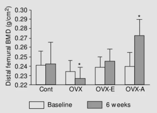
Bo ne m ine ral de nsity
No significant differences in baseline BMD were observed between groups. After 6 weeks, no significant difference was ob-served in the control group compared with baseline; however, a remarkable BMD de-crease was observed in the distal femur of the OVX group. A significant increase in BMD was observed in the OVX-A group and a nonsignificant increase was observed in the OVX-E group. At the end of the experimental period, BMD was significantly lower in OVX and higher in OVX-A than in the other groups (Figure 1).
Histo m o rpho m e try
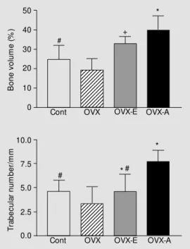
The static histomorphometric parameters of the distal femur showed lower trabecular bone volume (BV/TV) and trabecular number
D
is
ta
l f
em
ur
al
B
M
D
(g
/c
m
2) 0.30
0.29 0.28 0.27 0.26 0.25 0.24 0.23 0.22
Cont OVX OVX-E OVX-A
*
*
Baseline 6 w eeks
The OVX group presented signs of high bone turnover indicated by elevated osteoid volume and surface, increased erosion, and increased osteoblast and osteoclast surface without an increase in mineral apposition rate.
Treatment with 17ß-estradiol and alen-dronate produced a reduction in bone turn-over mainly by changes in bone formation parameters. This effect was particularly remarkable in the animals receiving alen-dronate. No mineralization impairment was noted in the alendronate group (Table 1).
Bio che m istry
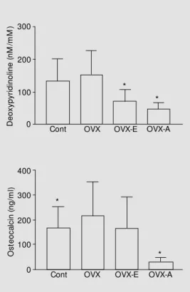
Bone resorption and formation markers measured by determination of Dpyr cross-links and osteocalcin, respectively, were in-creased in the OVX group compared to con-trol, but the difference was significant only in the osteocalcin assay. The 17ß-estradiol and alendronate treatments were associated with reduced Dpyr cross-link excretion and serum osteocalcin when compared to ovari-ectomized untreated rats (Figure 3).
Table 1. Histomorphometric variables of trabecular bone in the distal femur.
Cont (N = 10) OVX (N = 12) OVX-E (N = 11) OVX-A (N = 8) P value
BV/TV (% ) 24.8 ± 7.2*# 19.4 ± 5.8 32.8 ± 3.9*#+ 40.0 ± 7.3* <0.0001
Tb.Th (µm) 53.7 ± 8.9 58.1 ± 6.3 63.3 ± 4.8 52.1 ± 8.8 0.0137
Tb.Sp (µm) 181.1 ± 79.3*# 262.8 ± 87.1 131.0 ± 18.0*# 80.5 ± 21.1* <0.0001
Tb.N/mm 4.6 ± 1.2*# 3.3 ± 0.8 4.6 ± 1.8*# 7.7 ± 1.2* <0.0001
Es/Bs (% ) 6.6 ± 4.2 10.1 ± 7.3 8.3 ± 2.8 8.0 ± 3.6 0.5180
Oc.s/Bs (% ) 2.2 ± 2.0 2.4 ± 2.4 1.6 ± 0.8 1.8 ± 0.8 0.7470
OV/BV (% ) 0.2 ± 0.2* 0.6 ± 0.4 0.2 ± 0.1* 0.03 ± 0.03* <0.0001
O.Th (mm) 2.6 ± 0.7 3.0 ± 1.1 2.8 ± 0.7 1.6 ± 1.3 0.3147
Os/Bs (% ) 2.3 ± 1.2* 6.1 ± 4.4 2.1 ± 1.4* 0.4 ± 0.4* 0.0002
Ob.s/Bs (% ) 1.2 ± 0.8* 3.6 ± 2.8 1.6 ± 1.3* 0.3 ± 0.3* 0.0014
M AR (µm/day) 0.3 ± 0.1* 0.2 ± 0.1 0.1 ± 0.0* 0.2 ± 0.0* 0.0004
Cont, sham-operated control rats; OVX, untreated ovariectomized rats; OVX-E, ovariectomized rats + 17ß-estradiol; OVX-A, ovariectomized rats + alendronate. Bone histomorphometric variables for the distal femur are abbreviated as follow s: BV/TV = trabecular bone volume; Tb.Th = trabecular thickness; Tb.Sp = trabecular separation; Tb.N = trabecular number; Es/Bs = eroded surface; Oc.s/Bs = osteoclastic surface; OV/BV = osteoid volume; O.Th = osteoid thickness; Os/Bs = osteoid surface; Ob.s/Bs = osteoblastic surface; M AR = mineral apposition rate. *vs OVX; #vs OVX-A; +vs Cont.
in OVX compared with control. This reduc-tion was significantly inhibited by 17ß-estra-diol and alendronate treatment. The BV/TV ratio and trabecular number measured in OVX-A were significantly higher than in OVX-E (Figure 2). In contrast, trabecular thickness was higher in OVX-E than in OVX-A.
12345678 12345678 12345678 12345678 12345678 12345678 12345678 12345678 12345678 12345678 12345678
12345678 12345678 12345678 12345678 12345678 12345678 12345678 12345678 12345678 12345678
B
on
e
vo
lu
m
e
(%
)
50
40
30
20
10
0
*
*# #
Cont OVX OVX-E OVX-A
Cont OVX OVX-E OVX-A
#
Tr
ab
ec
ul
ar
n
um
be
r/
m
m
10.0
7.5
5.0
2.5
0.0
*
Figure 2. Bone volume and tra-becular number in the distal fe-mur in the four groups: sham control (Cont), ovariectomized (OVX), ovariectomized treated w ith 17ß-estradiol for 6 w eeks (OVX-E), and ovariectom ized treated w ith alendronate for 6 w eeks (OVX-A). Data are re-ported as means ± SD. P<0.05: *vs OVX; #vs OVX-A; +vs control (ANOVA).
Co rre latio n be twe e n BMD and
histo m o rpho m e tric param e te rs
BMD measurements at the distal femur were positively correlated with BV/TV and trabecular number. In contrast, BMD was negatively correlated with trabecular separa-tion (Figure 4).
D iscussio n
Bone loss induced by ovariectomy in rats has been widely used as a model of post-menopausal osteoporosis and has been vali-dated as a clinically relevant model of this condition in humans. In our study, bone loss induced by ovariectomy was observed in untreated rats 6 weeks after extraction. The site imaged was the distal femur metaphysis, an area that represents cancellous bone and loses bone rapidly after ovariectomy. Sev-eral studies have described similar findings
D
eo
xy
py
rid
in
ol
in
e
(n
M
/m
M
) 300
200
100
0 Cont OVX OVX-E OVX-A
O
st
eo
ca
lc
in
(n
g/
m
l)
400
200
100
0 Cont OVX OVX-E OVX-A
* *
* *
300
B
V
/T
V
(%
)
60
50
40
30
20
10
0
* * * * *
*
**
r = 0.74 P<0.0001
0.175 0.200 0.225 0.250 0.275 0.300 0.325 Distal femur BM D (g/cm2)
Tb
.N
/m
m
12
10
8
6
4
2
0
* * * * * * * *
0.175 0.200 0.225 0.250 0.275 0.300 0.325 r = 0.74 P<0.0001
Distal femur BM D (g/cm2)
*
Cont OVX OVX-E OVX-A *
*** ** ** r = -0.74 P<0.0001
Tb
.S
p
(µ
m
)
500
400
300
200
100
0
0.175 0.200 0.225 0.250 0.275 0.300 0.325 Distal femur BM D (g/cm2)
Figure 3. Deoxypyridinoline and osteocalcin measured by ELISA during the 6th w eek: control (Cont), ovariectomized (OVX), ovariectomized treated w ith 17ß-estradiol for 6 w eeks (OVX-E), and ovariectomized treated w ith alendronate for 6 w eeks (OVX-A). Bars represent the mean ± SD. * P<0.05 compared to OVX (ANOVA).
related to estrogen deficiency lasting a few days to several weeks (23,24).
We chose invasive and noninvasive tech-niques to analyze changes caused by ova-riectomy and the effects of 17ß-estradiol and alendronate. There has been little documen-tation analyzing correlations between bone parameters using these techniques in rats (25,26). In the present study, distal femur BMD was correlated with histomorphomet-ric parameters. Although histomorphometry has higher resolution and permits the quanti-fication of both dynamic and static bone parameters, DXA demonstrated practical advantages by permitting longitudinal skel-etal quantification in vivo (27-31). Our
anal-ysis confirms that both methods are indeed useful for the measurement of bone.
Histomorphometric analysis confirmed that alendronate increased bone mass more effectively than 17ß-estradiol. An interest-ing findinterest-ing was that both groups showed higher bone mass restoration than the non-ovariectomized group. Similar findings were observed by other authors. Seedor et al. (32) administered alendronate and observed a bone mass increase. The same findings were obtained by Belena et al. (33) in nonhuman primates. Similarly, Kalu et al. (34) adminis-tered 17ß-estradiol to ovariectomized ani-mals and found an increase in BV/TV com-pared to the non-ovariectomized group. Tobias and Compston (35) suggested that estrogen can stimulate osteoblast function and perhaps perform an anabolic action. They suggested that antireabsorptive drugs such as estrogen would be interesting to use in high doses in certain relatively short-term situations, such as the development of os-teoporosis in the early postmenopausal pe-riod.
Other studies have shown that these drugs were effective in increasing BV/TV; how-ever, comparisons cannot be made with our protocol due to the great variation in doses and average treatment time used in these trials. In our study, we have used minimum
doses with maximum effects (32,36,37). There is no correlation between doses in the present study and in humans.
We believe that the bone loss observed with acute estrogen deficiency represents the increase in remodeling space that occurs when the bone turnover rate accelerates. In the treatment with estrogen or bisphospho-nates there is generally a reduction of bone turnover to premenopausal levels or below, when assessed by histomorphometric meas-urements. This decrease is thought to be associated with the maintenance of bone mass in postmenopausal women and in ani-mal studies. Our findings agree with those reported by others (16,20,23,26,36).
Alendronate had no significant effect on histomorphometric bone resorption param-eters, including eroded surface and osteo-clast surface. In contrast, alendronate sig-nificantly inhibited urinary excretion of Dpyr. The absence of a decrease in osteoclastic surface suggests reduction of bone resorp-tion through a slowing-down funcresorp-tion (4,38), contrary to other studies which have sug-gested a decrease in osteoclastogenesis or an increase in osteoclast apoptosis (7,19).
Mineralization defects have been reported in etidronate, the first bisphosphonate used for osteoporosis treatment (36). High doses of alendronate, however, did not produce increased surface, volume or osteoid thick-ness, nor did they significantly change the mineral apposition rate.
correla-tion between the biochemical parameters (e.g., pyridinoline) and histomorphometric parameters measured on the iliac crest in patients with osteoporosis.
Because of the little documentation avail-able comparing 17ß-estradiol and alendro-nate in preventing bone loss in the same experiment, our study has proven to be an interesting investigative experiment. BMD analysis demonstrated that treatment with high doses of 17ß-estradiol and alendronate prevents ovariectomy-induced bone loss in female rats. We encountered only one study that compared the effects of both drugs in the same experiment. Lumbar and proximal tibiae were examined by computed tomography
and by histomorphometry. The authors ob-served that 17ß-estradiol was more effective than alendronate at the doses of 0.03 mg/kg, subcutaneously, twice weekly and 100 µg/ kg, orally, respectively (26).
We concluded that at the doses used in the present study, both drugs are effective in preventing bone loss after ovariectomy with alendronate producing a better outcome when compared with 17ß-estradiol.
Ackno wle dgm e nts
The authors are grateful to Mr. Wagner Vasques Dominguez for statistical assistance.
Re fe re nce s
1. Lindsay R (1995). Estrogen deficiency. In: Riggs BL & M elton III LJ (Editors), Os-teoporosis: Etiology, Diagnosis and M an-agement. Raven Press, New York. 2. Kanis JA, Gertz BJ, Singer F & Ortolani S
(1995). Rationale for the use of alendro-nate in osteoporosis. Osteoporosis Inter-national, 5: 1-13.
3. Beek EV, Low ik C, Pluijm GDP & Papoulos S (1999). The role of geranylgeranylation in bone resorption and its suppression by bisphosphonates in fetal bone. Explants in vitro: a clue to the mechanism of action of nitrogen-containing bisphosphonates. Journal of Bone and M ineral Research, 14: 722-729.
4. Colucci S, M inielli V, Zambonin G, Cirulli N, M ori G, Serra M , Patella V, Zallone AZ & Grano M (1998). Alendronate reduces adhesion of human osteoclast-like cells to bone and bone protein-coated surfaces. Calcified Tissue International, 63: 230-235.
5. Giuliani N, Pedrazzoni M , Passeri G & Gi-rasole G (1998). Bisphosphonates inhibit IL-6 production by human osteoblast-like cells. Scandinavian Journal of Rheumatol-ogy, 27: 38-41.
6. Ito M , Amizuka N, Nakajima T & Ozaw a H (1999). Ultrastructural and cytochemical studies on cell death of osteoclasts in-duced by bisphosphonate treatment. Bone, 25: 447-452.
7. Nishikaw a M , Akatsu T, Katayama Y, Yasutomo Y, Kado S, Kugai N, Yamamoto
M & Nagata N (1996). Bisphosphonates act on osteoblastic cells and inhibit osteo-clast formation in mouse marrow cul-tures. Bone, 18: 9-14.
8. Plotkin LI, Weinstein RS, Parfitt AM , Roberson PK, M anologas SC & Bellido T (1995). Prevention of osteocyte and os-teoblast apoptosis by bisphosphonates and calcitonin. Journal of Clinical Investi-gation, 104: 1363-1374.
9. Rodan GA & Fleisch HÁ (1996). Bisphos-phonates: M echanisms of action. Journal of Clinical Investigation, 97: 2692-2696. 10. Sahni M , Guenther HL, Fleisch H, Collin P
& M artin J (1993). Bisphosphonates act on rat bone resorption through the media-tion of osteoblasts. Journal of Clinical In-vestigation, 91: 2004-2011.
11. Sato M , Grasser W, Endo N, Akins R, Simmons H, Thompson DD, Golub E & Rodan GA (1991). Alendronate localiza-tion in rat bone and effects on osteoclast ultrastructure. Journal of Clinical Investi-gation, 88: 2095-2105.
12. Vitté C, Fleisch H & Guenther HL (1996). Bisphosphonates induce osteoblasts to secrete an inhibitor of osteoclast-medi-ated resorption. Endocrinology, 137: 2324-2333.
13. M iller SC & Bow man Jee WSS (1995). Available animal models of osteopenia -small and large. Bone, 17 (Suppl 4): 117-123.
14. M osekilde LI (1995). Assessing bone qual-ity animal models in preclinical
osteoporo-sis research. Bone, 17 (Suppl 4): 343-352. 15. Rodgers BJ, Faugere M CM & M alluche H (1993). Animal models for the study of bone loss after cessation of ovarian func-tion. Bone, 14: 369-377.
16. Thompson DD, Simmons HA, Pirie CM & Ke HZ (1995). FDA guidelines and animal models for osteoporosis. Bone, 17 (Suppl 4): 125-133.
17. M ohamed S, Shapira D, Leichter I, Rez-nick A & Silbermann M (1998). Ability of different techniques of measuring bone loss in aging female rats. Calcified Tissue International, 42: 375-382.
18. Frost HM & Jee WSS (1991). On the rat model of human osteopenias and os-teoporosis. Bone and M ineral, 18: 227-236.
19. Kalu DN (1991). The ovariectomized rat model of postmenopausal bone loss. Bone and M ineral, 15: 175-192. 20. Wronski TJ, Low ry PL, Walsh CC &
Ignaszew ski A (1985). Skeletal alterations in ovariectomized rats. Calcified Tissue International, 37: 324-328.
21. Yamazaki I & Yamaguchi H (1989). Char-acteristics of an ovariectomized osteo-penic rat model. Journal of Bone and M in-eral Research, 4: 13-22.
23. Lane NE, Haupt D, Kimmel DB, M odin G & Kinney JH (1999). Early estrogen re-placement therapy reverses the rapid loss of trabecular bone volume and prevents further deterioration of connectivity in the rat. Journal of Bone and M ineral Re-search, 14: 206-214.
24. Pastoriau P, Chomel A & Bonnet J (1995). Specific evaluation of localized bone mass and bone loss in the rat using dual-energy absorptiometry subregional analysis. Os-teoporosis International, 5: 143-149. 25. Cosman F, Schnitzer M B, M cCann PD,
Parisien M V, Dempster DW & Lindsay R (1992). Relationships betw een quantita-tive histological measurements and non-invasive assessments of bone mass. Bone, 13: 237-242.
26. Sato M , Bryant HU, Iversen P, Helterbrand J, Smietana F, Bemis K, Higgs R, Turner CH, Ow an I, Takano Y & Burr DB (1996). Advantages of raloxifene over alendronate or estrogen on nonreproductive and re-productive tissues in the long-term dos-ing of ovariectomized rats. Journal of Pharmacology and Experimental Thera-peutics, 279: 298-305.
27. Ammann P, Rizzolo R, Slosman D & Bonjour JP (1992). Sequencial and pre-cise in vivo measurement of bone mineral density in rats using dual energy X-ray absorptiometry. Journal of Bone and M in-eral Research, 7: 311-316.
28. M itlak BH & Sato M (1997). Bone mineral measurements by DXA in animals. In: Arnett TR & Henderson B (Editors), M
eth-ods in Bone Biology. Chapman & Hall, London.
29. Griffin M G, Kimble R, Hopper W & Pacifici R (1993). Dual-energy X-ray absorptio-metry of the rat: Accuracy, precision and measurement of bone loss. Journal of Bone and M ineral Research, 8: 795-800. 30. Rozemberg S, Vandromme J, Neve J,
Aguilera A, M uregancuro A, Peretz A, Kinthaert J & Ham H (1995). Precision and accuracy of in vivo bone mineral measure-ment in rats using dual-energy X-ray ab-sorptiometry. Osteoporosis International, 5: 47-53.
31. Yamaguchi H, Kushida K, Yamazaki K & Imove T (1995). Assessment of spine bone mineral density in ovariectomized rats using DXA. Journal of Bone and M in-eral Research, 10: 1033-1039.
32. Seedor JG, Quarruccio HÁ & Thompson DD (1991). The bisphosphonate alendro-nate (M K-217) inhibits bone loss due to ovariectomy in rats. Journal of Bone and M ineral Research, 6: 339-346.
33. Belena R, Toolan BC, Shea M , M arkatos A, M eyers ER, Lee SC, Opas EE, Seedor JG, Klein H, Frankenfield D, Quartuccio H, Floravanti C, Clair J, Brow n E, Hayes WCM & Rodan GA (1993). The effects of 2-year treatment w ith the aminobisphos-phonate alendronate on bone metabo-lism, bone histomorphometry, and bone strength in ovariectomized nonhuman pri-mates. Journal of Clinical Investigation, 92: 2577-2586.
34. Kalu DN, Liu CC, Salermo E, Hollis B,
Echon R & Ray M (1991). Skeletal re-sponse of ovariectomized rats to low and high doses of 17 ß-estradiol. Bone and M ineral, 14: 175-187.
35. Tobias JH & Compston JE (1999). Does estrogen stimulate osteoblast function in postmenopausal w omen? Bone, 24: 121-124.
36. Wronski TJ, Yen F & Scott KS (1991). Estrogen and diphosphonate treatment provides long-term protection against osteopenia in ovariectomized rats. Jour-nal of Bone and M ineral Research, 6: 387-394.
37. Wronski TJ, Cintrón M , Doherty AL & Dann LM (1988). Estrogen treatment pre-vents osteopenia and depresses bone turnover in ovariectomized rats. Endocri-nology, 123: 681-686.
38. Chavassieux PM , Arlot M E, Roux JP, Portero N, Daifotis A, Yates AJ, Hamdy NAT, M alice M P, Freedholm D & M eunier PJ (2000). Effects of alendronate on bone quality and remodeling in glucocorticoid-induced osteoporosis: A histomorpho-metric analysis of transiliac biopsies. Jour-nal of Bone and M ineral Research, 15: 754-762.