BASIC RESEARCH
Protective effects of Tualang honey on bone structure
in experimental postmenopausal rats
Siti Sarah Mohamad Zaid,ISiti Amrah Sulaiman,IINor Hayati Othman,IIIIma-Nirwana Soelaiman,IVAhmad Nazrun Shuid,IVNorazlina Mohamad,IVNorliza MuhamadIV
IUniversiti Putra Malaysia, Faculty of Environmental Studies, Department of Environmental Sciences, Selangor, Malaysia.IIUniversiti Sains Malaysia, School
of Medical Sciences, Health Campus, Department of Pharmacology, Kelantan, Malaysia. IIIUniversiti Sains Malaysia, School of Medical Sciences, Department of Pathology, Health Campus, Kubang Kerian, Kelantan, Malaysia.IVUniversiti Kebangsaan Malaysia, Faculty of Medicine, Department of Pharmacology, Kuala Lumpur, Malaysia.
OBJECTIVE:The objective of this study was to evaluate the effects of Tualang honey on trabecular structure and compare these effects with those of calcium supplementation in ovariectomized rats.
METHODS:Forty female, Sprague-Dawley rats were randomly divided into five groups (n = 8): four controls and one test arm. The control arm comprised a baseline control, sham-operated control, ovariectomized control, and ovariectomized calcium-treated rats (receiving 1% calcium in drinking water ad libitum). The test arm was composed of ovariectomized, Tualang honey-treated rats (received 0.2 g/kg body weight of Tualang honey). Both the sham-operated control and ovariectomized control groups received vehicle treatment (deionized water), and the baseline control group was sacrificed without treatment.
RESULTS:All rats were orally gavaged daily for six weeks after day one post-surgery. The bone structural analysis of rats in the test arm group showed a significant increase in the bone volume per tissue volume (BV/TV), trabecular thickness (Tb.Th) and trabecular number (Tb.N) and a significant decrease in inter-trabecular space (Tb.Sp) compared with the ovariectomized control group. The trabecular thickness (Tb.Th) in the test arm group was significantly higher compared with the ovariectomized-calcium treated group, and the inter-trabecular space (Tb.Sp) in the test arm group was significantly narrower compared with the ovariectomized-calcium treated group.
CONCLUSION: In conclusion, ovariectomized rats that received Tualang honey showed more improvements in trabecular bone structure than the rats that received calcium.
KEYWORDS: Tualang honey; Ovariectomy; Osteoporosis; Trabecular; Structural.
Zaid SSM, Sulaiman SA, Othman NH, Soelaiman IN, Shuid AN, Mohamed N, et al. Protective effects of Tualang honey on bone structure in experimental postmenopausal rats. Clinics. 2012;67(7):779-784.
Received for publication onNovember 7, 2011;First review completed onFebruary 14, 2012;Accepted for publication onMarch 2, 2012 E-mail: [email protected]
Tel.: 609-7672350
INTRODUCTION
In women, bone loss progresses much more rapidly after menopause due to estrogen deficiency (1). Declining serum estradiol levels can increase the lifespan of osteoclasts, leading to bone resorption; however, the lifespan of osteoblasts decreases, leading to less bone being formed. Ovariectomized rats are widely used as an animal model for the menopausal state because of the similarities in bone changes (2). In postmenopausal women and ovariectomized rats, there is negative bone balance, greater loss of cancellous than the cortical regions of long bones and reductions in bone mineral density due to declining estrogen levels (3).
Following ovariectomy, bone perforations are primarily seen at the trabecular plates of cancellous bone (4,5), and its strength depends on bone volume and bone structures, which consist of connected bony plates and bone matrix (6). After ovariectomy, the area that is initially affected is the secondary spongiosa (central metaphyseal area) of long bones (7). One of the agents used for osteoporosis treatment is calcium supplementation (8). Other alternative treatments use natural products. Polyphenols in fruits and vegetables have demonstrated beneficial effects on bone in rats (9,10). Natural antioxidant vitamins, such as palm tocotrienols, have been shown to prevent bone loss in many osteoporosis-induced rat models (11). Tualang honey (AgroMas, Malaysia) is a wild, multi-floral honey produced by bees (Apis dorsata) that form hives on the branches of giant trees named Tualang in the Malaysian rainforest. Tualang honey has higher phenolic content and greater radical scavenging activity compared with other honey sources (12). Honey contains approximately 200 substances, including a mixture of sugars (fructose, glucose, maltose, and sucrose) and small amounts Copyrightß2012CLINICS– This is an Open Access article distributed under
the terms of the Creative Commons Attribution Non-Commercial License (http:// creativecommons.org/licenses/by-nc/3.0/) which permits unrestricted non-commercial use, distribution, and reproduction in any medium, provided the original work is properly cited.
of other constituents, such as minerals, proteins, vitamins, organic acids, flavonoids, phenolic acids, enzymes, and other phytochemicals (13). A study on postmenopausal women taking Tualang honey at 20 mg/day for four months was found to have similar bone densitometry findings when compared with hormone replacement therapy (14). Our preliminary study showed the positive effects of Tualang honey on the reproductive system and bones (15). The present study was conducted to further evaluate the effects of Tualang honey on trabecular structure and to compare these effects with those of calcium supplementation in ovariecto-mized rats.
MATERIALS AND METHODS
The study was conducted on 40 three-month-old, virgin, female, Sprague-Dawley rats weighing between 220 and 240 g and obtained from the Laboratory Animal Research Unit (LARU), Health Campus, Universiti Sains Malaysia. After acclimatization for two weeks, the rats were divided into five groups (n = 8 per group): four controls and one test arm. The four control arms were 1) baseline control (BC), 2) sham-operated (SHAM group), 3) ovariectomized-control (OVX group), and 4) ovariectomized-calcium treated (PC). The ovariectomy and sham operation were conducted under ketamine anesthesia. The rats were anesthetized using 90 mg/kg ketamine (Troy Laboratories, Pty Ltd, Australia) and 10 mg/kg xylazil (Troy Laboratories, Pty Ltd, Australia). The BC group did not undergo ovariectomy or a sham operation but were sacrificed at the start of the experiment to obtain the baseline histomorphometric para-meters. The PC group was given 1% calcium in drinking water, which was suppliedad libitum. The calcium supple-ment was prepared according to previous methods (16) by adding 1 g of lactic acid hemicalcium salt (Sigma, St. Louis, Missouri, USA) to 99 ml of deionized water to make a concentration of 1%. Calcium supplementation was given to the positive control group to compare the effects of honey administration with calcium supplementation. The control rats (SHAM and OVX) were given 0.5 ml of distilled water. The rats in the test arm (TH group) also had bilateral ovariectomy under ketamine anesthesia. The rats in the TH group were given 0.2 g/kg body weight of Tualang honey. Tualang honey was freshly prepared every morning by dissolving the honey in deionized water. The administration of test and control materials was started at day 14 after post-surgery day 1 by oral gavage (9:00-10:00 am daily for six weeks). The Tualang honey used was supplied by the Fede-ral AgricultuFede-ral Marketing Authority (FAMA), Ministry of Agriculture and Agro-Based Industry, Malaysia. The honey was filtered to remove solid particles, concentrated in an oven at 40
˚
C and subjected to c irradiation at 25 kGy at Sterilgamma (M) Sdn. Bhd. (Selangor, Malaysia). Through-out the treatment period, a daily vaginal smear was taken, while body weights and total food intake were recorded weekly. The vaginal smear cytology was performed by a licensed cytologist according to a previously described method (17).After six weeks, the rats were sacrificed 24 hours after the last dose of treatment under excess ketamine anesthesia (100 mg/kg). The right femur was harvested, and the distal section was longitudinally cut and then fixed in 10% formalin. The bone was processed without decalcification in methyl methacrylate (BDH, Poole, England) medium. The
bone was dehydrated with a series of ethanol solutions (70%, 90%, and 100%) and then infiltrated in methyl methacrylate. The bone was sectioned at 9 mm thickness using a microtome (Leica RM2155, Nussloch, Germany). The sections were stained with von Kossa and subjected to histomorphometry examination using a light microscope (Leica DMRXA2, Wetzlar, Germany) attached to an image analyzer with VideoTest-Master software (VT, St. Peters-burg, Russia). All histomorphometric parameter measure-ments were performed randomly at the metaphyseal region, which was located 3-7 mm from the lowest point of the growth plate and 1 mm from the lateral cortex, excluding the endocortical region (Figure 1). All measurements and calculations were performed according to the American Society for Bone and Mineral Research (ASBMR) nomen-clature and guidelines (18). Structural parameters were analyzed using an image analyzer (Leica DMRXA2, Wetzlar, Germany) with VideoTest-Master software (VT, St. Petersburg, Russia). First, total trabecular bone area, total tissue area and bone perimeters (primary data) were measured using this software. Then, these primary data were expressed in three-dimensional form as follows: total trabecular bone volume (bone volume, BV), total tissue volume (tissue volume, TV) and total bone surface (bone surface, BS). Finally, the structural parameters were calcu-lated based on these primary data, as shown in Table 1.
The sample size was calculated using PS Power and Sample Size Calculation version 2.1.31 and based on a
previous study conducted by Hermizi et al. (19). The power of the study was set at 80%, with a 95% confident interval, while the standard deviation (s) observed was 1.4 of
trabecular thickness, and the difference in population means (d) was set at 3.0 of the trabecular thickness. The calculated sample size was eight per group. The data analysis was completed using the Statistical Package for Social Sciences (SPSS version 12.0.1) software. The Kolmogorov-Smirnov test was used to test for normality. For the non-parametric statistics, the Kruskal-Wallis test for multiple comparisons was used due to the lack of a normal distribution for all data, followed by the Mann-Whitney U test to compare differences between the two groups. The results are expressed as the median and interquartile range (IR: the difference between the 75th and 25th percentile).P-values of less than 0.05 were considered statistically significant.
The protocol used in this study was approved by the Animal Ethics Committee, Universiti Sains Malaysia (approval no. PPSG/07(A)/044).
RESULTS
The results show that providing Tualang honey had positive effects on bone structures (Table 2) but not on the body weight of ovariectomized rats (Table 3). The ovar-iectomized rats treated with Tualang honey (TH group) had a significant increase in bone volume per tissue volume (BV/TV); the trabecular thickness (Tb.Th), trabecular number (Tb.N), and inter-trabecular space (Tb.Sp) were significantly lower in the TH group compared with the OVX group (Table 2). In contrast, 1% calcium treatment in the PC group did not significantly improve these parameters when compared with the OVX group. The trabecular thickness (Tb.Th) in the TH group was higher compared with the PC group, and the inter-trabecular space (Tb.Sp) was narrower compared with the PC group. The histomorphometrical analysis of bone structures showed that the bone volume per tissue volume (BV/TV), trabecular thickness (Tb.Th) and trabecular number (Tb.N) were significantly decreased in the OVX group compared with the SHAM and baseline
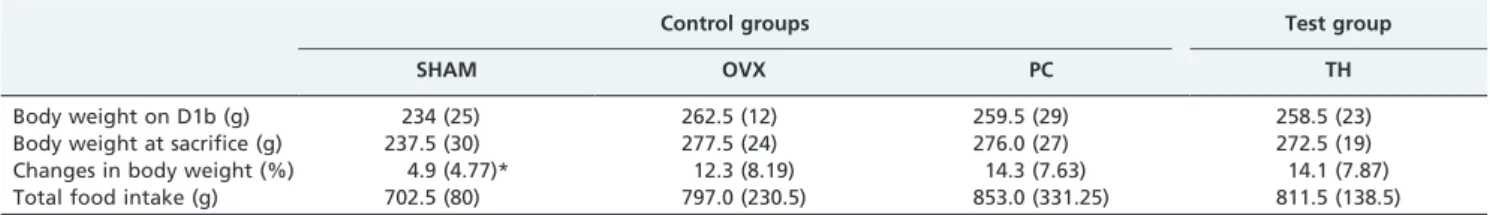
control (BC) groups (Table 2 and Figure 2). Only the inter-trabecular space (Tb.Sp) was significantly increased in the OVX group compared with the SHAM and BC groups. The SHAM group showed significantly lower changes in body weight compared with the OVX, TH, and PC groups (Table 3). Rats treated with Tualang honey showed the same body weight changes as rats in the OVX control groups. No changes in femur weight were observed in the TH and PC groups compared with the OVX group (Table 4). However, there was a significant decrease in femur weight for the OVX, TH, and PC groups compared with the SHAM group.
DISCUSSION
Our study shows that Tualang honey improved the trabecular structure of the femur bone in ovariectomized rats, an animal model for the menopausal state. The effect of Tualang honey was better than that observed in the rats that received calcium, indicating that honey supplementation could be used to prevent osteoporosis during menopause. The histomorphometrical analysis of bone structures at the secondary spongiosa in the present study showed that the trabecular bone volume (BV/TV), trabecular thickness (Tb.Th) and trabecular number (Tb.N) were decreased, while the inter-trabecular space (Tb.Sp) was increased in the OVX control group compared with the SHAM group. The changes in trabecular bone structure parameters in the OVX control group were due to an imbalance in the normal remodeling process. We observed that Tualang honey prevented this change. The improvements observed in all structural parameters indicate that Tualang honey is effective in the prevention of ovariectomy-induced bone loss due to bone resorption. Tualang honey appears to be more efficacious than calcium supplementation.
Honey is a natural beehive product well known for its high phytochemical content (phenolic compounds or polyphenols that contribute to positive physiological effects). Honey is rich in antioxidants, such as flavonoids and phenolic acids (20-23). Flavonoids constitute the most important class of
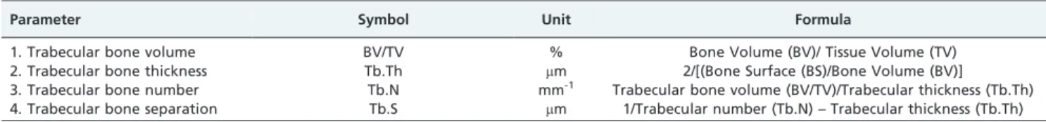
Table 1 -Formula for calculating the structural parameter values.
Parameter Symbol Unit Formula
1. Trabecular bone volume BV/TV % Bone Volume (BV)/ Tissue Volume (TV)
2. Trabecular bone thickness Tb.Th mm 2/[(Bone Surface (BS)/Bone Volume (BV)]
3. Trabecular bone number Tb.N mm-1 Trabecular bone volume (BV/TV)/Trabecular thickness (Tb.Th)
4. Trabecular bone separation Tb.S mm 1/Trabecular number (Tb.N) – Trabecular thickness (Tb.Th)
Table 2 -Effects of Tualang honey on bone histomorphometry.
Control groups Test group
BC SHAM OVX PC TH
BV/TV (%) 56.15 (2.34)**,#,ˆˆˆ 38.24 (5.62)**, ˆ ˆ 21.41 (16.72) 25.31 (14.81)## 29.13 (9.22)*,# Tb.Th (mm) 68.79 (7.01)***,##,ˆˆˆ 54.75 (9.30)***,ˆˆ ˆ 27.29 (8.66) 30.66 (7.75)###
38.64 (7.35)**,###,ˆ ˆ
Tb.N (mm-1) 13.01 (7.02)** 13.52 (4.11)**, ˆ 7.50 (4.31) 9.00 (6.01)# 12.51 (5.10)**
Tb.Sp (mm) 90.96 (18.77)**,#,ˆˆˆ 117.35 (4.46)*, ˆˆˆ 220.27 (63.92) 230.45 (40.75)###
157.78 (32.85)*,###,ˆˆˆ
BC = Baseline control group; SHAM = Sham-operated control group; OVX = Ovariectomized control group; TH = Tualang honey- treated group at 0.2 g/ kg/bw; PC = Positive control group (1% calcium).
All values are expressed as the median (IR).
aKruskal–Wallis test. *
p,0.05, **p,0.01 and ***p,0.001 vs. OVX control group;#
p,0.05,##
p,0.01 and###
phenolic compounds in honey and can be categorized as flavonols, flavavones, flavones, anthocyanidins and isofla-vones. Flavonoids found in honey include pinocembrin, pinobanksin, chrysin, galangin, luteolin, quercetin, and kaempferol (24). The use of flavonoids to improve post-menopausal osteoporosis is well documented. Quercetin and kaempferol are a subfamily of flavonoids (flavonols) reported to have a potential role in protection against postmenopausal osteoporosis in ovariectomized rats (25). Honey and other plants share similar phenolic compounds or polyphenols. Flavonols, such as quercetin and kaempferol, have been shown to directly induce apoptosis of mature osteoclasts, thus inhibiting bone resorption and decreasing intracellular reactive oxygen species (ROS) in osteoclasts by a mechanism involving estrogen receptors (ER) (25). A study on kaemp-ferol alone indicates that it prevents ovariectomized-induced bone loss by promoting osteoblast function (26).
The mechanisms of action of both flavonols on bone are still not completely understood. Flavanols may interact
directly with estrogen receptors ER-ß and ER-a(25). ER-ß is more abundant than ER-ain trabecular bone compared with cortical bone (27); thus, a greater proportion of flavonols might bind to trabecular bone, resulting in a greater preservation of trabecular bone than cortical bone. Studies on postmenopausal animals have shown that oxidative stress has an important impact on the differentiation and function-ing of osteoclasts (31,32); oxidative stress also induces lipid peroxidation and H2O2 production and a decrease in
enzymatic antioxidants, such as Superoxide Dismutase (SOD) and Glutathione Peroxidase (GPx) (31). Phenolic glycosides have potential antioxidant activity (32) and immunostimulatory effects (33). The composition and inter-action of polyphenols in honey make it a unique and excellent source of stable, free radical scavengers among polyphenol-containing foods (34). Another component of honey that has been shown to increase calcium absorption is gluconic acid. Gluconic acid is a major organic acid in honey and one of the products of the enzymatic glucose oxidase
Table 3 -Effects of Tualang honey on body weight and food intake during six-week administration.
Control groups Test group
SHAM OVX PC TH
Body weight on D1b (g) 234 (25) 262.5 (12) 259.5 (29) 258.5 (23)
Body weight at sacrifice (g) 237.5 (30) 277.5 (24) 276.0 (27) 272.5 (19)
Changes in body weight (%) 4.9 (4.77)* 12.3 (8.19) 14.3 (7.63) 14.1 (7.87)
Total food intake (g) 702.5 (80) 797.0 (230.5) 853.0 (331.25) 811.5 (138.5)
SHAM = Sham-operated control group; OVX = Ovariectomized control group;
PC = Positive control group (1% calcium); TH = Tualang honey-treated group at 0.2 g/kg/bw. All values are expressed as the median (IR).
aKruskal–Wallis test. bD1: First day of treatment. *p
,0.05 vs. OVX control group (Mann-Whitney U test).
reaction (35). It is significantly useful in the modulation of intestinal calcium absorption calcium (36). When ingested in food, gluconic acid is fermented by lactic acid bacteria (Lactobacillus reuteriandL. mucosae) to produce lactate and acetate. Acid-utilizing bacteria (Megasphaera elsdenii and
Mitsuokella multiacida) then convert these products to form butyrate, a type of short-chain fatty acid (SCFA) that is rapidly absorbed by the mucosa of the large intestine (37). The greater efficacy of Tualang honey compared with calcium supplementation is likely due to the enhancement in intestinal calcium absorption caused by honey.
Tualang honey did not produce any adverse effects on body weight or food intake. Although not statistically significant, we observed that the body weight of the ovariectomized rats treated with Tualang honey was lower than that of the ovariectomized rats not given honey. This observation, if translated to humans, indicates that honey has the potential to reduce health problems related to overweight and obesity seen in the menopausal state, such as chronic osteoarthritis, cardiovascular diseases and some cancers. The limitation in the interpretation of this finding is the short study duration, which was only six weeks. It would be interesting to observe the effect of honey over a longer study period. Our findings were similar to the results of several previous studies on natural products. Treatment with kaempferol, which is highly abundant inGinkgo biloba
extracts, did not suppress increases in body weights in ovariectomized rats (27). Although the study period was longer [10 weeks], the duration of the study was similar to ours, as treatment was started at week 4 after ovariectomy. In conclusion, Tualang honey improved the trabecular structure of bone in ovariectomized rats. The effect of Tualang honey in rats is better than the effects observed in rats that received calcium. These findings suggest that Tualang honey is better than calcium supplementation in preventing osteoporosis in the menopausal state. Therefore, Tualang honey could potentially be used as an alternative to calcium supplementation in osteoporosis treatment.
ACKNOWLEDGMENTS
The authors would like to acknowledge the Federal Agriculture Marketing Authority (FAMA), the Ministry of Agriculture and Agro-Based Industry Malaysia for supplying the Tualang honey (Agromas, Malaysia) for the present research, the Universiti Sains Malaysia for providing the research grant (No: 304/PPSP/6131505), the University Putra Malaysia for financial assistance to the first author, Siti Sarah Mohamad Zaid, and
the University Kebangsaan Malaysia for the use of lab facilities for this study.
AUTHOR CONTRIBUTIONS
Zaid SM conducted all the lab work and data analysis and drafted the manuscript. Sulaiman SB led the design of the study, served as the main supervisor to Zaid SM, was the principal investigator of the grant study, provided guidance in the data analysis and revised the manuscript. Mohamad N contributed in the study design and provided guidance in the histomorphometry part of the study. Soelaiman IN, Shuid AN and Muhamad N provided guidance in the histomorphometry part of the study. Othman NH is the pathologist who evaluated the bone histology and finalized the revised manuscript.
REFERENCES
1. Pacifici R. Estrogen deficiency, T cells and bone loss. Cellular Immunology. 2008;252(1-2):68-80, http://dx.doi.org/10.1016/j.cellimm.2007.06.008. 2. Goss PE, Qi S, Josse RE, Pritzker KPH, Mendes M, Hu H, et al. The
steroidal aromatase inhibitor exemestane prevents bone loss in ovar-iectomized rats. Bone. 2004;34(3):384-92, http://dx.doi.org/10.1016/ j.bone.2003.11.006.
3. Kalu DN. The ovariectomized rat model of postmenopausal bone loss. Bone Miner. 1991;15(3):175-91.
4. Wronski TJ, Dann LM, Qi H, Yen CF. Skeletal effects of withdrawal of estrogen and diphosphonate treatment in ovariectomized rats. Calcif Tissue Int. 1993;53(3):210-6, http://dx.doi.org/10.1007/BF01321840. 5. Yamaura M, Nakamura T, Kanou A, Miura T, Ohara H, Suzuki K. The
effect of 17 beta-estradiol treatment on the mass and the turnover of bone in ovariectomized rats taking a mild dose of thyroxin. Bone Miner. 1994;24(1):33-42, http://dx.doi.org/10.1016/S0169-6009(08)80129-8. 6. Mellish RW, Ferguson Pell MW, Cochran GV, Lindsay R, Dempster DW.
A new manual method for assessing two-dimensional cancellous bone structure: comparison between iliac crest and lumbar vertebra. Bone Miner Res. 1991;6(7):689-96.
7. Bagi CM, Miller SC. Comparison of osteopenic changes in cancellous bone induced by ovariectomy and/or immobilization in adult rats. Anat Rec. 1994;239(3):243-54, http://dx.doi.org/10.1002/ar.1092390303. 8. Mackerras D, Lumley T. First- and second-year effects in trials of calcium
supplementation on the loss of bone density in postmenopausal women. Bone. 1997;21(6):527-33, http://dx.doi.org/10.1016/S8756-3282(97)00181-6. 9. Muhlbauer RC, Lozano A, Reinli A. Onion and a mixture of vegetables, salads, and herbs affect bone resorption in the rat by a mechanism independent of their base excess. J Bone Miner Res. 2002;17(7):1230-6, http://dx.doi.org/10.1359/jbmr.2002.17.7.1230.
10. Devareddy L, Hoosmand S, Collins JK, Lucas EA, Chai SC, Arjmandi BH. Blueberry prevents bone loss in ovariectomized rat model of postmenopausal osteoporosis. The Journal of Nutritional Biochemistry. 2008;19(10):694-9, http://dx.doi.org/10.1016/j.jnutbio.2007.09.004. 11. Ahmad NS, Khalid BA, Luke DA, Ima Nirwana S. Tocotrienol offers
better protection than tocopherol from free radical-induced damage of rat bone. Clin Exp Pharmacol Physiol. 2005;32(9):761-70, http:// dx.doi.org/10.1111/j.1440-1681.2005.04264.x.
12. Krishna Kihsore R, Halim AS, Nurul Syazna MS, Sirajudeen KNS. Tualang honey has higher phenolic content and greater radical scavenging activity with other honey sources. Nutrition Research. 2011;31(4):322-5.
Table 4 -Effects of Tualang honey on weight of femur reported as relative organ weight (mg/g body weight).
Control groups Test group
BC SHAM OVX PC TH
Absolute weight (mg)
808.15 (48.10) 884.91 (86.40) 876.51 (108.80) 919.51 (72.51) 877.50 (155.20)
Relative weight 3.38 (0.11) *,# 3.64 (0.23) 3.05 (0.22)## 3.13 (0.21)### 3.04 (0.55)## (mg/g body
weight)
Absolute weight = Wet weight of tissue.
Relative weight = Wet weight of tissue normalized to final body weight.
BC = Baseline control group; SHAM = Sham-operated control group; OVX = Ovariectomized control group; PC = Positive control group (1% calcium); TH = Tualang honey-treated group at 0.2 g/kg body weight.
All values are expressed as the median (IR).
aKruskal–Wallis test. *p
,0.05 vs. OVX control group;#
p,0.05,##
p,0.01 and###
13. Aljadi AM and Kamaruddin MY. Evaluation of the phenolic contents and antioxidant capacities of two Malaysian floral honeys. Food Chemistry. 2004;85:513-8, http://dx.doi.org/10.1016/S0308-8146(02)00596-4. 14. Lily Husniata Y. The effects of Tualang honey on postmenopausal
women. In 2nd International conference on the medicinal use of honey. 2010; Renaissance Hotel, Kota Bharu, Kelantan, Malaysia.
15. Zaid SSM, Sulaiman AS, Sirajudeen KNM, Othman NH. The effects of Tualang honey on female reproductive organs, tibia bone and hormonal profile in ovariectomised rats-animal model for menopause. BMC Complement Altern Med. 2010;10:82-8, http://dx.doi.org/10.1186/ 1472-6882-10-82.
16. Norazlina M, Ima Nirwana S, Khalid BAK. Calcium supplementation improves bone and lean mass in vitamin E deficient rats. JAFES. 2001;19(1-2):23-9.
17. Marcondes FK, Bianchi FJ, Tanno AP. Determination of the estrous cycle phases of rats: some helpful considerations. Brazil Journal of Biology. 2002;62(4A):609-14, http://dx.doi.org/10.1590/S1519-69842002000400008. 18. Parfitt AM, Drezner MK, Glorieux FH, Kanis JA, Malluche H, Meunier PJ, et al. Bone histomorphometry: standardization of nomenclature, symbols and units. Reports of the ASBMR Histomorphometry Nomenclature Committee. J Bone Miner Res. 1987;2(6):595-610. 19. Hermizi H, Faizah O, Ima-Nirwana S, Ahmad Nazrun S, Norazlina M.
Beneficial effects of tocotrienol and tocopherol on bone histomorpho-metric parameters in sprague-dawley male rats after nicotine cessation. Calcif Tissue Int. 2009;84(1):65-74, http://dx.doi.org/10.1007/s00223-008-9190-x.
20. Aljadi AM, Kamaruddin MY. Evaluation of the phenolic contents and antioxidant capacities of two Malaysian floral honeys. Food Chemistry. 2004;85:513-518, http://dx.doi.org/10.1016/S0308-8146(02)00596-4. 21. Al-Mamary M,Al-Meeri A, Al-Habori M. Antioxidant activities and total
phenolics of different types of honey. Nutrition Research. 2002;22(9):1041-7, http://dx.doi.org/10.1016/S0271-5317(02)00406-2.
22. Phrzynska K, Biesaga M. Analysis of phenolic acids and flavonoids in honey. Analytical Chemistry. 2008;28(7):893-902.
23. Gheldof N, Wang XH, Engeseth NJ. Identification and quantification of antioxidant components of honeys from various floral sources. J Agric Food Chem. 2007;50(21):5870-7.
24. Gheldof N, Wang XH, Engeseth NJ. Buckwheat honey increases serum antioxidant capacity in humans. J Agric Food Chem. 2003;51(5):1500-5, http://dx.doi.org/10.1021/jf025897t.
25. Wattel A, Kamel S, Mentaverri R, Lorget F, Prouillet C, Petit JP, et al. Potent inhibitory effect of naturally occurring flavonoids quercetin and
kaempferol on in vitro osteoclastic bone resorption. Biochem Pharmacol. 2003.65(1):35-42, http://dx.doi.org/10.1016/S0006-2952(02)01445-4. 26. Trivedi R, Kumar S, Kumar K, Siddiqui JA, Swarnkar G, Gupta V, et al.
Kaempferol has osteogenic effect in ovariectomized adult Sprague-Dawley rats. Mol Cell Endocrinol. 2008;289(1-2):85-93, http://dx.doi.org/10.1016/ j.mce.2008.02.027.
27. Kuiper GG, Carlsson B, Grandien K, Enmark E, Haggblad J, Nilsson S, et al. Comparison of the ligand binding specificity and transcript tissue distribution of estrogen receptors alpha and beta. Endocrinology 1997;138(3):863-70.
28. Onoe Y, Miyaura C, Ohta H, Nozawa S, Suda T. Expression of estrogen receptor beta in rat bone. Endocrinology. 1997;138(10):4509-12, http:// dx.doi.org/10.1210/en.138.10.4509.
29. Zhang J, Munger RG, West NA, Cutler DR, Wengreen HJ, Corcoran CD. Antioxidant intake and risk of osteoporotic hip fracture in Utah: an effect modified by smoking status. Am J Epidemiol. 2006;163(1):9-17. 30. Ha BJ, Lee SH, Kim HJ, Lee JY. The role of Salicornia herbacea in
ovariectomy-induced oxidative stress. Biol Pharm Bul. 2006;29(7):1305-9, http://dx.doi.org/10.1248/bpb.29.1305.
31. Ozgocmen S, Kaya H, Fadillioglu E, Aydogan R, Yilmaz Z. Role of antioxidant systems, lipid peroxidation, and nitric oxide in postmeno-pausal osteoporosis. Mol Cell Biochem. 2007;295(1-2):45-52, http:// dx.doi.org/10.1007/s11010-006-9270-z.
32. Wu Q, Fu DX, Hou AJ, Lei GQ, Liu ZJ, Chen JK, et al. Antioxidative phenols and phenolic glycosides from Curculigo orchioides. Chem Pharm Bull. 2005;53(8):1065-7, http://dx.doi.org/10.1248/cpb.53.1065. 33. Lakshmi V, Pandey K, Puri A, Saxena RP, Saxena KC. Immunostimulant
principles from Curculigo orchioides. J Ethnopharmacol. 2003;89(2-3):181-4, http://dx.doi.org/10.1016/S0378-8741(03)00160-0.
34. Prior R, Martin A, Sofic E, McEwan J, O’Brien C, Lischner N. Antioxidant capacity as influenced by total phenolic and anthocyanin content, maturity and variety of Vaccinium species. Journal Agriculture Food Chemistry. 1998;46(7):2686-93, http://dx.doi.org/10.1021/jf980145d. 35. Jeffrey AE, Echazareta CM. Medicinal uses of honey. Revista Biomedica.
1996;7:43-9.
36. Fournier P, Dupius Y. Modulation of intestinal absorption of calcium. J Physiol (Paris). 1975;70:479–1.