online | memorias.ioc.fiocruz.br
Increased frequency of micronuclei in the lymphocytes of patients
chronically infected with hepatitis B or hepatitis C virus
Samantha Therezinha Almeida Pereira Leite1, Marilene Borges da Silva2, Marco Andrey Pepato1,
Francisco José Dutra Souto1, Raquel Alves dos Santos3, Carmen Lucia Bassi-Branco1/+
1Faculdade de Medicina 2Instituto de Biociências, Universidade Federal de Mato Grosso, Cuiabá, MT, Brasil 3Universidade de Franca, Franca, SP, Brasil
In this study, we analysed the frequency of micronuclei (MN), nucleoplasmic bridges (NPBs) and nuclear buds (NBUDs) and evaluated mutagen-induced sensitivity in the lymphocytes of patients chronically infected with hepati-tis B virus (HBV) or hepatihepati-tis C virus (HCV). In total, 49 patients with chronic viral hepatihepati-tis (28 HBV-infected and 21 HCV-infected patients) and 33 healthy, non-infected blood donor controls were investigated. The frequencies (‰) of MN, NPBs and NBUDs in the controls were 4.41 ± 2.15, 1.15 ± 0.97 and 2.98 ± 1.31, respectively. The frequencies of MN and NPBs were significantly increased (p < 0.0001) in the patient group (7.01 ± 3.23 and 2.76 ± 2.08, respec-tively) compared with the control group. When considered separately, the HBV-infected patients (7.18 ± 3.57) and infected patients (3.27 ± 2.40) each had greater numbers of MN than did the controls (p < 0.0001). The HCV-infected patients displayed high numbers of NPBs (2.09 ± 1.33) and NBUDs (4.38 ± 3.28), but only the HBV-HCV-infected patients exhibited a significant difference (NPBs = 3.27 ± 2.40, p < 0.0001 and NBUDs = 4.71 ± 2.79, p = 0.03) in comparison with the controls. Similar results were obtained for males, but not for females, when all patients or the HBV-infected group was compared with the controls. The lymphocytes of the infected patients did not exhibit sensi-tivity to mutagen in comparison with the lymphocytes of the controls (p = 0.06). These results showed that the lym-phocytes of patients who were chronically infected with HBV or HCV presented greater chromosomal instability.
Key words: hepatitis B - hepatitis C - lymphocytes - micronucleus
Hepatitis B virus (HBV) and hepatitis C virus (HCV) infections affect millions of individuals worldwide. Ap-proximately 10-15% of HBV-infected patients and 70-80% of HCV-infected patients develop the chronic form of their respective diseases. The primary problem asso-ciated with persistent HBV and HCV infections is the development and progression of liver disease, which ranges in severity from minimal lesions throughout the liver parenchyma to severe fibrosis, cirrhosis and hepa-tocellular carcinoma (HCC) (Szabó et al. 2004).
Both HBV and HCV may promote potentially mu-tagenic cellular processes, such as integration into the host DNA (in the case of HBV) and the generation of oxidative stress as a consequence the immune response (Farinati et al. 1995, Bartsch & Nair 2004, Saigo et al. 2008, Sung et al. 2012).
The mutagenic effects of HBV and HCV infections are visible in hepatocytes and lymphocytes, as demonstrated by the presence of the oxidative promutagenic DNA le-sion 8-oxo-7,8-dihydro-2’-deoxyguanosine (8-oxodG), which is primarily found in the lymphocytes of HCV-infected patients (Hagen et al. 1994, Farinati et al. 1995, 1999, Cardin et al. 2001). In addition, the leukocytes of HCV-infected patients and HBV-infected patients have
doi: 10.1590/0074-0276140183
Financial support: CNPq, CAPES (to STAPL and CLB-B), FAPE-MAT (737379/2008)
+ Corresponding author: [email protected] Received 2 April 2013
Accepted 11 October 2013
been demonstrated to exhibit increased chromosomal instability (Machida et al. 2010).
The DNA damage response (DDR) includes various downstream pathways that coordinate cell cycle arrest with the repair of damaged DNA. Alternatively, the DDR can mediate the clearance of affected cells that are be-yond repair through apoptosis or senescence (Heijink et al. 2013). Cells that are inefficient in these mechanisms generally exhibit higher sensitivity to mutagenic agents and it has been suggested that these cells may be pref-erentially selected during tumour development (Jackson & Bartek 2009). The in vitro mutagen sensitivity assay in lymphocytes has been used to examine the individ-ual response to mutagen exposure because the cells of individuals with suboptimal DNA repair capacities are predicted to accumulate higher levels of DNA damage than those of individuals with more efficient DNA le-sion repair (Wu et al. 2007). However, although evidence suggests that chronic HCV and HBV infections contrib-ute to the development of HCC in addition to lympho- proliferative diseases (Zuckerman et al. 1997, Huang et al. 2012), few studies have investigated the mutagen sen-sitivity of the lymphocytes of patients that are chroni-cally infected with HBV or HCV (Wu et al. 1998).
SUBJECTS, MATERIALS AND METHODS
Study population - In total, 49 patients with chronic viral hepatitis, consisting of 28 HBV-infected patients and 21 HCV-infected patients, and 33 healthy, non-infected controls were included in the study. The patients were recruited from the Clinic of Infectious Diseases of Julio Muller Hospital (Federal University of Mato Grosso, Cui-abá, state of Mato Grosso, Central-West Brazil). Chronic HBV infection was confirmed by persistent HBV surface antigenemia lasting more than six months. Chronic HCV infection was confirmed by the presence of HCV RNA in blood tests. Cirrhosis was diagnosed by liver biopsy or based on clinical observations, laboratory tests or ul-trasonographic evidence. Non-infected subjects were re-cruited from a group of healthy blood donors at the Public Blood Bank of Mato Grosso State. The absence of infec-tion in the control group was confirmed by laboratory tests. Information about alcohol consumption, tobacco use, ethnicity and age was obtained from medical records and from an interviewer-administered questionnaire, which also included questions about exposure to muta-gens and any history of cancer in the individual.
Cytokinesis-block MNs (CBMN) assay - The CBMN assay was performed as described by Fenech and
Mor-ley (1985), with minor modifications. In total, 5 μL of
venous blood was collected in heparin-Vacutainer tubes (Becton & Dickinson, Franklin Lakes, NJ, USA) and lymphocyte cultures (2 per subject) were established us-ing 0.3 mL of whole blood added to RPMI-1640 medium (Sigma-Aldrich, St. Louis, MO, USA) supplemented with 20% foetal calf serum (Cultilab, Campinas, SP, Brazil), 0.001% penicillin (Vetec, Duque de Caxias, RJ, Brazil), 0.0005% streptomycin (Sigma-Aldrich) and 2% phytohemagglutinin (Cultilab). After the cultures were incubated for 44 h at 37ºC in a BOD incubator (Eletro-lab, São Paulo, SP, Brazil), cytochalasin B (Sigma-Al-drich) was added to the cultures (6 µg/mL). The cells were harvested by centrifugation at 72 h after the cul-ture was initiated. The lymphocytes were treated with a hypotonic solution (1% sodium citrate w/v) and fixed in a solution of methanol:acetic acid (3:1 v/v); in both cases, the solutions were ice cold and freshly prepared. The cell suspension was dropped onto a pre-cooled mi-croscope slide and air dried before being stained for 5-7 min with 5% Giemsa in Sorensen phosphate buffer (0.06 M Na2HPO4 and 0.06 M KH2PO4, pH 6.8). Microscopic analysis was performed with a light microscope (Nikon, Melville, NY, USA) at 400X magnification. For each individual, 2,000 binucleated cells were analysed for the presence of MN, NPBs and NBUDs in accordance with previously established criteria (Fenech et al. 2003). We calculated the frequency of each biomarker (num-ber in 1,000, ‰) using the following formula: ‰ of X = (number of X/2,000) x 1,000, where X is MN, NPBs or NBUDs (Montero et al. 2006).
Mutagen sensitivity evaluation - To determine mu-tagen sensitivity, the well-established mumu-tagen DXR was used to treat lymphocytes from patients and non-infected subjects at 44 h after the culture was initiated. The cells were treated with 0.15 µg/mL DXR (Bergamo,
Taboão da Serra, SP, Brazil) diluted in sterile distilled water for 28 h, which brought the total culture time to 72 h. The experimental conditions for the DXR treatment were previously established in preliminary experiments. The conditions for cell harvesting and slide preparation were described in the previous section. Sensitivity to DXR was expressed as induced DNA damage: [(mean MN after DXR)-(basal mean MN)].
Statistical analysis - An age comparison between groups was performed using one-way ANOVA and the Bonferroni post-hoc test. When the data exhibited un-equal variance, the median numbers of MN, NPBs and NBUDs were compared between the groups using the non-parametric Kruskal-Wallis test followed by Dunn’s post-hoc test. Similarly, the median numbers of MN ob-tained in the mutagen sensitivity test were compared be-tween the groups using the Mann-Whitney U test. The mean measurements of the induced DNA damage were compared using the Student’s t test. The G-test or the χ2
testwas used to compare ethnicity, sex, alcohol intake and tobacco intake between the groups. A linear regres-sion model was constructed using the Stata 8.2 software programme (StataCorp, College Station, TX, USA) to verify the independence of the frequencies of MN, NPBs and NBUDs from the virus type, sex, age, alcohol intake, tobacco intake and the use of antiviral drugs. A value of
p ≤ 0.05 was used as the criterion for significance. The
statistical analyses were performed using the statistical software programme Bioestat 5.0 (Ayres et al. 2012).
Ethics - This study was approved by the Ethical Re-search Board of Julio Muller Hospital (protocol 439/ CEP-HUJM/07) and informed consent to voluntarily participate was given by all of the subjects.
RESULTS
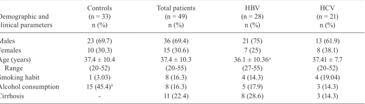
Of the 49 patients included in the study, 28 were chron-ically infected with HBV (21 males and 7 females) and had a mean age of 36.1 years (ranging from 21-56 years), while 21 were chronically infected with HCV (13 males and 8 females) and had a mean age of 44.1 years (ranging from 27-55 years). The non-infected control group con-sisted of 23 males and 10 females with a mean age of 37.4 years (ranging from 20-52 years). No differences were ob-served between the non-infected group and the HBV or HCV-infected patients with regard to their sex (p = 0.97) and smoking habits (p = 0.16). The mean age was similar between the infected patients and the non-infected con-trol subjects; however, the mean age of the HCV-infected patients was significantly higher than that of either the chronically HBV-infected group or the controls (p < 0.05). Alcohol consumption among the study subjects ranged from 0-8 g/day and was more frequently reported by the non-infected control subjects (45.45%) than by the HBV-infected patients (17.86%) or the HCV-HBV-infected patients (14.28%) (p = 0.003). Cirrhosis was present in eight HBV-infected patients (28.6%) and three HCV-HBV-infected patients (14.29%) (p = 0.31) (Table I). All patients with cirrhosis were males. Seven patients (6 HBV-infected and 2 HCV-infected patients) were undergoing antiviral therapy at the time of the study (1 lamivudine and tenofovir, 2 ribavirin
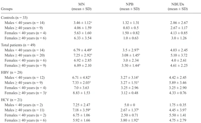
The numbers of MN, NPBs and NBUDs observed in the patient group were 7.01 ± 3.23, 2.76 ± 2.08 and 4.57 ± 2.98, respectively. The frequencies of MN and NPBs, but not NBUDs, were significantly increased (p < 0.0001) compared with the frequencies in the controls (4.41 ± 2.15 and 1.15 ± 0.97 for MN and NPBs, respectively, in the controls). Considering the HBV-infected patients and the HCV-infected patients separately, the numbers of MN in the HBV-infected patients (7.18 ± 3.57) and HCV-infected patients (6.78 ± 2.80) were significantly higher than in the non-infected group (4.41 ± 2.15) (p < 0.0001). The num-bers of NPBs and NBUDs in the HBV-infected patients (3.27 ± 2.40 and 4.71 ± 2.79, respectively) and in patients infected with HCV (2.09 ± 1.33 and 4.38 ± 3.28, respec-tively) were increased relative to those of the control group (1.15 ± 0.97 and 2.98 ± 1.31, respectively). However, only the difference between the HBV-infected group and the control group was statistically significant (p = 0.03) (Ta-ble II). The results were independent of age, sex, alcohol consumption, tobacco intake and the presence of cirrhosis for MN (R = 0.22, p = 0.02), NPBs (R = 0.23, p < 0.001) and NBUDs (R = 0.15, p = 0.03). The antiviral therapy did not significantly influence any of the analysed param-eters. The frequency of MN was significantly influenced by sex only for males aged < 40 years in the control group, who showed a significantly lower frequency of MN (3.46 ± 1.12) than did females (5.63 ± 1.60) of the same age (p = 0.01). The frequency of MN and NPBs was signifi-cantly higher in males, among all patients and within the HBV-infected group in comparison with the controls (p < 0.01) (Table III). The frequency of NPBs was signifi-cantly higher in females aged > 40 years in comparison with the controls (p < 0.01). The frequency of MN, NPBs and NBUDs was not significantly higher in male patients with cirrhosis in comparison with male patients without cirrhosis (Table IV).
Cells from 15 chronically infected patients (9 HBV-in-fected and 6 HCV-inHBV-in-fected patients) and 14 non-inHBV-in-fected individuals were tested for DXR sensitivity. The numbers of MN were not significantly different between the HCV-infected and the HBV-HCV-infected patients; therefore, the patients were grouped together for comparison with the
non-infected individuals to improve the statistical analy-sis. The patients exhibited significantly higher numbers of MN in both untreated (6.76 ± 3.03, p = 0.03) and treated (15.03 ± 4.94, p = 0.001) cells compared with the cells of non-infected individuals (4.25 ± 1.45 and 9.39 ± 3.39 in untreated and treated cells, respectively). DXR treatment significantly increased the number of MN in both the con-trol group (p = 0.0002) and the patient group (p < 0.0001). The mean measurements of the induced DNA damage were higher in the patient group (8.30 ± 5.40) than in the non-infected control group (5.18 ± 3.45). However, this difference was not found to be statistically significant, which was possibly due to a type 2 error (p = 0.06).
DISCUSSION
Chronic hepatotropic virus (HBV and HCV) infec-tions are characterised by potentially mutagenic cellular events, such as an increase in oxidative stress and vi-ral integration into the DNA of the host cell (Farinati et al. 1995, Bartsch & Nair 2004, Saigo et al. 2008, Sung et al. 2012). In the present study, we demonstrated that the lymphocytes of HBV-infected patients and HCV-in-fected patients exhibited an increased frequency of MN compared with lymphocytes from healthy, non-infected individuals. This result is in contrast to findings
previ-TABLE I
Demographic and clinical characteristics of patients chronically infected with hepatitis B virus (HBV) or hepatitis C virus (HCV) and of control subjects
Demographic and clinical parameters
Controls (n = 33)
n (%)
Total patients (n = 49)
n (%)
HBV (n = 28)
n (%)
HCV (n = 21)
n (%)
Males 23 (69.7) 36 (69.4) 21 (75) 13 (61.9)
Females 10 (30.3) 15 (30.6) 7 (25) 8 (38.1)
Age (years) Range
37.4 ± 10.4 (20-52)
37.4 ± 10.3 (20-55)
36.1 ± 10.36a (27-55)
37.41 ± 7.7 (20-52)
Smoking habit 1 (3.03) 8 (16.3) 4 (14.3) 4 (19.04)
Alcohol consumption 15 (45.4)b 8 (16.3) 5 (17.9) 3 (14.3)
Cirrhosis - 11 (22.4) 8 (28.6) 3 (14.3)
a: ANOVA and Bonferroni post-hoc test, p < 0.05; b: G test, p = 0.004.
TABLE II
Frequency of micronucleus (MN), nucleoplasmatic bridges (NPB) and nuclear buds (NBUDs) in hepatitis B virus (HBV)
or hepatitis C virus (HCV)-infected patients and controls
Groups
MN (mean ± SD)
NPB (mean ± SD)
NBUDs (mean ± SD)
Controls (n = 33) 4.41 ± 2.15 1.15 ± 0.97 2.98 ± 1.31 Total patients (n = 49) 7.01 ± 3.23a 2.76 ± 2.08a 4.57 ± 2.98 HBV (n = 28) 7.18 ± 3.57a 3.27 ± 2.40a 4.71 ± 2.79b HCV (n = 21) 6.78 ± 2.80a 2.09 ± 1.33 4.38 ± 3.28
ously reported by Ozkal et al. (2005). However, these authors reported a higher frequency of chromosome breaks, leading to the formation of acentric chromo-some/chromatid fragments that ultimately contributed to the formation of MN. Furthermore, the MN may have re-sulted from an inability of whole chromosomes to travel to the spindle poles during mitosis (Fenech et al. 2011). A
higher frequency of chromosome gaps, aneuploidy and poly-ploidy in the peripheral blood mononuclear cells (PBMCs) of HCV-infected patients was reported previously (Machida et al. 2010). The present results suggest that chronic infection with HBV or HCV accounts for chromosomal instability in lymphocytes and this phenomenon is characterised by the formation of acentric fragments and/or aneuploidy.
We also observed increased numbers of NPBs and NBUDs in HBV-infected patients. NPBs may occur when dicentric chromosomes originating from chro-mosome breaks or telomere-to-telomere end fusions are pulled to opposite poles of the cell during mitosis. NBUDs are primarily considered to be formed from am-plified DNA that is being eliminated from chromosomes (Fenech et al. 2011). Although gene amplifications (MYC
and ERBB2) were demonstrated in HCC tissue samples that were infected with HBV or HCV (Al-Qahtani et al. 2010), no reports have described this genetic alteration in PBMCs from individuals with viral hepatitis. Dicentric Y chromosomes have also been reported in the PBMCs of HCV-infected patients (Machida et al. 2010). Togeth-er with our results, these data suggest that the chromo-somal instability in lymphocytes that results from HBV infection (and likely also from chronic HCV infection) is characterised by the presence of dicentric chromosomes and gene amplification.
TABLE III
The effect of age and sex on frequency of micronucleus (MN), nucleoplasmatic bridges (NPB) and nuclear buds (NBUDs) n hepatitis B virus (HBV) or hepatitis C virus (HCV)-infected patients and controls
Groups
MN (mean ± SD)
NPB (mean ± SD)
NBUDs (mean ± SD)
Controls (n = 33)
Males < 40 years (n = 14) 3.46 ± 1.12a 1.32 ± 1.31 2.86 ± 2.67
Males ≥ 40 years (n = 9) 4.06 ± 1.59 0.83 ± 0.5 2.67 ± 1.17
Females < 40 years (n = 4) 5.63 ± 1.60 1.50 ± 0.82 4.13 ± 0.85
Females ≥ 40 years (n = 6) 6.33 ± 3.54 1.0 ± 0.63 3.0 ± 1.26
Total patients (n = 49)
Males < 40 years (n = 14) 6.79 ± 4.49b 3.5 ± 2.97b 4.03 ± 2.45
Males ≥ 40 years (n = 20) 7.25 ± 2.92b 3.08 ± 1.45b 5.10 ± 3.72
Females < 40 years (n = 6) 6.92 ± 2.85 3.0 ± 2.34 4.0 ± 2.61
Females ≥ 40 years (n = 9) 6.89 ± 2.10 3.50 ± 1.44b 4.61 ± 2.25
HBV (n = 28)
Males < 40 years (n = 12) 6.71 ± 4.82b 3.27 ± 3.14b 4.42 ± 2.45
Males ≥ 40 years (n = 9) 7.33 ± 2.03b 3.27 ± 1.51b 5.89 ± 3.46
Females < 40 years (n = 4) 7.0 ± 3.63 3.25 ± 2.96 3.25 ± 2.90
Females ≥ 40 years (n = 3)c 8.83 ± 1.53 3.12 ± 0.48 4.33 ± 0.76
HCV (n = 21)
Males < 40 years (n = 2) 7.25 ± 2.47 5.0 ± 0 1.75 ± 0.35
Males ≥ 40 years (n = 11) 7.18 ± 3.59b 2.67 ± 1.37b 4.45 ± 3.97
Females < 40 years (n = 2) 6.75 ± 1.06 2.50 ± 0.71 5.50 ± 1.41
Females ≥ 40 years (n = 6) 5.92 ± 1.66 3.80 ± 1.92b 4.75 ± 2.79
a: statistically significant in comparison to females of the same age-class (p = 0.01, Mann-Whitney U test); b: statistically signifi-cant in relationship to controls of the same age class (p < 0.01, ANOVA, post-test Student t or Kruskal-Wallis, post test Student-Newman-Keuls); c: groups with n < 4 were not included in statistical analysis; SD: standard deviation.
TABLE IV
The effect of cirrhosis on frequency of micronucleus (MN), nucleoplasmatic bridges (NPB) and nuclear buds (NBUDs) in males hepatitis B virus (HBV) or hepatitis C virus
(HCV)-infected patients
Groups
Cirrhosis (n)
MN (mean ± SD)
NPB (mean ± SD)
NBUDs (mean ± SD)
Total patients No (23) 6.82 ± 4.10 2.98 ± 2.53 3.67 ± 1.75 Yes (11) 7.54 ± 2.27 2.68 ± 1.58 5.00 ± 2.59 HBV No (15) 6.56 ± 4.36 3.26 ± 2.91 4.13 ± 1.72 Yes (8) 8.25 ± 2.23 3.06 ± 1.68 5.25 ± 2.39 HCV No (9) 7.31 ± 3.80 2.43 ± 1.63 2.81 ± 1.53 Yes (3) 7.00 ± 2.90 1.66 ± 0.76 4.33 ± 3.54
In this study, females and subjects aged ≥ 40 years ex -hibited more MN than did males and subjects < 40 years, respectively, among both controls and HBV-infected pa-tients. Although these differences were not statistically sig-nificant, the results are in accordance with the well-known influence of sex and age on MN frequencies, as reported before (Fenech 1998, Bonassi et al. 2001, Fenech & Bo-nassi 2011). Reports on the effects of sex and age on NPBs and NBUDs are conflicting in the literature (Donmez-Al-tuntas & Bitgen 2012, Nefic & Handzic et al. 2013).
Considering the patient group, we found statistically significantly higher frequencies of MN in males than in females; however, females did not present cirrhosis in our sample. It is known that DNA damage levels in the leukocytes of HBV-infected patients and HCV-infected patients significantly correlate with the presence of liver lesions (Farinati et al. 1999, Cardin et al. 2001, Grossi et al. 2008, Hoare et al. 2013). In fact, in the current study, the frequencies of MN (in HBV-infected patients) and NBUDs (in HBV-infected patients and HCV-infected patients) were increased in cirrhotic males, but this dif-ference was not significant. Whether the extension of liver lesions in male patients is correlated with the fre-quency of MN found in lymphocytes is not completely clear in the sample investigated here. Even considering that the statistical analysis may have been influenced by the small size of our sample, these results should be con-sidered with caution, especially because chromosome damage detected by the MN assay is an important bio-marker for cancer prediction (Bonassi et al. 2007).
HBV and HCV may contribute to increased chro-mosomal aberrations in infected cells by direct and in-direct pathways. Regarding the inin-direct pathway, it has been shown that the presence of reactive oxygen species (ROS) resulting from cytokine activity during chronic inflammation has a potent mutagenic effect (Yan et al. 2006). A relationship between infection with HBV or HCV and increased production of ROS, chromosomal aberrations and other DNA damage has been reported previously (Hagen et al. 1994, Machida et al. 2010). Furthermore, leukocytes from patients who are chroni-cally infected with HBV or HCV exhibit higher levels of 8-OHdG, which is the most frequent ROS-induced base lesion (Farinati et al. 1999, Cardin et al. 2001).
Viral integration is considered to have direct muta-genic potential in hepatocytes. The integration of HBV into the human genome affects the expression of genes located near the site of insertion and also causes more widespread alterations of chromosomal stability (Saigo et al. 2008, Sung et al. 2012). Because viral genome inte-gration into the host DNA also frequently occurs in the PBMCs of chronically HBV-infected patients (Murakami et al. 2004), this process may contribute to genomic in-stability in these cells. Recently, it was demonstrated that in chronic HCV infection, the presence of double strand breaks occurs concomitantly with shortened telomeres in T lymphocytes. This phenomenon is associated with the level of fibrosis and may influence the response to treat-ment (Hoare et al. 2013).
The frequency of mutations is directly influenced by the efficiency of the DNA repair mechanisms because failure to remove a lesion can facilitate mutational
fixa-tion. Extensive evidence has demonstrated that proteins produced by HBV and HCV interact with the proteins of the DNA repair machinery and inhibit their functions in host cells (Chen et al. 2008, Machida et al. 2010, Pal et al. 2010). Therefore, it is possible that the cells of infected patients exhibit less efficient DNA repair mechanisms due to the effects of the viral proteins, which may con-tribute to the elevated frequency of DNA damage de-tected in this study.
Au et al. (2010) reported that when the cells of exposed populations (in this case, HCV-infected patients or HBV-infected patients) are challenged with a DNA-damaging agent in vitro, the in vivoexposure-induced repair ciency is dramatically amplified. Additionally, the defi-ciency will be detectable in a challenge assay as an in-crease in the number of chromosomal aberrations, MN or unrepaired DNA strand breaks.
Less effective DNA repair may also result in a higher sensitivity to mutagens. It has been shown that B lympho-cytes infected with HCV in vitro exhibit increased sen-sitivity to bleomycin due to the action of the NS3 protein and the core viral protein (Machida et al. 2010). Further-more, it was demonstrated that lymphocytes from HBV or HCV-infected HCC patients exhibit an increased sensitiv-ity to bleomycin and benzo(a)pyrene-diol-epoxide, which is associated with an increased risk of cancer development (Wu et al. 1998). The results obtained in the present study showed that the lymphocytes of HBV-infected patients and HCV-infected patients are not more sensitive to DXR than the lymphocytes of non-infected subjects. However, a challenge assay with other substances, such as bleomy-cin, needs to be performed to allow more definite conclu-sions about mutagen sensitivity in these patients.
In summary, the present study demonstrated that the lymphocytes of patients who are chronically infected with HBV or HCV exhibit greater chromosomal instability, characterised by the presence of MN, NPBs and NBUDs. Although we did not observe a statistically significant re-sult, a possible influence of cirrhosis on these parameters should be considered for further investigation.
ACKNOWLEDGEMENTS
To Gevanil Arruda, Nilson Santana Botelho and Maria Celina da Penha, for providing technical assistance.
REFERENCES
Al-Qahtani A, Al-Hazzani T, Al-hussain T, Al-Ghamdi A, Al-Mana H, Al-Arifi S, Al-Ahdal M, Aly M 2010. Correlation between clinical characteristics, survival and genetic alterations in pa-tients with hepatocellular carcinoma from Saudi Arabia. Cancer Genet Cytogenet 203: 269-277.
Au WW, Giri AK, Ruchirawat M 2010 Challenge assay: a functional biomarker for exposure-induced DNA repair deficiency and for risk cancer. Int J Hyg Environ Health 213: 32-39.
Ayres M, Ayres Jr M, Ayres DL, dos Santos AAS 2012. BioEstat 5.0. Available from: mamiraua.gov.br.
results with the cytokinesis-block micronucleus assay in human lymphocytes: I. Effect of laboratory protocol, scoring criteria and host factors on the frequency of micronuclei. Environ Mol Mutagen 37: 31-45.
Bonassi S, Znaor A, Ceppi M, Lando C, Chang WP, Holland N, Kirsch-Volders M, Zeiger E, Ban S, Barale R, Bigatti MP, Bo-lognesi C, Cebulska-Wasilewska A, Fabianova E, Fucic A, Hag-mar L, Joksi EG, Martelli A, Migliore L, Mirkova E, Scarfi MR, Zijno A, Norppa H, Fenech M 2007. An increased micronucleus frequency in peripheral blood lymphocytes predicts the risk of cancer in humans. Carcinogenesis 28: 625-631.
Cardin R, Saccoccio G, Masutti F, Bellentani S, Farinati F, Tiribelli C 2001. DNA oxidative damage in leukocytes correlates with the severity of HCV-related liver disease: validation in an open popu-lation study. J Hepatol 34: 587-592.
Chen HY, Tang NH, Lin N, Chen ZX, Wang XZ 2008. Hepatitis B virus X protein induces apoptosis and cell cycle deregulation through interfering with DNA repair and checkpoint responses.
Hepatol Res 38: 174-182.
Donmez-Altuntas H, Bitgen N 2012. Evaluation of the genotoxicity and cytotoxicity in the general population in Turkey by use of the cytokinesis-block micronucleus cytome assay. Mutat Res 748: 1-7.
Farinati F, Cardin R, De Maria N, Della Libera G, Marafin C, Lecis E, Burra P, Floreani A, Cecchetto A, Naccarato R 1995. Iron stor-age, lipid peroxidation and glutathione turnover in chronic anti-HCV positive hepatitis. J Hepatol 22: 449-456.
Farinati F, Cardin R, Degan P, De Maria N, Floyd RA, Van Thiel DH, Naccarato R 1999. Oxidative DNA damage in circulating leuko-cytes occurs as an early event in chronic HCV infection. Free Radic Biol Med 27: 1284-1291.
Fenech M 1998. Important variables that influence base-line micronu-cleus frequency in cytokinesis-blocked lymphocytes-a biomarker for DNA damage in human populations. Mutat Res 404: 155-165.
Fenech M, Bonassi S 2011. The effect of age, gender, diet and lifestyle on DNA damage measured using micronucleus frequency in hu-man peripheral blood lymphocytes. Mutagenesis 26: 43-49.
Fenech M, Chang WP, Kirsch-Volders M, Holland N, Bonassi S, Zeiger E 2003. HUman MicroNucleus project. HUMN project: detailed description of the scoring criteria for the cytokinesis-block micronucleus assay using isolated human lymphocyte cul-tures. Mutat Res 534: 65-75.
Fenech M, Kirsch-Volders M, Natarajan AT, Surralles J, Crott JW, Parry J, Norppa H, Eastmond DA, Tucker JD, Thomas P 2011. Molecular mechanisms of micronucleus, nucleoplasmic bridge and nuclear bud formation in mammalian and human cells. Mu-tagenesis 26: 125-132.
Fenech M, Morley A 1985. Solutions to the kinetic problem in the micronucleus assay. Cytobios 43: 233-246.
Grossi S, Sumberaz A, Gosmar M, Mattioli F, Testino G, Martelli A 2008. DNA damage in peripheral blood lymphocytes of patients with cirrhosis related to alcohol abuse or to hepatitis B and C viruses. Eur J Gastroenterol Hepatol 20: 22-25.
Hagen TM, Huang S, Curnutte J, Fowler P, Martinez V, Wehr CM, Ames BN, Chisari FV 1994. Extensive oxidative DNA damage in hepatocytes of transgenic mice with chronic active hepatitis destined to develop hepatocellular carcinoma. Proc Natl Acad Sci USA 91: 12808-12812.
Heijink AM, Krajewska M, van Vugt MA 2013. The DNA damage response during mitosis. Mutat Res 750: 45-55.
Hoare M, Shankar A, Shah M, Rushbrook S, Gelson W, Davies S,
Akbar A, Alexander GJ 2013. γ-H2AX+CD8+ T lymphocytes
can-not respond to IFN-α, IL-2 or IL-6 in chronic hepatitis C virus
infection. J Hepatol 58: 868-874.
Huang B, Li J, Zhou Z, Zheng D, Liu J, Chen M 2012. High preva-lence of hepatitis B virus infection in multiple myeloma. Leuk Lymphoma 53: 270-274.
Jackson SP, Bartek J 2009. The DNA-damage response in human biol-ogy and disease. Nature 461: 1071-1078.
Machida K, McNamara G, Cheng KT, Huang J, Wang CH, Comai L, Ou JH, Lai MM 2010. Hepatitis C virus inhibits DNA damage repair through reactive oxygen and nitrogen species and by inter-fering with the ATM-NBS1/Mre11/Rad50 DNA repair pathway in monocytes and hepatocytes. J Immunol 185: 6985-6998.
Montero R, Serrano L, Araujo A, Dávila V, Ponce J, Camacho R, Mo-rales E, Méndez A 2006. Increased cytogenetic damage in a zone in transition from agricultural to industrial use: comprehensive analysis of the micronucleus test in peripheral blood lympho-cytes. Mutagenesis 21: 335-342.
Murakami Y, Minami M, Daimon Y, Okanoue T 2004. Hepatitis B vi-rus DNA in liver, serum and peripheral blood mononuclear cells after the clearance of serum hepatitis B virus surface antigen.
J Med Virol 72: 203-214.
Nefic H, Handzic I 2013. The effect of age, sex and lifestyle factors on micronucleus frequency in peripheral blood lymphocytes of the Bosnian population. Mutat Res 753: 1-11.
Ozkal P, Ilgin-Ruhi H, Akdogan M, Elhan AH, Kaçar S, Sasmaz N 2005. The genotoxic effects of hepatitis B virus to host DNA.
Mutagenesis 20: 147-150.
Pal S, Polyak SJ, Bano N, Qiu WC, Carithers RL, Shuhart M, Gretch DR, Das A 2010. Hepatitis C virus induces oxidative stress, DNA damage and modulates the DNA repair enzyme NEIL1. J Gastro-enterol Hepatol 25: 627-634.
Saigo K, Yoshida K, Ikeda R, Sakamoto Y, Murakami Y, Urashima T, Asano T, Kenmochi T, Inoue I 2008. Integration of hepatitis B virus DNA into the myeloid/lymphoid or mixed-lineage leukemia (MLL4) gene and rearrangements of MLL4 in human hepatocel-lular carcinoma. Hum Mutat 29: 703-708.
Sung WK, Zheng H, Li S, Chen R, Liu X, Li Y, Lee NP, Lee WH, Ariyaratne PN, Tennakoon C, Mulawadi FH, Wong KF, Liu AM, Poon RT, Fan ST, Chan KL, Gong Z, Hu Y, Lin Z, Wang G, Zhang Q, Barber TD, Chou WC, Aggarwal A, Hao K, Zhou W, Zhang C, Hardwick J, Buser C, Xu J, Kan Z, Dai H, Mao M, Reinhard C, Wang J, Luk JM 2012. Genome-wide survey of recurrent HBV integration in hepatocellular carcinoma. Nat Genet44: 765-769.
Szabó E, Páska C, Kaposi Novák P, Schaff Z, Kiss A 2004. Similari-ties and differences in hepatitis B and C virus induced hepatocar-cinogenesis. Pathol Oncol Res 10: 5-11.
Wu X, Gu J, Patt Y, Hassan M, Spitz MR, Beasley RP, Hwang LY 1998. Mutagen sensitivity as a susceptibility marker for human hepatocellular carcinoma. Cancer Epidemiol Biomarkers Prev 7: 567-570.
Wu X, Gu J, Spitz MR 2007. Mutagen sensitivity: a genetic predispo-sition factor for cancer. Cancer Res 67: 3493-3495.
Yan B, Wang H, Rabbani ZN, Zhao Y, Li W, Yuan Y, Li F, Dewhirst MW, Li CY 2006. Tumor necrosis factor-alpha is a potent en-dogenous mutagen that promotes cellular transformation. Cancer Res 66: 11565-11570.