∗Correspondence: B. Yilmaz. Department of Analytical Chemistry. Fac-ulty of Pharmacy. Ataturk University. 25240 - Erzurum, Turkey. E-mail: [email protected]
A
vol. 51, n. 2, apr./jun., 2015 http://dx.doi.org/10.1590/S1984-82502015000200005
Differential pulse voltammetric determination of diclofenac in
pharmaceutical preparations and human serum
Bilal Yilmaz
*, Selcuk Kaban, Bilge Kagan Akcay, Ulvihan Ciltas
Department of Analytical Chemistry, Faculty of Pharmacy, Ataturk University, Erzurum, Turkey
This article describes a differential pulse voltammetric (DPV) method for the determination of diclofenac in pharmaceutical preparations and human serum. The proposed method was based on electro-oxidation of diclofenac at platinum electrode in 0.1 M TBAClO4/acetonitrile solution.The well-deined two
oxidation peaks were observed at 0.87 and 1.27 V, respectively. Calibration curves that obtained by using current values measured for second peak were linear over the concentration range of 1.5-17.5 μg mL-1 and 2-20 μg mL-1 in supporting electrolyte and serum, respectively. Precision and accuracy were
also checked in all media. Intra- and inter-day precision values for diclofenac were less than 3.87, and accuracy (relative error) was better than 4.12%. The method developed in this study is accurate, precise and can be easily applied to Diclomec, Diclolam and Voltaren tablets as pharmaceutical preparation. In addition, the proposed technique was successfully applied to spiked human serum samples. No electro-active interferences from the endogenous substances were found in human serum.
Uniterms: Diclofenac/determination. Differential pulse voltammetry/quantitative analysis. Pharmaceutical formulations/analysis. Human serum/analysis
Este artigo descreve um método de voltametria de pulso diferencial (VPD) para a determinação de diclofenaco em preparações farmacêuticas e em soro humano. O método proposto foi baseado em eletroxidação de diclofenaco no eléctrodo de platina em solução 0,1 M TBAClO4/acetonitrila. Dois picos de oxidação bem deinidos foram observados em 0,87 e 1,27 V, respectivamente. As curvas de calibração obtidas utilizando-se valores de corrente medidos por segundo pico foram lineares no intervalo de concentração de 1,5-17,5 μg mL-1 e 2-20 μg mL-1 em eletrólito suporte e soro, respectivamente. Precisão e exatidão também foram veriicadas em todos os meios. Valores de precisão intra- e inter-dia para o diclofenaco foram inferiores a 3.87 e a precisão (erro relativo) foi melhor do que 4,12%. O método desenvolvido neste estudo é exato, preciso e pode ser facilmente aplicado a Diclomec, Diclolam e comprimidos Voltaren, como preparação farmacêutica. Além disso, a técnica proposta foi aplicada com sucesso em amostras de soro humano. Não se observaram interferências das substâncias endógenas no soro humano.
Unitermos: Diclofenaco/determinação. Voltametria de pulso diferencial/análise quantitativa. Formulações farmacêuticas/análise. Soro humano/análise.
INTRODUCTION
Diclofenac (Figure 1), [o-(2,6-dichloroanilino)
phenyl] acetate), is a non-steroidal anti-inflammatory drug (NSAID) with strong pyretic, analgesic and
anti-inlammatory properties (Iliiescu, Baia, Miclaus, 2004).
It is widely used in clinical medicine for the treatment
of inlammatory conditions such as rheumatoid arthritis,
osteoarthritis and ankylosing spondylitis (Gostick et al.,
1990;Crowley et al., 1990). The eficacy of diclofenac
equals that of many newer and established NSAIDs. As an analgesic, it has a fast onset and a long duration of action. Compared to other NSAIDs, diclofenac is well tolerated and rarely produces gastrointestinal ulcerations or other serious side effects. Thus, diclofenac can be considered as
choice used in the treatment of acute and chronic, painful
and inlammatory conditions (Roskar, Kmetec, 2003). To date, several methods for the determination of diclofenac have been reported. These include potentiometry (Shamsipur, Jalali, Ershad, 2005; Santini,
Pezza, Pezza, 2006; Hassan et al., 2005), capillary zone
electrophoresis (Jin, Zhang, 2000), high performance
liquid chromatography (HPLC) (González, Yuln, Volonté,
1999; Kasperek, 2008; Arcelloni et al., 2001), high-performance liquid chromatography-mass spectrometry
(HPLC-MS) (Abdel-Hamid, Novotny, Hamza, 1990),
spectrophotometry (Botello, Perez-Caballero,1995),
spectroluorometry (Arancibia, Boldrini, Escandar, 2000;
Damiani et al., 1999; Carreira et al., 1995),thin layer
chromatography (Sun, Fabre, 1994), gas chromatography
(Siou, Pommier, Godbillon, 1991; Yilmaz, 2010),
polarographic analysis (Xu, Chen, Song, 2004),
spectrophotometry (De Souza, Tubino,2005;
Agatonovic-Kustrin et al., 1997; Matin, Farajzadeh, Joyuban, 2005;
Sastry, Prasad-Tipirneni, Suryanarayana, 1989; Sena et
al., 2004) andelectrochemistry methods (Blanco-Lopez et al., 2003; Goyal, Chatterjee, Agrawal, 2010; Manea et al.,
2010). In all electrochemical methods, modiied electrodes
were used due to the gradual passivation or contamination
of unmodiied solid working electrodes occurred during the diclofenac oxidation. The development of modiied
electrodes is certainly one of the most extensive areas of research in analytical chemistry. However, it should
be emphasized that electrode modiication is frequently
considered tedious and laborious process that can limit the use of these types of electrodes in routine analysis. Carbon-paste electrodes, due to their ease of construction, renewability and compatibility with various types of modifiers, have been widely used as a suitable matrix
for the preparation of modiied electrodes. Further, they
show rather low background current compared to solid graphite or noble metal electrodes (Valentini et al., 2003).
In recent years, applications of carbon nanotube modiied
carbon paste electrodes have provided considerable improvements in the electrochemical behaviour of
biologically important compounds (Antiochia et al., 2004;
Norouzi et al., 2007). Metal nanowires such as dysprosium
showed behaviour like carbon nanotubes. A carbon-paste electrode containing 10% (w/w) of dysprosium nanowire/carbon paste electrode, in comparison with carbon-paste electrodes without nanowire, showed a very effective catalytic activity in the electrochemical oxidation of diclofenac. The reported methods were
inluenced by interference of endogenous substances and
potential loss of drugs in the re-extraction procedure and involving lengthy, tedious and time-consuming plasma sample preparation and extraction processes requiring a sophisticated and expensive instrumentation.
The development of a new method capable of determining the drug amount in pharmaceutical dosage forms is important. Electro-analytical techniques have been used for the determination of a wide range of drug compounds with the advantages that there are, in most, instances no need for derivatization and that these techniques are less sensitive to matrix effects than other analytical techniques. Additionally, application of electrochemistry includes the determination of
electrode mechanism. Redox properties of drugs can
give insights into their metabolic fate or their in vivo
redox processes or pharmacological activity (Kissenger,
Heineman, 1996; Kauffmann, Vire, 1993; Ozkan, Uslu,
Senturk, 2004). Despite the analytical importance of the
electrochemical behaviour and oxidation mechanism of diclofenac, no report has been published on the voltammetric study of the electrochemical oxidation of diclofenac in non-aqueous media. Diclofenac is not soluble in water. It can be dissolved in water containing 30% methanol. Methanol has a narrow potential range. Therefore, it was dissolved in acetonitrile which had wide potential range.
It is well known that the experimental and instrumental parameters directly affect the electrochemical process and voltammetric response of drugs. Consequently, it would be interesting to investigate the oxidation process of diclofenac in aprotic media. Therefore, the goal of this work was the development of new DPV method for the direct determination of diclofenac in pharmaceutical preparations and spiked human serum samples without any time-consuming extraction or evaporation steps prior to drug assay. This paper describes fully validated, simple, rapid, selective and sensitive procedures for the determination of diclofenac employing DPV at the platinum disc electrode. Besides, the proposed method
might be an alternative to the chromatographic methods in therapeutic drug monitoring.
MATERIAL AND METHODS
Chemicals and reagents
Diclofenac was obtained from Sigma (St. Louis, MO,
USA). Acetonitrile (Fluka for HPLC analysis) was puriied
by drying with calcium hydride, followed by distillation from phosphorus pentoxide. Tetrabutylammonium
perchlorate (TBAClO4) were purchased from Fluka
and used as received without further purification.
Diclomec, Diclolam and Voltaren tablets were obtained
from pharmacy (Erzurum, Turkey). Human serum was obtained from Yakutiye blood bank, Erzurum, Turkey.
Electrochemical instrumentation
Electrochemical experiments were performed on a Gamry Potentiostat Interface 1000 controlled with software PHE 200 and PV 220. All measurements were carried out in a single-compartment electrochemical cell with a standard three-electrode arrangement. A platinum
disk with an area of 0.72 cm2 and a platinum wire were used
as the working and the counter electrodes, respectively. The working electrode was successively polished with 1.0, 0.3 and 0.05 µm alumina slurries (Buehler) on microcloth pads (Buehler). After each polishing, the electrode was washed with water and sonicated for 10 min in acetonitrile. Then, it was immersed into a hot piranha solution (3:1, H2SO4, 30% H2O2) for 10 min, and rinsed copiously with water. All potentials were reported
versus Ag/AgCl/KCl (3.0 M) reference electrode (BAS
Model MF-2078) at room temperature. The electrolyte solutions were degassed with purified nitrogen for 10 min before each experiment and bubbled with nitrogen during the experiment. Operating conditions for DPV were pulse amplitude 50 mV, pulse width 50 ms and scan rate 20 mV s-1.
Preparation of the standard and quality control solutions
The stock standard solution of diclofenac was
prepared in 0.1 M TBAClO4/acetonitrile to a concentration
of 100 µg mL-1 and stored at 4 °C. Working standard
solutions were prepared from the stock solution. A calibration graph was constructed in the range of 1.5, 2.5, 5, 7.5, 10, 12.5, 15 and 17.5 µg mL-1 for diclofenac (n=6).
For quality control (QC) samples containing concentration
4, 8 and 16 µg mL-1 of diclofenac, the stock solution was
diluted with 0.1 M TBAClO4/acetonitrile.
Procedure for pharmaceutical preparations
A total 10 tablets of diclofenac (Diclomec, Diclolam
and Voltaren) were accurately weighed and powdered. An amount of this powder corresponding to one tablet diclofenac content was weighed and accurately transferred
into 100 mL calibrated lask and 50 mL of 0.1 M TBAClO4/
acetonitrile was added and then the lask was sonicated to 10 min at room temperature. The lask was illed to volume
with 0.1 M TBAClO4/acetonitrile. The resulting solutions
in both cases were iltered through Whatman ilter paper no 42 and suitably diluted to get inal concentration within
the limits of linearity for the respective proposed method. The drug content of diclofenac tablets was calculated from the current potential curve.
Recovery studies
To study the accuracy and reproducibility of the proposed technique, recovery experiments were carried out using the standard addition method. In order to know whether the excipients show any interference with the analysis, known amounts of pure diclofenac were added to the pre-analysed tablet formulation and the mixtures
were analysed by the proposed method. After ive repeated
experiments, the recovery results were calculated using the calibration equation.
Analysis of spiked serum samples
Drug-free human blood, obtained from healthy volunteers (after obtaining their written consent), was centrifuged (5000 rpm) for 30 min at room temperature and separated serum samples were stored frozen until
assay. An aliquot volume of serum sample was fortiied with diclofenac dissolved in acetonitrile to achieve inal
concentration of 100 µg mL-1. Acetonitrile removes serum
proteins more effectively, as the addition of 1.0 mL volume
of serum is suficient to remove the proteins. After vortexing
for 30 s, the mixture was then centrifuged for 5 min at 5000 rpm for getting rid of serum protein residues and the supernatant was taken carefully. Appropriate volumes of
this supernatant were transferred into the volumetric lask.
The concentration of diclofenac was varied in the range of
2-20 µg mL-1 in human serum samples. Quantiications were
RESULTS AND DISCUSSION
Voltammetric behaviour of diclofenac
The electrochemical behaviour of diclofenac was investigated at the Pt disc electrode in anhydrous
acetonitrile solution containing 0.1 M TBAClO4 as the
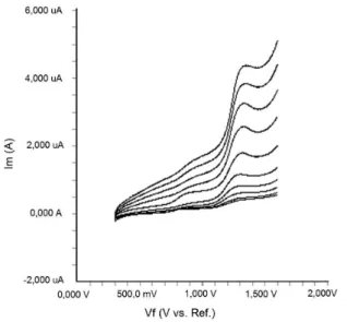
supporting electrolyte by using cyclic voltammetry (CV). Figure 2 shows a typical cyclic voltammogram of
20 μg mL-1 diclofenac recorded under these conditions
for the scan rate of 0.2 V s-1. In the anodic sweep, two
oxidation peaks are seen at about the potentials of 0.87 and 1.27 V, respectively.
In order to gain a deeper insight into the voltammetric waves, the effect of scan rate on the anodic peak currents
(I) and peak potentials (E) was studied in the range of
0.01-1 V s-1 of the potential scan rates in acetonitrile
solution containing 20 μg mL-1 concentration of diclofenac (Figure 3).
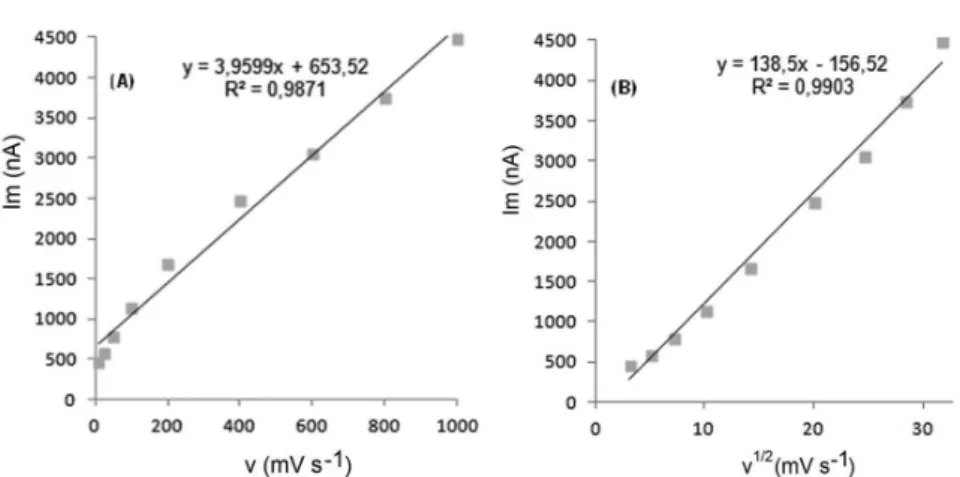
The representative linear sweep voltammograms
obtained at Pt electrode for 20 μg mL-1 diclofenac as a function of the scan rate are presented in Figure 4. Scan rate dependency experiments show that the peak currents vary
linearly with the scan rate (ν) (Figures 4a,b), which points
out the adsorption-controlled process. However, the plots of logarithm of peak currents versus logarithm of scan rates for
20 μg mL-1 concentration of diclofenac display straight lines with 0.497 slope (Figure 4c), which are close to theoretical value of 0.5 expected for an ideal diffusion-controlled
electrode process (Laviron, Roullier, Degrand, 1980).
Log I-log ν curve is more eligible for this aim,
therefore, a diffusional process for peak should be
considered. These results suggest that the redox species are diffusing freely from solution and not precipitating onto the electrode surface. The reason for this behaviour may be due to the solubility of the intermediate species in acetonitrile or poor adherence of products on the electrode surface.
The oxidation peak potential (E) for the peaks shifts toward more positive values with increasing scan rate. The relationship between the peak potential and scan rate is described by the following equation,
E = E0’ + RT/[(1 – )n
aF][0.78 + ln(D 1/2k
s -1) –
0.5 lnRT/[(1 – )naF]] + RT/[(1 – )naF]/2 lnv
and from the variation of peak potential with scan rate
αna can be determined, where α is the transfer coeficient
and na is the number of electrons transferred in the rate
determining step. According to this equation, the plots of
the peak potentials versus ln ν for the oxidation peak show linear relationship. The slope indicates the value of αna is
0.38 for peak. On the basis of above results, na is 1 then
the value of α is calculated as 0.38, which is reasonable
for most irreversible electrode processes. Based on above discussions, the oxidation process of diclofenac is controlled by the diffusion step and one electron and one proton are involved in the reaction.
Analytical applications and validation of the proposed method
In order to develop a voltammetric procedure for the
FIGURE 2 - Cyclic voltammogram for the oxidation of 20 μg mL-1 diclofenac in acetonitrile containing 0.1 M TBAClO
4
at Pt disk electrode, scan rate: 0.2 V s-1.
FIGURE 3 - Linear sweep voltammograms for the oxidation of 20 μg mL-1 diclofenac in acetonitrile containing 0.1 M TBAClO
4
FIGURE 4 - A, B, C: Dependence of peak current on the scan rate (20 μg mL-1).
determination of drug, we selected the DPV technique,
since the peaks were sharper and better deined at lower
concentration of diclofenac than those obtained by cyclic and linear sweep voltammetry with a lower background current, resulting in improved resolution. DPV is an effective and rapid electro-analytical technique with well-established advantages, including good discrimination against background currents and low detection limits
(Wang,1996; Kissenger,Heineman, 1996).
Calibration graphs from the standard solution of diclofenac according to the procedures described above were constructed by using DPV. A linear relation in the
concentration range between 1.5-17.5 μg mL-1 was found, indicating that the response was diffusion controlled in this range (Figure 5).
Above this concentration (17.5 μg mL-1), a loss of linearity was probable due to the adsorption of diclofenac on the electrode surface. The characteristics of the calibration plots are summarized in Table I.
The precision of the DPV method was determined by repeatability (intra-day) and intermediate precision
(inter-day). Repeatability was evaluated by analysing QC
samples six times per day, at three different concentrations which were QC samples. The intermediate precision was evaluated by analysing the same samples once daily
for two days. The RSD of the predicted concentrations
from the regression equation was taken as precision. The accuracy of this analytic method was assessed as the percentage relative error. For all concentrations studied, intra- and inter-day relative standard deviation values
were ≤3.87% and for all concentrations of diclofenac the
relative errors were ≤4.12%.
The limit of detection (LOD) and the limit of
quantiication (LOQ) were calculated on the peak current
using the following equation:
LOD = 3 s/m; LOQ = 10 s/m
FIGURE 5 - DPV voltammograms obtained for determination in supporting electrolyte (blank, 1.5, 2.5, 5, 7.5, 10, 12.5, 15
and 17.5 µg mL-1).
where s is the standard deviation of the peak currents (three runs) and m is the slope of the calibration curve. The LOD and LOQ values were also shown in Table I.
Repeating ive experiments on 15 μg mL-1 diclofenac for DPV technique tested the repeatability and reproducibility of peak potential and peak currents. The results were
shown also in Table I. Repetition of the sample analysis after 72 h period did not show any signiicant change in
the results of the analyses.
Determination of diclofenac in tablets
pharmaceutical preparations, using the related calibration
straight lines without any sample extraction or iltration
and after adequate dilutions. The results show that the proposed method was successfully applied for the assay of diclofenac in its pharmaceutical dosage forms (Table II). The accuracy of the method was determined by its
recovery during spiked experiments. Recovery studies
were carried out after the addition of known amounts of the pure drug to various pre-analysed formulation of diclofenac. According to the results, excipients presented in tablet do not interfere with the analysis (Table II).
There is no oficial method in any pharmacopoeias
(e.g. USP, BP or EP) or literature method related to pharmaceutical dosage forms of diclofenac. To prove the absence of interferences by excipients, recovery studies were carried out. The results demonstrate the validity of the proposed method for the determination of diclofenac in tablets. These results reveal that both methods had adequate precision and accuracy and consequently can be applied to the determination of diclofenac in pharmaceuticals without any interference from the excipients.
Birajdar, Meyyanathan and Suresh (2010)developed
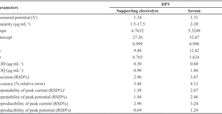
TABLE I - Regression data of the calibration lines for the quantitative determination of diclofenac
Parameters DPV
Supporting electrolyte Serum
Measured potential (V) 1.34 1.31
Linearity (μg mL-1) 1.5-17.5 2-20
Slope 6.7632 5.3249
Intercept 27.26 32.67
R 0.999 0.998
Sa 9.48 12.42
Sb 0.765 1.624
LOD (μg mL-1) 0.30 0.60
LOQ (μg mL-1) 0.90 1.80
Precision (RSD%) 2.96 3.87
Accuracy (% relative error) 3.46 4.12
Repeatability of peak current (RSD%)a 1.38 2.67
Repeatability of peak potential (RSD%) 1.94 2.46
Reproducibility of peak current (RSD%) 2.96 3.24
Reproducibility of peak potential (RSD%) 0.69 1.24
RSD: Relative standard deviation, aAverage of six replicate determinations, Sa: Standard deviation of intercept of regression line,
Sb: Standard deviation of regression line slope, R: Correlation coeficient, LOD: Limit of detection, LOQ: Limit of quantiication
TABLE II - Recovery of the diclofenac in pharmaceutical preparations
Diclomec Diclolam Voltaren
Labelled claim (mg) 100 50 75
Amount found (mg)a 99.4 50.2 75.4
RSD% 2.43 1.84 2.73
Bias% -0.60 0.40 0.53
Added (μg mL-1) 10 10 10
Found (μg mL-1) 9.94 10.04 10.09
Recovery% 99.4 100.4 100.9
RSD% of recovery 2.52 1.92 2.32
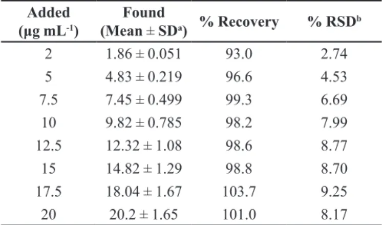
TABLE III - Recovery of diclofenac in human serum
Added
(μg mL-1)
Found
(Mean ± SDa) % Recovery % RSD b
2 1.86 ± 0.051 93.0 2.74
5 4.83 ± 0.219 96.6 4.53
7.5 7.45 ± 0.499 99.3 6.69
10 9.82 ± 0.785 98.2 7.99
12.5 12.32 ± 1.08 98.6 8.77
15 14.82 ± 1.29 98.8 8.70
17.5 18.04 ± 1.67 103.7 9.25
20 20.2 ± 1.65 101.0 8.17
SDa:Standard deviation of six replicate determinations,
RSD: Relative standard deviation, bAverage of six replicate
determinations a reverse phase HPLC method for the simultaneous
determination of rabeprazole and diclofenac in tablet. The method was based on HPLC separation of both drugs in
reverse phase mode using Phenomenox C18 column with
Waters HPLC system by using mobile phase composition of acetonitrile and 50 mM ammonium acetate buffer
(pH 3.6) (60:40 v/v) at flow rate 1mL min-1. Detection
wavelength used at 254 nm. Linearity was obtained in the concentration range of 1.0-3.2 µg mL-1 for rabeprazole and 6.0-16.0 µg mL-1 diclofenac.
Sastry, Prasad-Tipirneni and Suryanarayana
(1989) described a spectrophotometric method for
the determination of diclofenac sodium in bulk s a m p l e s a n d p h a r m a c e u t i c a l p r e p a r a t i o n s w i t h
p-N,N-dimethylphenylenediamine as solvent and
maximum absorbance at 670 nm. The reaction is sensitive
enough to permit the determination of 2.0-24 µg mL-1.
Agrawal Shivramchandra(1991) described two
methods for the determination of diclofenac. In the irst
method diclofenac reduces iron(III) to iron(II) having a maximum absorbance at 520 nm. The reaction obeys
Beer’s law for the concentrations of 10-80 µg mL-1. In
the second method, diclofenac is treated with methylene blue in the presence of phosphate buffer (pH 6.8) and the complex is extracted with chloroform. The complex has a maximum absorbance at 640 nm and linearity was in the range 5-40 µg mL-1.
T h o n g c h a i e t a l . ( 2 0 0 6 ) d e v e l o p e d a h i g h performance thin layer chromatographic method for the determination of diclofenac sodium in pharmaceutical formulations. The drug was extracted from the sample then various aliquots of this solution were spotted automatically by means of Camag Linomat IV on a silica gel 60 F254 aluminium plate, using a mixture of toluene: ethyl acetate: glacial acetic acid (60:40:1, v/v/v) as mobile phase. The
spot areas were quantiied by densitometry at 282 nm.
Linear calibration curve was obtained over the range 5-80 µg mL-1.
For DPV measurements, calibration curve was
linear over the concentration range of 1.5-17.5 μg mL-1 for diclofenac, which is as good as or superior to that reported
in other papers (Birajdar, Meyyanathan, Suresh, 2010;
Sastry, Prasad-Tipirneni, Suryanarayana, 1989; Agrawal
et al., 1991; Thongchai et al., 2006).
Determination of diclofenac in human serum samples
The optimized procedure was successfully applied for the determination of diclofenac in protein-free spiked human serum samples. Acetonitrile and methanol were
tried as the serum precipitating agents. The best results were obtained with acetonitrile. Hence, acetonitrile was used for the following studies. The measurements of diclofenac in serum samples were performed as described in the analysis of spiked serum samples section. For the applicability of the proposed method to the human serum, the calibration equation was obtained in spiked human serum. Calibration equation parameters and validation data are shown in Table I. Obtained recovery results of spike human serum samples are given in Table III.
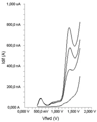
Using the proposed DPV technique, no sample pre-treatment was required other than precipitation and dilution steps. The recovery results of diclofenac (Table III) in serum samples were calculated from the related linear regression equations, which are given in Table I. Good recoveries of diclofenac were achieved from this type of matrix. Analysis of serum samples by DPV involved only protein precipitation and centrifugation, no time-consuming extraction and evaporation steps are required. In this study, the serum proteins and endogenous substances in serum samples are precipitated by the addition of acetonitrile, which is centrifuged at 5000 rpm, and the supernatant was taken and diluted with the supporting electrolyte and directly analysed. Typical DPV curve of diclofenac examined in serum samples are shown in Figure 6.
As can be seen in Figure 6, no oxidation compounds and no extra noise peaks present in biological material peak occurred in the potential range where the analytical peak appeared.
Stability of serum samples kept in refrigerator
(+4 °C) was tested by making ive consecutive analyses of
no signiicant changes in the peak currents and potentials between the irst and last measurements.
CONCLUSIONS
In this work, a new DPV technique has been developed for the determination of diclofenac in pharmaceutical preparations and human serum samples. The results obtained show that the above-described methods are useful not only for diclofenac determination in conventional electrolytes, but also in more complex matrices such as dosage forms and human serum samples. The principal advantages of DPV technique over other techniques are that they may be applied directly to the analysis of pharmaceutical dosage forms and biological samples without the need for separation or complex sample preparation, since there was no interference from the excipients and endogenous substances. The method is rapid, requiring less than 5 min to run sample.
ACKNOWLEDGEMENTS
This study was supported by a Grant from Ataturk
University Research Foundation (Project no: 2012/78). The authors have no conlicts of interest that are directly
relevant to the content of this study.
REFERENCES
ABDEL-HAMID, M.E.; NOVOTNY, L.; HAMZA, H.J. Determination of diclofenac sodium, flufenamic acid,
indomethacin and ketoprofen by LC-APCI-MS. Pharm.
Biomed. Anal., v.24, p.587-594, 2001.
AGATONOVIC-KUSTRIN, S.; ZIVANOVIC, L.J.; ZECEVIC, M.; RADULOVIC, D. Spectrophotometric study of
diclofenac-Fe(III) complex. J. Pharm. Biomed. Anal. v.16,
p.147-153, 1997.
A G R A W A L , Y. K . ; S H I V R A M C H A N D R A , K . S p e c t r o p h o t o m e t r i c d e t e r m i n a t i o n o f
diclofenac sodium in tablets. J. Pharm. Biomed. Anal., v.9,
p.97-100, 1991.
ANTIOCHIA, A.; LAVAGNINI, I.; MAGNO, F.; VALENTINI, F.; PALLESCHI, G. Single-wall carbon nanotube paste electrodes: a comparison with carbon paste, platinum and glassy carbon electrodes via cyclic voltammetric data.
Electroanalysis, v.16, p.1451-1458, 2004.
ARANCIBIA, J.A.; BOLDRINI, M.A.; ESCANDAR, G.M. Spectrofluorimetric determination of diclofenac in the
presence of alpha-cyclodextrin. Talanta, v.52, p.261-268,
2000.
ARCELLONI, C.; LANZI, R.; PEDERCINI, S.; MOLTENY, G. High-performance liquid chromatographic determination of
diclofenac in human plasma after solid-phase extraction. J.
Chromatogr., v.763, p.195-200, 2001.
BLANCO-LOPEZ, M.C.; LOBO-CASTANON, M.J.; MIRANDA-ORDIERES, A.J.; TUNON-BLANCO, p.Voltammetric response of diclofenac-molecularly imprinted ilm modiied carbon electrodes. Anal. Bioanal. Chem., v.377, p.257-261, 2003.
BOTELLO, J.C.; PÉREZ-CABALLERO, G. Spectrophotometric determination of diclofenac sodium with methylene-blue.
Talanta, v.42, p.105-108, 1995.
BIRAJDAR, A.S.; MEYYANATHAN, S.; SURESH, B. A RP-HPLC method for determination of diclofenac with
rabeprazole in solid dosage form. Pharm. Sci. Monitor.,
v.2 p.171-178, 2010.
FIGURE 6 - DPV voltammograms obtained for the determination
CARREIRA, L. A,; RIZK, M.; EL-SHABRAWY, Y.; ZAKHARI, n.A.; TOUBAR, S. S. Europium(III) ion probe spectroluorometric determination of diclofenac sodium. J. Pharm. Biomed. Anal., v.13, p.331-1337, 1995.
CROWLEY, B.; HAMILL, J.J.; LYNDON, S.; MCKELLICAN, J.F.; WILLIAMS, P.; MILLER, A.J. Controlled-release indomethacin and sustained-release diclofenac sodium in the treatment of rheumatoid-arthritis - a comparative
controlled clinical-trial. Curr. Med. Res. Opin., v.12,
p.143-150, 1990.
DAMIANI, p.C.; BEARZOTTI, M.; CABEZON, M.A.; OLIVIERI, A.C. Spectrofluorometric determination of
diclofenac in tablets and ointments. J. Pharm. Biomed.,
v.20, p.587-590, 1999.
DE SOUZA, R.L.; TUBINO, M. Spectrophotometric determination of diclofenac in pharmaceutical preparations.
J. Brazil. Chem. Soc., v.16, p.1068-1073, 2005.
GONZÁLEZ, L., YULN, G., VOLONTÉ, M.G. Determination of cyanocobalamin, betamethasone, and diclofenac sodium in pharmaceutical formulations, by high performance liquid
chromatography. J. Pharm. Biomed. Anal., v.20, p.487-492,
1999.
GOSTICK, N.; JAMES, I.G.; KHONG, T.K.; ROY, P.; SHEPHERD, P.R.; MILLER, A.J. Controlled-release indomethacin and sustained-release diclofenac sodium in the treatment of osteoarthritis - a comparative controlled
clinical-trial in general-practice. Curr. Med. Res. Opin.,
v.12, p.135-142, 1990.
GOYAL, R.N.; CHATTERJEE, S.; AGRAWAL, B. Electrochemical investigations of diclofenac at edge plane pyrolytic graphite electrode and its determination in human
urine. Sens. Actuators B, v.145, p.743-748, 2010.
HASSAN, S.S.M.; MAHMOUD, W.H.; ELMOSALLANY, M.A.F.; ALMAZZOOQI, M.H. Iron(II)- phthalocyanine as a novel recognition sensor for selective potentiometric
determination of diclofenac and warfarin drugs. J. Pharm.
Biomed., v.39, p.315-321, 2005.
ILIIESCU, T.; BAIA, M.; MICLAUS, v.A raman spectroscopic study of the diclofenac sodium-betacyclodextrin interaction.
Eur. J. Pharm. Sci., v.22, p.487-495, 2004.
JIN, W.; ZHANG, J. Determination of diclofenac sodium by capillary zone electrophoresis with electrochemical
detection. J. Chromatogr., v.868, p.101-107, 2000.
KAUFFMANN, J.M.; VIRE, J.C. Pharmaceutical and biomedical
applications of electroanalysis. Anal. Chim. Acta, v.273,
p.329-336, 1993.
KASPEREK, R. Determination of diclofenac sodium and
papaverine hydrochloride in tablets by hplc method. Acta
Poloniae Pharm., v.65, p.403-408, 2008.
KISSENGER, P.T.; HEINEMAN, W.R. (Eds.) Laboratory techniques in electroanalytical chemistry, second ed., New York: Marcel Dekker, 1996.
LAVIRON, E.; ROULLIER, L.; DEGRAND, C. A multilayer model for the study of space distributed redox modiied electrodes. Theory and application of linear potential sweep
voltammetry for a simple reaction. J. Electroanal Chem.,
v.112, p.11-23, 1980.
MANEA, F.; IHOS, M.; REMES, A.; BURTICA, G.; SCHOONMAN, J. Electrochemical determination of diclofenac sodium in aqueous solution on cu-doped
zeolite-expanded graphite-epoxy electrode. Electroanalysis, v.22,
p.2058-2063, 2010.
MATIN A.A.; FARAJZADEH, M.A.; JOYUBAN, A. A simple spectrophotometric method for determination of sodium
diclofenac in pharmaceutical formulations. Farmaco, v.60,
p.855-861, 2005.
NOROUZI, P.; SHIRVANI-ARANI, S.; DANESHGAR, P.; GANJALI, M.R. Sub-second adsorptive fast fourier transform coulometric technique as a novel method for the determination of nanomolar concentrations of sodium valproate in its pharmaceutical preparation in flowing
solution systems. Biosens. Bioelectron., v.22, p.1068-1074,
2007.
OZKAN, S.A.; USLU, B.; SENTURK, Z. Electroanalytical characteristics of amisulpride and voltammetric determination of the drug in pharmaceuticals and biological
media. Electroanalysis, v.16, p.231-237, 2004.
ROSKAR, R.; KMETEC, v.Liquid chromatographic
determination of diclofenac in human synovial fluid. J.
SANTINI A. O.; PEZZA, H. R.; PEZZA, L. Determination of diclofenac in pharmaceutical preparations using a potentiometric sensor immobilized in a graphite matrix.
Talanta, v.68, p.636-642, 2006.
SASTRY, C.S.P.; PRASAD-TIPIRNENI, A.S.R.; SURYANARAYANA, M.V. Spectrophotometric analysis of some anthranilic acid-derivatives and their pharmaceutical
preparations. Microchem. J., v.39, p.277-289, 1989.
SENA, M.M.; CHAUDHRY, Z.F.; COLLINS, C.H.; POPPI, R.J. N-way PLS applied to simultaneous spectrophotometric determination of acetylsalicylic acid, paracetamol and
caffeine. J. Pharm. Biomed. Anal., v.36, p.743-749, 2004.
SHAMSIPUR, M.; JALALI, F.; ERSHAD, S. Preparation of a diclofenac potentiometric sensor and its application to pharmaceutical analysis and to drug recovery from biological luids. J. Pharm. Biomed., v.37, p.943-947, 2005.
SIOU, A.; POMMIER, F.; GODBILLON, J. Determination of diclofenac in plasma and urine by capillary gas-chromatography mass-spectrometry with possible simultaneous determination of deuteriumlabeled diclofenac.
J. Chromatogr., v.571, p.87-100, 1991.
SUN, S.W.; FABRE, H. Practical approach for validating the tlc assay of an active ingredient in a pharmaceutical
formulation. J. Liq. Chromatogr., v.17, 433-445, 1994.
THONGCHAI, W.; LIAWRUANGRATH, B.; THONGPOON, C . ; M A C H A N , T. H i g h p e r f o r m a n c e t h i n l a y e r chromatographic method for the determination of
diclofenac sodium in pharmaceutical formulations. Chiang
Mai J.Sci., v.33 p.123-128, 2006.
VALENTINI, F.; AMINE, A.; ORLANDOCCI, S.; TERRANOVA, M.L.; PALLESCHI, G. Carbon nanotube puriication: Preparation and characterization of carbon
nanotube paste electrodes. Anal. Chem., v.75, p.5413-5421,
2003.
WANG, J. (Ed.) Electroanalytical techniques in clinical chemistry and laboratory medicine, New York: VCH Publishers, 1996.
XU, M.T.; CHEN, L.F.; SONG, J.F. Polarographic behaviors of diclofenac sodium in the presence of dissolved oxygen
and its analytical application. Anal. Biochem., v.329,
p.21-27, 2004.
YILMAZ, B. GC-MS determination of diclofenac in human
plasma. Chromatographia, v.71, p.549-551, 2010.
Received for publication on 30th January 2014