OR
IGI
N
A
L
R
E
S
E
A
R
C
H
Mailing address: – Carlos Henrique Fachin Bortoluci – Centro de Ciências da Saúde, Universidade do Sagrado Coração (USC) – Rua Irmã Arminda, 10-50 – Bauru (SP), Brazil – CEP: 17011-160 – E-mail: [email protected] – Financing source: Nothing to declare – Conlict of interest: Nothing to declare – Presentation: Jan. 2016 – Accepted for publication: Mar. 2016 – Approved by the Institutional Ethics Comittee: Protocol nº 034/2012.
1Professor from the Center for Health Sciences and Dean of Research and Graduate Studies at the University of Sagrado Coração –
Bauru (SP), Brazil.
2Student from the Physical Therapy Course at the University of Sagrado Coração – Bauru (SP), Brazil.
3Master in Physical Therapy at the Botucatu Faculty of Medicine (FMB) of Universidade Estadual Paulista (UNESP) – Bauru (SP), Brazil. 4Student from the Dentistry Course at the University of Sagrado Coração – Bauru (SP), Brazil.
5Professor from Health Sciences Center at the University of Sagrado Coração – Bauru (SP), Brazil.
ABSTRACT | The peripheral nerve injuries occur frequently and generally cause functional loss impacting negatively on patient’s life. The objective this study was to verify the eiciency of the combination of laser therapy and swimming in rats afected by axonotmesis. The sample was comprised of 50 Wistar rats and it was divided into 05 groups: Control Group; Surgical Control Group; Laser Experimental Group; Swimming Experimental Group and Laser Experimental combined with Swimming Group. The nerve was crushed into a 5 mm-long segment next to the sciatic nerve trifurcation with a pair of forceps for 60 seconds. The GaAs infrared laser (904nm) was used with energy radiated 0,4J the irst week, the second week 0,8J and 1,2J in the third and fourth week. For functional (FCI) evaluation, the animals were immobilized and the plantar region of their paws were painted with ink stamp. The procedure was repeated twice to each animal. The nerve morphometry (areas, diameters and thicknesses of the ibers, axons and myelin sheath) was performed with the measurement of 220 ibers per animal in each group . We can see that the GEL and GEN groups , obtained the best results when compared with the other groups (GC, GCC and GELAN) in all morphometric variables studied, but no statistically signiicant diference
12
was found between them. In functional analysis, it was observed that the gelan group obtained the best results when compared with the other groups (GCC , GEN and GEL) and when the GEL and GEN groups were compared, there was no statistically signiicant diference between them. Was conclued the GEL and GEN groups havd the best morphometric results, while the GELAN showed the best functional outcome. Therefore, it can be concluded that the combination of these features favoured the functional recovery of the animals
Keywords | Swimming; Laser Therapy; Rats, Wistar; Regeneration; Physiotherapy.
RESUMO | As lesões de nervos periféricos ocorrem frequentemente e, de modo geral, causam perda funcional impactando de forma negativa na vida do paciente. O objetivo do estudo foi veriicar a eiciência da associação da laserterapia e natação em ratos acometidos por axonotmeses. A amostra foi composta por 50 ratos da linhagem Wistar. Foram divididos em 5 grupos, sendo: grupo controle (GC); grupo controle cirúrgico (GCC); grupo experimental laser (GEL); grupo experimental natação (GEN) e grupo experimental laser associado à natação (GELAN). O nervo foi esmagado em um segmento de 5
Efect of the laser therapy in association with
swimming for a morphological nerve repair and
functional recovery in rats submitted to sciatic
axonotmesis
Efeito da associação da laserterapia com a natação no reparo morfológico
do nervo isquiático e na recuperação funcional de ratos submetidos à axonotmese
Efecto de la asociación entre la laserterapia y la natación en la recuperación morfológica
del nervio isquiático y en la recuperación funcional de ratas sometidas a axonotmesis
Geraldo Marco Rosa Junior1, Raiza Maiara Gutierrez Magalhães2, Vívian Cristina Rosa3,
mm de comprimento próximo a trifurcação do nervo isquiático, feito com uma pinça durante 60 segundos. Foi utilizado o laser infravermelho AsGa (904nm) com energia irradiada de 0,4 J na primeira semana, 0,8 J na segunda semana e 1,2 J na terceira e quarta semana. Para avaliação funcional (IFC), os animais foram imobilizados, e a região plantar das patas foram pintadas com tinta de carimbo. Esse procedimento foi repetido duas vezes com cada animal. Foi realizada a morfometria (áreas, diâmetros e espessuras das ibras, axônios e bainha de mielina) dos nervos com mensuração de 220 ibras por animal de cada grupo. Pudemos observar que os grupos GEL e GEN, em todas as variáveis morfométricas estudadas, obtiveram os melhores resultados, quando comparados com os outros grupos (GC, GCC e GELAN), mas não apresentou diferença estatisticamente signiicante entre eles. Na análise funcional observou-se que o grupo GELAN obteve o melhor resultado quando comparado com os outros grupos (GCC, GEL e GEN) e quando comparados os grupos GEL e GEN entre eles não houve diferença estatisticamente signiicante. A conclusão foi que os grupos GEL e GEN obtiveram os melhores resultados morfométricos, enquanto o GELAN apresentou o melhor resultado funcional. Portanto, pode-se concluir que a associação destes recursos favoreceu a recuperação funcional desses animais.
Descritores | Natação; Terapia a Laser; Ratos Wistar; Regeneração; Fisioterapia.
RESUMEN | Las lesiones de los nervios periféricos frecuentemente ocurren, y generalmente ocasionan pérdida funcional, lo que les causa daño a la vida de los pacientes. En este artículo se propone a veriicar la eicacia de la asociación de la laserterapia
y de la natación en ratas sometidas a axonotmesis. El muestro se compuso de 50 ratas Wistar. Se las dividieron en 5 grupos: grupo control (GC); grupo control quirúrgico (GCQ); grupo experimental láser (GEL); grupo experimental natación (GEN) y grupo experimental láser asociado con la natación (GELAN). Se aplastó con una pinza durante 60 segundos el nervio en un segmento de 5 mm de extensión cerca de la trifurcación del nervio isquiático. Se empleó el láser infrarrojo AsGa (904nm) con energía irradiada de 0,4 J en la primera semana, 0,8 J en la segunda y 1,2 J en la tercera y cuarta semanas. Para la evaluación funcional (IFC), se los inmovilizaron los animales y se los pintó con tinta estampilla la región plantar de las patas. Se repitió dicho procedimiento dos veces en cada animal. Se realizó la morfometría (áreas, diámetros y espesuras de las ibras, axónios y vaina de mielina) de los nervios con mensuración de 220 ibras por cada animal de cada grupo. Se notó que los grupos GEL y GEN, en todas las variables morfométricas estudiadas, presentaron los mejores resultados, en comparación con los otros grupos (GC, GCQ y GELAN), sin embargo no presentó diferencia estadísticamente signiicativa entre ellos. En el análisis funcional se observó que el grupo GELAN tuvo el mejor resultado en comparación con otros grupos (GCQ, GEL y GEN), y al comparar los grupos GEL y GEN no presentaron diferencia estadísticamente signiicativa. Se concluyó que los grupos GEL y GEN tuvieron mejores resultados morfométricos, mientras que el GELAN presentó el mejor resultado funcional. Por lo que se concluye que la asociación de dichos recursos les favoreció la recuperación funcional de dichos animales.
Palabras clave | Natación; Terapia por Láser; Ratas Wistar; Regeneración; Fisioterapia.
INTRODUCTION
Peripheral nerve injuries are a common occurrence and generally cause the patient to sufer functional
loss1 that consequently has a negative efect on his/
her life2. he most common causes of peripheral nerve
injury are irearm projectiles, falls and penetrative or contusion trauma, however, the main causative factor
are automobile accidents3.
Peripheral nerve injury is more frequent in individuals aged between 25 and 40 years. his relatively young age may cause signiicant economic and social consequences due to functional disability occurring in early life, as most individuals are at the prime of their professional capacity during this
period. hus, following an injury, any treatment that leads to a more accelerated functional recovery of the peripheral nerves is extremely valuable to
society4. he degenerative process in the muscle is
known to start shortly after nerve injury, therefore, intervention to re-establish myoneural interaction
must be swift5.
here are several surgical techniques and treatments
that have been developed6, these help to regenerate the
peripheral nerves with the aim of morphological and
functional improvement in less time7,8.
Exercise is the preferred method out of the rehabilitation strategies that are currently used, mainly due to it being characterized as a non-invasive
mechanism for releasing neurotrophic factors, especially the mainly Brain-Derived Neurotrophic Factor (BDNF), which is considered paramount in terms of
mediating neuroplasticity10.11 .
Several authors have shown beneicial results by using exercise, namely through greater budding
and extension of the axons12, increased number
of myelinated nerve ibers, 13,14 and improved
functional recovery of the injured member15-17.
here have been extensive discussions in the
scientiic community18 regardingrecommendations
as per the type of exercise as well as its duration and intensity.
Another feature used with the objective of promoting functional improvement for the injured person is phototherapy. Studies from the late 80s saw the beginning of thorough investigatory eforts whose purpose was to evaluate the protocols used as a therapeutic resource for regenerating peripheral nerves,
these studies used crush injury19 as an experimental
model. In modern times these protocols are still used as a great therapeutic resource for peripheral nerve
regeneration20.
Laser therapy, as an efective complementary therapeutic resource for treating nerve injuries, has gained prominence in physical therapy intervention protocols, which is mainly due to it being a non-invasive treatment that provides positive results for both regeneration and functional recovery. Among these beneits are: an inlammatory and anti-oedematous efect, potential for wound healing, pain relief, increased mitochondrial respiration, ATP synthesis and proliferation of ibroblasts, stimulating the proliferation of Schwann cells that secrete neurotrophic factors for nerve regeneration, among
other factors21-24.
Based on the belief in the beneicial efects of laser therapy and physical exercise, while considering the importance of functional recovery and returning to daily activities, the objective of this research was to investigate the efectiveness of laser therapy and swimming regarding the morphofunctional characteristics of rats that have been subjected to axonotmesis. It is our belief that the association of these protocols, in addition to the laser energy being gradually increased over the time of the injury, makes it possible to trigger a more eicient regenerative process that can provide the individual with more efective functional improvement.
METHODOLOGY
his study was approved by the Research Ethics Committee under the protocol number 034/2012.
50 male, 80-day old, Wistar rats (Rattus norvegicus
albinus) from the central bioterium were used. he
animals were kept in cages and given ad libitum access to food and water in a controlled environment (temperature between 21 to 25°C with 12 hours in the light and 12 hours the dark) with no restrictions on movement.
he 50 animals were randomly divided in ive groups (n=10, for each group) for axonotmesis, these being:
Control group (CG), where the animals were not subject to any intervention and observed for 30 days.
Surgical Control Group (SCG), where the animals were put through the surgical intervention, with the induction of nerve injury through crushing, however, they were not subjected to any treatment protocol following the surgery.
Experimental Laser Group (ELG), in which the animals were put through the surgical intervention, with the induction of nerve injury through crushing, and, subsequent to surgery, were treated with the laser therapy protocol.
Experimental Swimming Group (ESG), in which the animals were put through the surgical intervention, with the induction of nerve injury through crushing, and, subsequent to surgery, were subjected to the swimming exercise protocol.
Experimental Laser together with Swimming Group (ELSG), in which the animals were put through the surgical intervention, with the induction of nerve injury through crushing, and, subsequent to surgery, were treated with laser therapy and subjected to the swimming exercise protocol.
In order to perform the surgical procedure on the nerve, the animals were anesthetized with a drug combination of Ketamine Hydrochloride (80 mg/kg) and Xylazine Hydrochloride (15 mg/kg). he surgical procedure involved a 5cm incision on the dorsolateral region of the left pelvic limb of the animal. Following the incision, the adjacent musculature was separated in order to expose the right sciatic nerve.
of three 20-second stages. The musculature and skin were sutured with 4-0 thread following the procedure.
he laser instrument used in the laser therapy groups was an AsGa-904nm (Arseneto de Gálio, ENDOPHOTON - KLD). he irradiation was performed on the skin of the animal’s right pelvic limb, on the injured segment of the nerve. he procedures used by previously published studies
with the same pattern were used25,26. he instrument
was properly calibrated before the applications were performed.
he laser was applied, three times a week, for exactly 8 seconds in the irst week, 16 seconds in the second week and 24 seconds in the third and fourth for a total treatment time of 4 weeks. he animals were held by hand during the application of the laser.
The following parameters used were: 904nm wavelength, 50mw power, 40J/cm² energy fluency or density in the first week, 80J/cm² in the second week and 120J/cm² in the third and fourth week, the radiated energy was 0.4J in the first week, 0.8J in the second week and 1.2J in the third and fourth week. The purpose of increasing the dose was to advance the stages of peripheral nerve injury from the acute to subacute phase, and later to the chronic phase.
For the groups being submitted to the swimming exercise, the animals were placed in a glass tank with warm water (32±2°C), illed to a 40cm depth. he swimming exercises were performed ive times a week, with Saturdays and Sundays being rest days. During the irst week, the animals swam exclusively for 20 minutes on the irst day, with10 extra minutes being added each day up to a 40 minute duration, a time that was kept constant until the experiment’s conclusion.
In order to evaluate the animals’ functional capability, they were immobilized and placed in an acrylic pathway to walk on. he animals were ilmed during their walk so that the footprints from their tracks could be recorded. his procedure was repeated with each animal twice. he distance between the footprints made by the hind limbs were evaluated according to
the equation as described by Bain27, based on studies
by De Medinacelli28. he videos were converted into
sequential photos while following a calibration pattern for each image. he Sciatic Functional Index was used to evaluate functional recovery. he measurements were taken using 4.6.2 Image Pro plus software, and the data
were subjected to statistical analysis according to the P<0.05 index for all samples.
In order to collect the samples, the animals were anesthetized with a drug combination of Ketamine Hydrochloride (80 mg/kg) and Xylazine Hydrochloride (15 mg/kg), which was applied via intramuscular injection into the dorsolateral region of the left pelvic limb of the animal. Following the surgical sample collections, the animals were given a lethal dose of 150 mg/kg sodium pentobarbital and 2% lidocaine (20 mg/mL), which was administered intraperitoneally.
During the treatment, the histological samples from the sciatic nerve were ixed in Karnovsky solution, included in historesin and stained with osmium tetroxide. he samples were processed on a microtome in order to obtain the 5µm thick histological cuts.
he morphometry of the nerves was performed by measuring 220 ibers per animal in each group, which was done using a microcomputer with image capturing and analysis software linked to an optical microscope. he morphometric variables that were studied in the nerves are as follows: area of the ibers, area of the axons, minimum diameter of the ibers, minimum diameter of the axons, area of the myelin sheath and thickness of the myelin sheath.
he analysis of variance test (ANOVA) was used to compare the groups, followed by the TUKEY test when there was a signiicant diference detected. A paired T-test was used to make comparisons between the experimental and normal sciatic nerves. A signiicance level of p <0.05 was adopted for all analyses.
RESULTS
Upon comparing the area and the diameter of the nerve iber, the ELG and ESG groups presented the best
results (area = 38.42 µm² and 37.56 µm2, respectively,
and diameter = 4.02 µm and 3.96 µm, respectively) compared with the SCG and ELSG groups, which had 21.47 µm² and 28.76 µm² for area and 2.87 µm and 3.35 µm for diameter, respectively. he CG presented the highest mean for area with 50.12 µm² and 8.95 µm for diameter.
Table 1. Mean values and standard deviation for the area of the nerve ibers (m²) and mean values and standard deviation of the smaller diameter of the nerve ibers (µm)
Area of the Nerve Fiber Diameter of the Nerve Fiber
Mean SD Mean SD
CG 50.12a 4.77 8.95a 0.22
SCG 21.47b 3.18 2.87b 0.14
ELG 38.42c 5.82 4.02c 0.20
ESG 37.56c 4.52 3.96c 0.21
ELSG 28.76d 3.27 3.35d 0.45
Diferent letters indicate a statistical diference (p <0.05)
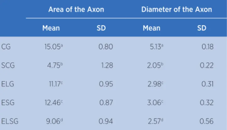
Based on the results from Table 2, it is possible to see that the area and the diameter of the axon from the ELG and ESG groups had the best results in the two variables analyzed (area = 11.17µm² and 12.46µm², and diameter = 2.98µm and 3.06µm, respectively) when compared with the SCG and ELSG groups, which had
4.75µm² and 9.06µm2 for the area of the axon and 2.05
µm and 2.57µm for the diameter of the axon. he CG showed the largest values with 15.05µm² of area and 5.1 µm of diameter.
Table 2. Mean values and standard deviation of the area of the axons (m²) and mean values and standard deviation of the smallest diameter of the axons (µm)
Area of the Axon Diameter of the Axon
Mean SD Mean SD
CG 15.05a 0.80 5.13a 0.18
SCG 4.75b 1.28 2.05b 0.22
ELG 11.17c 0.95 2.98c 0.31
ESG 12.46c 0.87 3.06c 0.32
ELSG 9.06d 0.94 2.57d 0.56
Diferent letters indicate a statistical diference (p <0.05)
According to the results from Table 3, it is possible to see that the area and the thickness of the sheath from the ELG and ESG groups showed the best results (area = 25.25µm² and 25.10 µm², respectively; thickness = 2.86µm and 2.72µm, respectively) when compared with the SCG and ELSG groups, which showed 7.72m² and 21.70µm ² in the area of the sheath, and 1.82µm and 2.28µm in the thickness of the sheath, respectively. he CG presented the highest values with 43.07µm² in the
area of the sheath and 3.82µm in the thickness of the sheath.
Table 3. Mean values and standard deviation of the areas of myelin sheaths (m²) and mean values and standard deviation of the thicknesses of the myelin sheaths (µm)
Area of Myelin Sheath Thickness of Myelin Sheath
Mean SD Mean SD
CG 43.07a 1.80 3.82a 0.33
SCG 7.72b 4.56 1.82b 0.25
ELG 25.25c 1.74 2.86c 0.37
ESG 25.10c 2.46 2.72c 0.45
ELSG 21.70d 2.93 2.28d 0.82
Diferent letters indicate a statistical diference (p <0.05)
Figure 1 shows the photomicrographs from the study groups and the morphological diferences presented by these groups.
(A) ELG; (B) ESG; (C) ELSG; (D) SCG and (E) CG
Figure 1. Plate of the micrographs from the groups showing the morphology of the nerve (1000x)
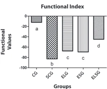
analysis result is the one closest to the control group. Diferent letters indicate statistical diference (p<0.05).
Functional Index
Groups CG SCG ELG ESG
ELSG
F
unc
tional
V
alues
Graphic 1. Graph showing the functional result of the groups. The ELSG Group showed a result closest to the CG
he ELSG groups had a result of -44.90, which was the best due to it being closest to the CG result (-12.11). he ESG and ELG groups did not show any statistical diference, reaching -69.32 and -67.51, respectively. As a result of being denervated, the SCG group had the worst result (-82.81).
DISCUSSION
Several authors have chosen to work experimentally
using crushing29 when studying peripheral nerve injuries,
whichareclassiied by Seddon30 as an axonotmesis. his
method partly preserves important support structures, such as: the endoneurum, the perineurium and the tubules from the Schwann cells, which function to guide the new axon in regenerating up to the target organ.
Several experiments involving denervated animals18
have described exercise as the factor that provides the
most budding and extension of the axons,12 the greatest
increase in the number of myelinated ibers13,14 and the
best improvement in functional recovery for the injured
member15. he results from this study are similar to those
found in the literature regarding evaluating functional recovery, due to the fact that the groups that performed exercise showed better results compared to groups who did not go through the swimming protocol. Other authors refer to the harmless efects of physical exercise
on the peripheral nerve regeneration process31,32.
Despite being somewhat controversial, some authors have suggested exercise as a complementary therapeutic
resource33 and, when combined with other therapeutic
practices such as electrotherapy34 or phototherapy6,19,
can result in a better recovery prognosis for peripheral nerve injuries. he ELSG group in this research, which combined laser therapy with swimming, presented a better functional result when compared to the ELG and ESG group, respectively.
Based on clinical and experimental research, there is evidence that some of the efects of laser therapy are an improvement in nerve function, increased metabolism of the neurons and greater production capacity of the myelin sheath. Due to laser therapy not being invasive, the ability to radiate injured nerves without intervention
is useful35.
Generally speaking, publications whose treatments involve a continuous laser output produced positive indings regarding peripheral nerve regeneration, however, the energy density used varies greatly, presenting data from 1.2J/cm² to 140J/cm². hus, a laser energy density that became gradually greater over time, during the course of the injury was chosen for this study, which ranged between 40J/cm², 80J/cm² and 120J/cm ² week by week in order to ind the ideal laser
dosage(PISTARINI et al., 2015; SOUSA et al., 2013;
LINS et al., 2010; NORONHA et al., 2004; LUCAS et
al., 2003)25,26,36-41.
Very little is known regarding the role of laser irradiation in terms of rehabilitating tissues of the locomotor system . However, laser irradiation is widely used to treat a variety of pathological conditions of the musculoskeletal system, including the peripheral
nerves25.
Light energy absorption by nerve tissue increases ATP synthesis and cell proliferation, which increases axonal energy metabolism, improving scarring in the regenerative process, and thereby, the expression of neurotrophic factors, such as: GAP-43 protein, TGF-1, expression of the GCRP gene, which increase the regeneration rate and directs the axon to the target organ. he increase of axonal budding is also described
as a result of the laser irradiation42,43.
During research performed by Reis35, the mean
According to Camargo26, who also used the AsGa
laser on peripheral nerve regeneration, the mean SFI was -47.71, which represents an even better result compared with the data from this study.
During a study conducted by Endo25,whoused
low-level laser therapy to accelerate the regeneration of peripheral nerves , there was a progressive improvement of the SFI, both in irradiated and control nerves (69% and 45%, respectively). According to
Endo25 , the iber density increased for the irradiated
nerves and decreased for the control nerves, showing a signiicant diference between the two (p=0.001). he authors concluded that low-level laser therapy efectively accelerates sciatic nerve regeneration in rats. his study also presented an increase in the area of the ibers, with the experimental laser group and the surgical control group obtaining values of 38.42µm² and 21.47µm², respectively.
According to a study performed by Oliveira31,
which involved using electrical stimulation and swimming for nerve regeneration and functional recovery during the acute phase of a axonotmesis in mice, the diameter of the axon was lower in the denervated groups, and that, when compared together, the group for which the best results were observed was the swimming group with the following values: 6.32±0.36 in the control group; 3.45±0.64 in the denervated group; 3.67±0.41 in the denervated + electro stimulation group; 4.34±0.69 in the denervated + swimming group; 4.04±0.38 in the denervated + swimming + electro stimulation group. Our study observed the same, the best result in relation to the diameter of the axon was in the swimming group.
During this study, the group in which there was an association between therapy and exercise, despite not showing good morphological results, presented an improvement in the functional analysis. We associate this improved functional result to the exercise, which requires the animal to release neurotrophic factors, and also to efect from the laser, which provides an anti-inlammatory, anti-oedematous and analgesic efect.
We believe that further research should be conducted in order to identify the expression of proteins involved in the peripheral nerve regenerative process, as well as to verify the muscular response to newly received innervation, which establishes a correlation between the mioneural interaction and possible readaptation by the motor plates
CONCLUSION
Based on the results presented here, the laser and isolated swimming were observed to promote a morphological improvement during the evaluation of nerve regenerative process, however, no statistically signiicant diference between them was found. However, the association of laser therapy and swimming was beneicial for functional recovery following peripheral nerve injury.
herefore, the conclusion is that laser therapy and swimming can promote the eicient morphological recovery of rats with peripheral nerve injury, and that associating these resources demonstrated a tendency for functional recovery. New swimming protocols should be therefore investigated with a view to establish a direct relationship between exercise intensity and functional recovery.
REFERENCES
1. Sunderland S. A classiication of peripheral nerve injuries producing loss of function. Brain. 1951;74:491-516
2. Rosberg HE, Carlsson KS, Hojgard S, Lindgren B, Lundborg G, Dahlin LB. Injury to the human median and ulnar nerves in the forearm – analysis of costs for treatmentand rehabilitation of 69 patients in southernsweden. J Hand Surg. 2005;30:35-9. 3. Kouyoumdjian JA. Peripheral nerve injuries: a retrospective
survey of 456 cases. Muscle Nerve. 2006;34:785-8.
4. Whitlock EL, Tufaha SH, Luciano JP, Yan Y, Hunter DA, Magill CK, Moore AM, Tong AY, Mackinnon SE, Borschel GH. Processed allografts and type I collagen conduits for repair of peripheral nerve gaps. Muscle Nerve. 2009;39(6):787-99. 5. Rosa-Junior GM, Bueno CRS, Heubel A, Bortoluci CHF,
Simionato LH, Daré LR, Silva MP, Dias DV. Efeito da corrente alternada simétrica sinusoidal na musculatura estriada esquelética desnervada experimentalmente. Salusvita. 2013;32(3):211-25.
6. Deumens R, Bozkurt A, Meek MF, Marcus MAE, Joosten EAJ, Weis J, Brook GA. Repairing injured peripheral nerves: bridging gap. Prog Neurobiol. 2010;92:245-76.
7. Gigo-Benato D, Geuna S, Rodrigues AC, Tos P, Fornaro M, Boux E, Battiston E, Giacobini-Robecchi MG. Low-power laser biostimulation enhances nerve repairafter end-to-side neurorrhaphy: a double-blind randomized studyin the rat median nerve model. Lasers Med Sci. 2004;19:57-65.
8. Rochkind S, El-Ani D, Nevo Z, Shahar A. Increase of neuronal sprouting and migration using 780nm laser phototherapy as procedure for cell therapy. Lasers Surg Med. 2009;41:277-81. 9. Kisner C, Colby LA. Therapeutic Exercise: Foundations and
Techniques. 5ª ed. Philadelphia; Davis Company, 2007. 10. Yarrow JF, White LJ, Mccoy SC, Borst SE. Training augments
derived neurotrophic factor (BDNF). Neurosci Letters. 2010;479:161-5.
11. Zoladz JA, Pilc A, Majerczak J, Grandys M, Zapart-Bukowska J, Duda K. Endurance training increases plasma brain-derived neurotrophic factor concentration in young healthy men. J Physiol Pharmacol. 2008;59(7):119-32.
12. Sabatier MJ, Redmon N, Schwartz G, English AW. Treadmill training promotes axon regeneration in injured peripheral nerves. Exp Neurol. 2008;211:489-93.
13. Ilha J, Araujo RT, Malysz T, Hermel EES, Rigon P, Xavier LL, Achaval M. Endurance and resistance exercise training programs elicit speciic efects on sciatic nerve regeneration after experimental traumatic lesion in rats. Neurorehabil Neural Repair. 2008;22(4):355-66.
14. Udina E, Puigdemasa A, Navarro X. Passive and active exercise improve regeneration and muscle reinnervation after peripheral nerve injury in the rat. Muscle Nerve. 2011;43:500-09.
15. Cobianchi S, Marinelli S, Florenzano F, Pavone F, Luvisetto S. Short-but not long-lasting treadmill running reduces allodynia and improves functional recovery after peripheral nerve injury. Neuroscience. 2010;168:273-87.
16. Marqueste T, Marqueste T, Alliez JR, Alluin O, Jammes Y, Decherchi P. Neuromuscular rehabilitation by treadmill running or electrical stimulation after peripheral nerve injury and repair. J Appl Physiol. 2004;96:1988-95.
17. Van Meeteren N, Brakkee JH, Hamers FP, Helders PJ, Gispen WH.Exercise training improves functional recovery and motor nerve conduction velocity after sciatic nerve crush lesion. Arch Phys Med Rehabil. 1997;78:70-7.
18. Udina E, Cobianchi S, Allodi I, Navarro X. Efects of activity-dependent strategies on regeneration and plasticity after peripheral nerve injuries. Ann Anat. 2011:1-7.
19. Gigo-Benato D, Geuna S, Rochkind S. Phototherapy for enhancing peripheral nerve repair: a review of the literature. Muscle Nerve. 2005;31:694-701.
20. Barbosa RI, Marcolino AM, Guirro RRJ, Mazzer N, Barbieri CH, Fonseca MCR. Efeito do laser de baixa intensidade (660nm) na regeneração do nervo isquiático lesados em ratos. Fisioter Pesqui. 2010;17(4):294-9.
21. Wang L, Hu L, Grygorczyk R, Shen X, Schwarz W. Modulation of extracellular atp content of mast cells and drg neurons by irradiation: studies on underlying mechanism of low-level-laser therapy. Mediators of Inlammation, Article ID 630361, 9 pages, 2015. doi:10.1155/2015/63036.
22. Martins F, Rennó ACM, Oliveira F, Minatel NP, Bortolin JA, Quintana HT, Aveiro MC. Low-level laser therapy modulates musculoskeletal loss in a skin burn model in rats. Acta Cirúrg Bras. 2015;30(2):94-9.
23. Wang CZ, Chen YJ, Wang YH, Yeh ML, Huang MH, Ho ML, Liang JI, Chen CH. Low-level laser irradiation improves functional recovery and nerve regeneration in sciatic nerve crush rat injury model. PloS One. 2014;9(8):103-348.
24. Huang Y.Y, Sharma SK, Carroll J, Hamblin MR. Biphasic dose response in low level light therapy-an update. Dose-Response. 2011;9(4):602-18.
25. Endo C, Barbieri CH, Mazzer N, Fasan VS. A laserterapia de baixa intensidade acelera a regeneração de nervos periféricos. Acta Ortop Bras. 2008;16(5):305-10.
26. Camargo VM, Costa J, André ES. Estudo comparativo entre dois tipos de raio laser de baixa potência e seus respectivos efeitos sobre a regeneração nervosa periférica. Fisioter Mov. 2006;9(2):127-34.
27. Bain JR, Mackinnon SE, Hunter DA. Functional evaluation of complete sciatic peroneal and posterior tibial nerve lesions in the rat. Plast Reconstr Surg. 1989;83(1):129-33.
28. De Medinaceli L, Freed WJ, Wyatt RJ. An index of the functional condition of rat sciatic nerve based on measurements made from walking tracks. Exp Neurol. 1982;77(3):634-43.
29. Câmara CN, Brito MV, Silveira EL, Silva DS, Simões VR, Pontes RWR. Histological analysis of low-intensity laser therapy efects in peripheral nerve regeneration in Wistar rats. Acta Cir Bras. 2011;26:12-8.
30. Seddon HJ. The use of autogenous grafts for the repair of large gaps in peripheral nerves. Brit J Surg. 1947;35:151-67. 31. Oliveira LS, Sobral LL, Takeda SYM, Betini J, Guirro RRJ,
Somazz MC, Teodori RM. Electrical stimulation and swimming in the acute phase of axonotmesis: their inluence on nerve regeneration and functional recovery. Rev Neurol. 2008;47:11-5.
32. Sobral LL, Oliveira LS, Takeda SYM, Somazz MC, Montebelo MIL, Teodori RM. Immediate versus later exercises for rat sciatic nerve regeneration after axonotmesis: histomorphometric and functional analyses. Rev Bras Fisioter. 2008;12(4):311-6. 33. Vaynman S, Gomez-Pinilla F. License to run: Exercise impacts
functional plasticity in the intact and injured central nervous system by using neurotrophins. Neurorehabil Neural Repair. 2005;19:283-95.
34. Asensio-Pinilla E, Udina E, Jaramillo J, Navarro X. Electrical stimulation combined with exercise increase axonal regeneration after peripheral nerve injury. Exp Neurol. 2009;219:258-65.
35. Reis FA, Belchior ACG, Nicolau RA, Fonseca TS, Carvalho PTC. Efeito da terapia com laser de arsenieto de gálio e alumínio (660nm) sobre a recuperação do nervo ciático de ratos após lesão por neurotmese seguida anastomose epineural: análise funcional. Ver Bras. Fisioter. 2008;12(3):215-21.
36. Barbosa RI, Marcolino AM, Guirro RRJ, Mazzer N, Barbieri CH, Fonseca MCR. Comparative efects of wavelengths of low-power laser in regeneration of sciatic nerve in rats following crushing lesion. Lasers Surge Med. 2010;42:673-82.
37. Pistarini LCY. Ação in vivo do potencial regenerative na degeneração walleriana de nervos periféricos com utilização de laser de baixa potência e composto polivitamínico
β-nerve®. São Paulo. Dissertação. [Mestre em ciências na área de tecnologia nuclear – materiais] – Instituto de Pesquisas Energéticas e Nucleares; 2015.
38. Sousa FA, Ribeiro TL, Fazan VPA, Barbieri CH. Lack of efectiveness of laser therapy applied to the nerve course and the correspondent medullary roots. Acta Ortop Bras. 2013;21(2): 92-7.
40. Noronha L, Chin EWK, Kimura LY, Graf R. Estudo morfométrico e morfológico da cicatrização após uso do laser erbium: YAG em tecidos cutâneos de ratos. J Bras Patol Med. 2004;40(1):41-8.
41. Lucas C, Gemert MJC, Haan RJ. Eicacy of low-level laser therapy in the management of stage III decubitus ulcers: a prospective, observer-blinded multicentre randomized clinical trial. Lasers Med Sci. 2003;18:72-7.
42. Hamblin MR, Demidova TN. Mechanisms of low level light therapy. Society of Photographic Instrumentation Engineers. 2006;61(40):1-12.