Expre ssio n o f e xtrace llular m atrix
co m po ne nts and the ir re ce pto rs in
the ce ntral ne rvo us syste m during
e xpe rim e ntal
Toxop lasm a gond ii
and
Tryp anosom a cruz i
infe ctio n
1Departamento de Imunologia, Instituto O swaldo Cruz, FIO CRUZ,
Rio de Janeiro, RJ, Brasil
2Departamento de Patologia, Universidade Federal Fluminense,
Niterói, RJ, Brasil A.A. Silva1,2,
E. Roffê1 and J. Lannes-Vieira1
Abstract
Alterations in extracellular matrix (ECM) expression in the central nervous system (CNS) usually associated with inflammatory lesions have been described in several pathological situations including neu-roblastoma and demyelinating diseases. The participation of fibronec-tin (FN) and its receptor, the VLA-4 molecule, in the migration of inflammatory cells into the CNS has been proposed. In Trypanosoma cruzi infection encephalitis occurs during the acute phase, whereas in
Toxoplasma infection encephalitis is a chronic persisting process. In immunocompromised individuals such as AIDS patients, T. cruzi or T. gondii infection can lead to severe CNS damage. At the moment, there are no data available regarding the molecules involved in the entrance of inflammatory cells into the CNS during parasitic encephalitis. Herein, we characterized the expression of the ECM components FN and laminin (LN) and their receptors in the CNS of T. gondii- and T. cruzi-infected mice. An increased expression of FN and LN was detected in the meninges, leptomeninges, choroid plexus and basal lamina of blood vessels. A fine FN network was observed involving T. gondii-free and T. gondii-containing inflammatory infiltrates. More-over, perivascular spaces presenting a FN-containing filamentous network filled with a4+ and a5+ cells were observed. Although an
increased expression of LN was detected in the basal lamina of blood vessels, the CNS inflammatory cells were a6-negative. Taken to-gether, our results suggest that FN and its receptors 4 and VLA-5 might be involved in the entrance, migration and retention of inflammatory cells into the CNS during parasitic infections.
Co rre spo nde nce J. Lannes-Vieira
Departamento de Imunologia Instituto O swaldo Cruz, FIO CRUZ Av. Brasil, 4365
21045-900 Rio de Janeiro, RJ Brasil
Fax: + 55-21-280-1589
E-mail: lannes@ gene.dbbm.fiocruz.br
Presented at the 5th Brazilian Symposium on Extracellular Matrix - SIMEC, Angra dos Reis, RJ, Brasil, September 7-10, 1998.
Research supported by CNPq, CAPES, IO C, and PAPES-Fiocruz.
Received January 22, 1999 Accepted February 2, 1999
Ke y wo rds
·Toxoplasm a gondii
·Trypanosom a cruzi
·Central nervous system
Intro ductio n
The central nervous system (CNS) is con-sidered to be an immunoprivileged site able to restrict the entry of inflammatory cells. In Chagas disease, a parasitic infection caused by Trypanosoma cruzi, meningoencephali-tis rarely occurs (1). It is more frequently found during the acute stage of infection in children under 2 years of age and in immu-nosuppressed transplanted or AIDS patients, the latter usually presenting symptoms com-patible with reactivation of the disease (1-4). The neuropathologic picture produced by experimental infection with T. cruzi in im-munocompetent animals showed amastigote forms and parasite antigens in neurons, glial and microglial cells. The presence of en-cephalitis in multiple foci of variable inten-sity and random distribution, with nodular arrangement of the inflammatory mono-nuclear cells, has been reported (5-7). Toxo-plasma gondii infection is another parasitic disease with involvement of the CNS. This infection is usually controlled by the host immune system, resulting in an asymptomat-ic chronasymptomat-ic infection maintained by dormant parasitic cysts, mainly in nervous tissue (8). In immunocompromised individuals the in-fection with T. gondii can lead to severe CNS damage (9).Also, intense inflamma-tory infiltrates with irregular distribution com-posed mainly of macrophage, CD8+ and CD4+ T cells (10,11) have been observed in the CNS during experimental toxoplasmic in-fection.
In the nervous system, extracellular ma-trix (ECM) components participate in vari-ous physiological processes regulating cell migration, proliferation and axonal pathfind-ing (12,13). Alterations in ECM expression in the CNS have been observed in several pathological situations including demyeli-nating diseases and neuroblastoma (12,14-16). In demyelinating diseases such as ex-perimental allergic encephalomyelitis (EAE), a fibronectin (FN)-containing network is
observed in the perivascular cuffs (16). In addition, recent studies have shown that in EAE the VLA-4 integrin, a ligand for FN and the adhesion molecule VCAM-1, is involved in the entrance of antigen-specific activated CD4+ T cells into the CNS leading to demy-elination (17,18). Increased expression of soluble fibronectin in serum (19) and alter-ations of ECM expression in cardiac tissue have been reported to occur during experi-mental T. cruzi infection (20; Santos PVA and Lannes-Vieira J, unpublished data). Moreover, a recent study showed that anti-bodies recognizing laminin (LN) and its re-ceptor the VLA-6 molecule are able to block the migration of splenic CD4+ T cells ob-tained from chronically T. cruzi-infected mice to cardiac tissue, suggesting the participa-tion of these molecules in the migraparticipa-tion of T cells into the inflamed myocardium during experimental chagasic infection (21). How-ever, there are no available data concerning the molecules involved in the entrance of inflammatory cells into the CNS during para-sitic infections. To address this question, we studied the expression of the ECM compo-nents and their receptors in the CNS of T. gondii- and T. cruzi-infected mice.
Mate rial and Me tho ds
Anim als
Female C3H/He and C57BL/6 mice (5-7 weeks old) were obtained from the Animal Facilities, Bio-Manguinhos, Fundação Oswaldo Cruz. Groups of 10 animals were kept in polypropylene cages with food and water ad libitum throughout the experiments.
T. cruz i infe ctio n
esti-mated according to Breners method (23), and employed as a parameter to establish the acute (42 days post-infection) and chronic (90 days post-infection) phases.
T. gond ii infe ctio n
Experimental toxoplasmosis was induced in female C57BL/6 mice by injecting intra-peritoneally 15 cysts of the ME-49 strain of
T. gondii. As previously determined (8), the animals were sacrificed during the acute (20 days post-infection) and chronic phases (46 days post-infection).
Antibo die s
Polyclonal antibodies specific for the ECM proteins FN and LN and monoclonal antibodies recognizing the a4, a5 and a6 chains of the VLA molecules were purchased from PharMingen (San Diego, CA, USA). The specific antibody recognizing T. cruzi
antigens was a gift from Dr. Rosa Teixeira de Pinho (Department of Immunology, Oswaldo Cruz Institute, Brazil). Biotinylated antibod-ies recognizing rat or rabbit immunoglobulin and the peroxidase-streptavidin complex were purchased from Amersham Interna-tional (Buckinghamshire, England). Appro-priate controls were prepared by replacing primary antibodies with purified rat immu-noglobulin or normal rabbit serum.
Histo patho lo gical studie s
Groups of 5 mice were submitted to car-diac perfusion with saline (200 ml per ani-mal) under anesthesia and sacrificed at vari-ous times post-infection. Groups of 3 age-matched control mice were sacrificed at the same times. The encephalon was removed, embedded in tissue-freezing medium (Tis-sue Tek, Miles Laboratories, Clifton, NJ, USA) and stored in liquid nitrogen. Serial sections 5-7 µm thick were prepared by sag-ittal cuts and fixed in cold acetone. These
sections were stained with hematoxylin and eosin or submitted to indirect immunoper-oxidase.
Im m uno histo che m istry
The indirect immunoperoxidase tech-nique was used as previously described (24). Briefly, serial cryostat sections were mounted on poly-L-lysine-covered glass slides and fixed for 10 min in cold acetone. Endoge-nous peroxidase and nonspecific antibody binding were blocked by incubating the speci-mens with PBS containing 0.1% sodium azide and normal goat serum (diluted 1/50). Next, we performed sequential incubations with primary unlabeled antibodies or species-matched control Igs, secondary biotinylated antibodies (goat rabbit Ig or goat anti-rat Ig) and the streptavidin-peroxidase com-plex. All incubations were performed for 1 h with antibodies diluted in 1% PBS-BSA and were followed by washes in PBS. The per-oxidase reaction was developed with amino-ethylcarbazole in the presence of hydrogen peroxide. The material was counterstained with Mayers hematoxylin and analyzed un-der the light microscope.
Re sults
C57BL/6 and C3H/He mice were infected with the ME-49 strain of T. gondii and the Colombian strain of T. cruzi, respectively, to analyze the expression of ECM components and their receptors in the CNS during para-sitic infections. Initially, the pathological alterations present in the CNS during these infections were characterized. Intense in-flammatory infiltrates localized mainly in areas of incomplete blood-brain barrier, me-ninges, leptomeme-ninges, choroid plexus and basal lamina of blood vessels were observed during the acute phase of experimental T. gondii and T. cruzi infection (data not shown). During chronic infection, isolated T. gondii
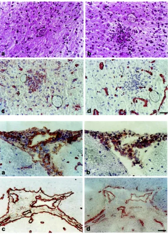
in-filtrates (Figure 1a) were frequently observed, although inflammatory infiltrates associated with cysts and T. gondii-free inflammatory infiltrates were also detected (Figure 1b). As previously reported, also in experimental chagasic infection the inflammatory infil-trates were frequently not related to the pres-ence of T. cruzi-antigen (Silva AA, Marino APM, SantosPVA, Roffê E, Quirico-Santos T, Paiva CN and Lannes-VieiraJ, unpub-lished data). In contrast to T. cruzi infection which presents a self-resolving acute en-cephalitis, the inflammatory infiltrates were more intense during the chronic phase of T. gondii infection (Table 1).
As a first attempt to determine which molecules are involved in the migration of inflammatory cells into the CNS during para-sitic infections, we performed immunohisto-chemical assays for the detection of ECM components, namely FN and LN. There was an increased expression of FN and LN in the CNS of both acute and chronically T. gondii -and T. cruzi-infected mice when compared to the pattern found in uninfected controls (Table 1). The increased FN expression was detected mainly in the meninges, leptome-ninges, cerebellum and basal lamina of blood
vessels in both infections (data not shown). Also, a fine FN filamentous network involv-ing T. gondii-free and T. gondii-containing inflammatory infiltrates was frequently ob-served in the CNS parenchyma (Figure 1c). Although an increased expression of LN was found in the basal lamina of blood vessels, choroid plexus, meninges and leptomenin-ges of acute and chronically T. gondii- and T. cruzi-infected mice, the expression of this molecule was not associated with the pres-ence of inflammatory infiltrates (Figure 1d). Immunohistochemical assays were also applied to investigate the expression of ECM receptors in the CNS. The blood vessels present in the brain tissue of acutely T.
gondii-and T. cruzi-infected mice showed intense expression of a4 and a5 molecules. Also, perivascular spaces presenting a FN-con-taining filamentous network filled with a 4-(Figure 2a,b)and a5-bearing cells were de-tected during acute infection. Although the expression of the LN and a6 molecules was detected in the endothelial layer of blood vessels, the CNS infiltrating mononuclear cells were a6-negative (Figure 2c,d). In T. gondii-infected mice the intense alterations in expression of a4and a5 molecules ob-served during acute infection persisted dur-ing the chronic phase. In contrast, durdur-ing chronic T. cruzi infection a slightly increased expression of a4and a5 molecules restricted to the endothelial layer of blood vessels was detected. These data are summarized in Table 1.
D iscussio n
In the present study we investigated the pattern of ECM components and their recep-tors in the central nervous system during parasitic infections. We observed an in-creased expression of FN and LN during the acute and chronic stages of experimental T. gondii and T. cruzi infection similar to that observed in the CNS in other inflammatory processes, such as tumors and autoimmune Table 1 - The presence of parasite antigens and inflammatory infiltrates, and the
expression of ECM components and their receptors in the CNS of T. gondii- and T. cruzi-infected mice.
The occurrence/intensity of the analyzed parameters w as scored as follow s: +++, many; ++, moderate amounts; +, few ; ±, very few ; -, negative staining. NT, Not tested; FN, fibronectin; LN, laminin.
Analyzed parameter Experimental groups
T. gondii T. cruzi
Uninfected Acute Chronic Uninfected Acute Chronic
T. gondii cysts - - ++ NT NT NT
T. cruzi antigen NT NT NT - +
-Inflammatory infiltrates - ++ +++ - +++ ±
FN + ++ ++ + ++ ++
a4 - ++ + ± + ±
a5 - ++ + ± + ±
LN ++ +++ +++ ++ +++ +++
diseases (14-16). During the acute phase of both T. gondii and T. cruzi infection, the appearance of a fine FN-containing network in the enlarged perivascular spaces coin-cided with the formation of mononuclear inflammatory infiltrates, suggesting the
par-ticipation of this ECM glycoprotein in the migration of inflammatory cells into the CNS during these infections. In multiple sclerosis and experimental allergic encephalomyelitis the altered expression of FN in the CNS was paralleled by the formation of inflammatory
Figure 1 - Isolated T. gondii cysts (a) or parasite cysts surrounded by inflammatory infiltrates (b) w ere observed in the CNS of chronically infected mice. Immu-nohistochemical assays revealed the expression of a fine FN fila-mentous netw ork involving mon-onuclear inflammatory cells (c). LN expression, how ever, w as restricted to blood vessels (d). T. gondii cysts are indicated by the dotted line. M agnification: 400X.
perivascular cuffs (15,16). These findings led to the proposal that ECM components participate in the establishment of chronic inflammation during autoimmune processes. Interestingly, the presence of a fine FN filamentous network was observed involv-ing T. gondii-free and T. gondii-containing inflammatory infiltrates. On the other hand, LN was not detected by our immunohisto-chemical assays in these inflammatory infil-trates, suggesting a more effective involve-ment of FN in the genesis of CNS inflamma-tory lesions during T. gondii infection. Con-cerning the origin of this FN, it is possible that nervous or mononuclear cells present in the inflammatory infiltrates could release ECM molecules in nervous tissue (25-27). Indeed, some studies have demonstrated that motor neurons, glial cells and astrocytes are able to secrete ECM molecules such as FN and LN when neuronal-glial interaction oc-curs and during the astrocyte proliferative response following injury (25,26).
The modulators and the mechanisms lead-ing to the alterations in ECM expression in the CNS during parasitic infections are un-known. We may speculate that corticoids or cytokines such as IFN-g and TNF-a, which have been shown to modulate ECM expres-sion (24,28,29) and to be systemically en-hanced during Chagas infection (30-32) and toxoplasmosis (11,33,34), could modulate ECM expression in the CNS during parasitic diseases besides contributing to damage to the blood-brain barrier. During the chronic stage of T. cruzi infection the CNS inflam-mation does not persist, although an increased expression of ECM was still observed in the basal lamina of blood vessels. It is possible that the soluble molecules such as cytokines produced systemically or locally by the few inflammatory cells restricted to areas of in-complete blood-brain barrier could account for the alterations in ECM expression. Also, the altered ECM expressed in the CNS may contribute to the generation/perpetuation of the observed encephalitis, stabilizing and
presenting cytokines to inflammatory cells (35,36).
ECM molecules play an important role in the host cell-parasite interaction (37,38). In this study we did not observe an association between the presence of parasite cysts or antigens and FN or LN expression in the infected tissues. However, in vitro studies showed that L929 fibroblasts and C2C12 muscle cells infected with T. cruzi or treated with shed parasite antigens present an in-creased expression of ECM proteins (Pinho RT and Lannes-Vieira J, unpublished re-sults). Thus, parasite antigens present in CNS lesions as a result of antigen shedding or parasite clearance by immune cells could induce inflammatory and/or nervous cells to produce ECM molecules. Further studies are required to examine this possibility.
Regarding the expression of ECM recep-tors, we showed that the enhancement of FN expression is accompanied by an increased expression of a4 and a5 and by the appear-ance of a4- and a5-bearing inflammatory cells during the acute phase of T. cruzi infec-tion and during acute and chronic Toxo-plasma encephalitis. Conversely, the in-creased expression of LN detected in the basal lamina of blood vessels was not paral-leled by the presence of a6 on the surface of infiltrating mononuclear cells. The presence of a4+ and a5+ mononuclear cells inside a FN-containing network suggests the partici-pation of these molecules in the migration of immune cells into the CNS leading to the development of inflammatory processes dur-ing parasitic diseases. In fact, this possibility is supported by the findings that antibodies recognizing the VLA-4 molecule, but not antibodies against other adhesion molecules, effectively prevented the accumulation of leukocytes in the CNS and the development of encephalomyelitis in Lewis rats. Also, in
Re fe re nce s
1. Pittella JEH (1993). Central nervous sys-tem in Chagas’ disease. An updating.
Revista do Instituto de M edicina Tropical, 35: 111-116.
2. Rocha A, M eneses ACO, Silva AM , Ferreira M S, Nishioca SA, Burgarelli M KN, Almeida E, Turcato-Jr G, M etze K & Lopes ER (1994). Pathology of patients w ith Chagas’ disease and acquired immunode-ficiency syndrome. American Journal of Tropical M edicine and Hygiene, 50: 261-268.
3. Prata A (1994). Chagas’ disease. Infec-tious Disease Clinics of North America, 8: 61-76.
4. Pentreath VW (1995). Trypanosomiasis and the nervous system. Pathology and immunology. Transactions of the Royal Society of Tropical M edicine and Hygiene, 89: 9-15.
5. Torres M & Villaça J (1919). Encefalite e mielite cauzadas por um Tripanozoma (T. cruzi). M emórias do Instituto Osw aldo Cruz, 11: 80-89.
6. Pittella JEH, M eneguette C, Barbosa AJA & Bambirra EA (1990). Histopathological and immunohistochemical study of the brain in the acute and chronic phases of experimental trypanosomiasis cruzi in dogs. Annals of Tropical M edicine and
Parasitology, 84: 615-621.
7. Pittella JEH (1991). Central nervous sys-tem involvement in experimental trypan-osomiasis cruzi. M emórias do Instituto Osw aldo Cruz, 86: 141-145.
8. Gazzinelli RT, Denkers EY & Sher A (1993). Host resistance to Toxoplasma gondii: model for studying the selective induction of cell-mediated immunity by intracellular parasites. Infectious Agents and Disease, 2: 139-149.
9. Hunt er CA & Rem ingt on JS (1994). Immunopathogenesis of toxoplasmic en-cephalitis. Journal of Infectious Diseases, 170: 1057-1067.
10. Schlüt er D, Deckert -Schlüt er M , Schw endemann G, Brunner H & Hof H (1993). Expression of major histocompat-ibility complex class II antigens and levels of interferon-g, tumour necrosis factor, and interleukin-6 in cerebrospinal fluid and serum in Toxoplasm a gondii-inf ect ed SCID and im m unocom pet ent C.B-17 mice. Immunology, 78: 430-435. 11. Gazzinelli RT, Brézin A, Li Q, Nussenblatt
RB & Chan CC (1994). Toxoplasma gondii: Acquired ocular toxoplasmosis in the mu-rine model, protective role of TNF-a and IFN-g. Experimental Parasitology, 78: 217-229.
12. Venstrom KA & Reichardt LF (1993). Ex-tracellular matrix II: Role of exEx-tracellular matrix molecules and their receptors in the nervous system. FASEB Journal, 7: 996-1003.
13. Gilat D, Cahalon L, Hershkoviz R & Lider O (1996). Interplay of T cells and cyto-kines in the context of enzymatically modi-fied extracellular matrix. Immunology To-day, 17: 16-20.
14. Pilkington GJ (1996). The role of the extra-cellular matrix in neoplastic glial invasion of the nervous system. Brazilian Journal of M edical and Biological Research, 29: 1159-1172.
15. Sobel RA & M itchell M E (1989). Fibronec-tin in multiple sclerosis lesions. American Journal of Pathology, 135: 161-169. 16. Shin T, Kojima T, Tanuma N, Ishihara Y &
M atsumoto Y (1995). The subarachnoid space as a site for precursor T cell prolif-eration and effector cell selection in ex-perimental autoimmune encephalomyeli-tis. Journal of Neuroimmunology, 56: 171-177.
17. Yednock TA, Cannon C, Fritz LC, Sanchez-M adrid F, Steinman L & Karin N (1992). Prevention of experimental autoimmune encephalomyelitis by antibodies against a4ß1 integrin. Nature, 356: 63-66. encephalomyelitis that after transmigration
throughout endothelial layer and migration into the brain parenchyma T cells lose or down-regulate the expression of VLA-4 (39). On this basis, we cannot exclude the possi-bility that the a6 molecule could play a role in the migration of inflammatory cells into the CNS, being lost or down-regulated after transmigration. This possibility is supported by the demonstration that LN and VLA-6 play an important role in the entrance of CD4+ cells obtained from chronically T. cruzi-infected mice into cardiac tissue (21). However, we should also consider the possi-bility that the inflammatory processes pres-ent in the CNS and myocardium of T. cruzi -infected mice have different origins, with different specificity and functional activity of the cell populations involved. Since the encephalitis is a self-resolving process and
the myocarditis is a chronic persisting in-flammation, then differences in the ECM receptor status of these cell populations are expected.
Taken together, our results suggest that ECM components and their receptors play a role in the extravasation and migration of inflammatory cells into the CNS during ex-perimental parasitic infections. The factors responsible for the entry and retention of lymphocytes within the CNS during para-sitic infections including cytokines and che-mokines are currently under investigation in our laboratory.
Ackno wle dgm e nts
18. Baron JL, M adri JA, Ruddle NH, Hashim G & Janew ay Jr CA (1993). Surface ex-pression of a4 integrin by CD4 T cells is required for their entry into brain paren-chyma. Journal of Experimental M edicine, 177: 57-68.
19. Truyens C, Rivera M T, Ouaissi A & Carlier Y (1995). High circulating levels of fibro-nectin and antibodies against its RGD ad-hesion site during mouse Trypanosoma cruzi infection: relation to survival. Experi-mental Parasitology, 80: 499-506. 20. Andrade SG, Grim aud JA &
Stocker-Guerret S (1989). Sequential changes of the connective matrix components of the myocardium (fibronectin and laminin) and evolution of cardiac fibrosis in mice in-fected w ith Trypanosoma cruzi. American Journal of Tropical M edicine and Hygiene, 40: 252-260.
21. Silva-Barbosa SD, Cotta-de-Almeida V, Riederer I, De M eis J, Dardenne M , Bonomo A & Savino W (1997). Involve-ment of laminin and its receptor in abro-gation of heart graft rejection by autoreac-tive T cells from Trypanosoma cruzi -in-fected mice. Journal of Immunology, 159: 997-1003.
22. Andrade SG (1974). Caracterização de cepas do Trypanosoma cruzi isoladas no recôncavo baiano. Revista de Patologia Tropical, 3: 65-121.
23. Brener Z (1962). Therapeutic activity and criterion of cure on mice experimentally infected w ith Trypanosoma cruzi. Revista do Instituto de M edicina Tropical, 4: 389-394.
24. Lannes-Vieira J, Van Der M eide PH & Savino W (1991). Extracellular matrix com-ponents of the mouse thymic microenvi-ronment. II. In vitro modulation of base-ment membrane proteins by interferon-g: relationship w ith thymic epithelial cell
pro-liferation. Cellular Immunology, 137: 329-340.
25. M asuda-Nakagaw a LM , M uller KJ & Nicholls JG (1993). Axonal sprouting and laminin appearance after destruction of glial sheaths. Proceedings of the National Academy of Sciences, USA, 90: 4966-4970.
26. Niquet J (1994). Proliferative astrocytes may express fibronectin-like protein in the hippocampus of epileptic rats. Neurosci-ence Letters, 180: 13-16.
27. Alito K, Hovi T & Vaheri A (1980). Fibro-nectin is produced by human macrophag-es. Journal of Experimental M edicine, 151: 602-613.
28. Lannes-Vieira J, Dardenne M & Savino W (1991). Extracellular matrix components of the mouse thymic microenvironment ontogenic studies and modulation by glu-cocorticoid hormones. Journal of His-tochemistry and CyHis-tochemistry, 39: 1539-1546.
29. Fabry Z, Raine CS & Hart M N (1994). Ner-vous tissue as an immune compartment: the dialect of the immune response in the CNS. Immunology Today, 15: 218-223. 30. Leit e-de-M oraes M C, Hont
ebeyrie-Joskow icz M , Leboulenger F, Savino W, Dardenne M & Lepault F (1991). Studies on the thymus in Chagas’ disease II. Thy-mocyte subset fluctuations in Trypanoso-ma cruzi-infected mice: Relationship to stress. Scandinavian Journal of Immunol-ogy, 33: 267-275.
31. Silva JS, M orrissey PJ, Grabstein KH, M ohler KM , Anderson D & Reed SG (1992). Interleukin 10 and interferon g regulation of experimental Trypanosoma cruzi infection. Journal of Experimental M edicine, 175: 169-174.
32. Starobinas N, Russo M , M inoprio P & Hontebeyrie-Joskow icz M (1991). Is TNFa
involved in early susceptibility of Trypano-soma cruzi C3H/He mice? Research in Immunology, 142: 117-122.
33. Chang HR, Grau GE & Pechere JC (1990). Role of TNF and IL-1 in infection w ith
Toxoplasma gondii. Immunology, 69: 33-37.
34. Langermanns JAM , van der Hust M EB, Nibbering PH, Hiemstra PS, Transen L & van Furth R (1992). IFN-g induced L-argi-nine dependent toxoplasmastatic activity in murine peritoneal macrophages is me-diated by endogenous tumour necrosis factor-a. Journal of Immunology, 148: 568-574.
35. Alon A, Cahalon L, Hershkoviz R, Elboz D, Reizis B, Wallach D, Akiyama SK, Yamada KM & Lider O (1994). TNF-a binds to the N-terminal domain of fibronectin and aug-ments the ß1-integrin-mediated adhesion of CD4+ T lymphocytes to the glycopro-tein. Journal of Immunology, 152: 1304-1322.
36. Hershkoviz R, Goldkorn I & Lider O (1995). Tumour necrosis factor-a interacts w ith laminin and functions as a pro-adhesive cytokine. Immunology, 85: 125-130. 37. Ouaissi M A (1988). Role of the RGD
se-quence in parasite adhesion to host cells.
Parasitology Today, 4: 169-173. 38. Vannier-Santos M A, Saraiva EM B, M artiny
A, Neves A & De Souza W (1992). Fibro-nectin shedding by Leishmania may influ-ence the parasite-macrophage interaction.
European Journal of Cell Biology, 59: 389-397.