Rogério José Tavares Ribeiro
HEPATIC PARASYMPATHETIC NERVE
DYSFUNCTION INVOLVED IN THE
DEREGULATION OF POSTPRANDIAL
WHOLE-BODY INSULIN SENSITIVITY:
A ROAD TO DIABETES
AND ASSOCIATED PATHOLOGIES
Faculdade de Ciências Médicas
Universidade Nova de Lisboa
Thesis submitted for the degree of Doctor of Philosophy (PhD)
on Life Sciences (Physiology), at the Faculdade de Ciências
“The most exciting phrase to hear in Science, the one that heralds new discoveries, is not ‘Eureka!’ but ‘That’s funny…’.”
The original results and conclusions presented in this thesis have been partially published in the following peer-reviewed papers:
Rogério T. Ribeiro, Ricardo A. Afonso, Maria P. Guarino, M. Paula Macedo (2008) “Loss of post-prandial insulin sensitization during aging”. J Gerontology A Biol Sci Med Sci 63(6): 560-565.
Rogério T. Ribeiro, Ricardo A. Afonso, M. Paula Macedo (2007) “Hepatic parasympathetic role in insulin resistance on an animal model of hypertension”. Metabolism 56: 227-233.
Ricardo A. Afonso, Rogério T. Ribeiro, M. Paula Macedo (2007) “Hepatic-dependent and –independent insulin action are impaired in the obese Zucker rat model”. Obesity. 15(2): 314-321.
Ricardo A. Afonso, Waine W. Lautt, Rogério T. Ribeiro, Douglas J. Legare, M. Paula Macedo (2007). “Insulin resistance in two models of obesity: A comparison of HISS-dependent and –inHISS-dependent insulin action in high-fat diet-fed and Zucker rats”. Proc.West. Pharmacol. Soc., 50: 110-114.
Rogério T. Ribeiro, Mark S. d’Almeida, Wayne W. Lautt, M. Paula Macedo (2005) “Insulin resistance induced by sucrose-feeding is due to an impairment of the hepatic parasympathetic nerves”. Diabetologia, 48: 976-983.
Ricardo A. Afonso, Rogério T. Ribeiro, M. Paula Macedo (2004). “Defective Hepatic Nitric Oxide Action Results in HISS-dependent Insulin Resistance in Spontaneously Hypertensive Rats”. Proc.West. Pharmacol. Soc., 47: 103-104.
Hepatic Insulin Sensitizing Substance in the Spontaneously Hypertensive Rat”. Proc. West. Pharmacol. Soc. 44: 27-28.
Rogério T. Ribeiro, Filipa Duarte-Ramos, M. Paula Macedo (2001) “The Fatty Zucker rat fa/fa shows a dysfunction of the HISS-dependent and –independent components of insulin action”. Proc. West. Pharmacol. Soc. 44: 29-30.
Dedico um agradecimento especial à Prof. Doutora Maria Paula Macedo. Muito além de ser a minha orientadora de doutoramento, soube nos momentos certos contagiar-me com a sua paixão pela Ciência; dando-me igualmente a oportunidade de participar na sua grande aventura, que é descobrir os elusivos caminhos da HISS.
Agradeço ao Prof. Doutor Pedro Freire Costa, com quem dei os primeiros passos na Faculdade de Ciências Médicas, sem desconfiar que as Gaiolas de Faraday que comecei por montar eram um bom prenúncio de que iria ficar “preso” à instituição durante vários anos.
Ao Prof. Doutor António Bensabat Rendas, agradeço o interesse, constante atenção e apoio com que acompanhou os trabalhos de investigação científica em que estive integrado.
Às Prof. Doutoras Maria Alexandra Ribeiro e Ana Isabel Santos; pela orientação e paciência nos meus primórdios científicos, e pela amizade que acredito irá sempre ser mantida.
To Prof. Wayne Lautt, from the Department of Pharmacology and Therapeutics, Faculty of Medicine, University of Manitoba, Canada, for his role in supporting my scientific evolution, and for the intellectually-rewarding get-togethers.
À Prof. Doutora Paula Videira, pela simpatia e pela confiança depositada ao me permitir a utilização do seu laboratório em horas impróprias. Também por ter sido a primeira a descortinar “nas estrelas” o Destino que me estava reservado.
Aos Profs. Drs. Manuel Ortigueira e Raul Rato, do Departamento de Engenharia Electrotécnica, da Faculdade de Ciências e Tecnologia da Universidade Nova de Lisboa, pelo crash-course em processamento de sinais e pela hospitalidade demonstrada
conselhos relacionados a alguns aspectos desta tese.
À FCT - Fundação para a Ciência e Tecnologia, e à APDP – Associação Protectora dos Diabéticos de Portugal, por apoiarem os projectos no âmbito dos quais este trabalho foi realizado.
À SPEDM – Sociedade Portuguesa de Endocrinologia, Diabetes e Metabolismo, e à SPD – Sociedade Portuguesa de Diabetologia, pela honra que me concederam ao premiarem trabalhos e artigos publicados durante a persecução desta tese de doutoramento.
Gostaria de agradecer aos colegas que comigo partilham, ou partilharam, laboratórios:
Ao Ricardo Afonso, com quem trabalhei em maior proximidade. Termos muitas vezes pontos de vista diferentes sobre o trabalho científico, e sobre a vida, nunca impediu que soubéssemos facilmente dialogar e chegar a conclusões produtivas.
À Ana Fernandes, uma força da natureza. O seu empenho na vida científica é uma inspiração, e a sua frontalidade de “Mulher do Norte” é um bálsamo.
À Rita Patarrão, uma presença sempre bem-disposta e opinativa.
À Jackie, pelo companheirismo que punha em ebulição o laboratório de Electrofisiologia.
Ao Pedro, pelo saber enciclopédico que gerava conversas sempre enriquecedoras, e pela amizade.
Fisiopatologia têm contribuído para que o meu trabalho decorra da melhor maneira: Sofia Ramos, Dra. Mafalda Nascimento, Mariana Lavajo, Gracinda Menezes e Isabel Ribeiro da Silva.
Ainda na Faculdade de Ciências Médicas, um cumprimento especial para o Hélio, Zélia, Sílvia, Inês, Sofia e Joana.
Por detrás da vida científica, existem aqueles que escoram a nossa dimensão humana:
À Lupe, por tudo. Pelo apoio inigualável durante a escrita desta dissertação; pelo que vivemos nestes últimos meses, emoções espalhadas por três continentes; pelos planos que ambicionamos concretizar juntos; pelo amor e pela compreensão. Eu avisei, por tudo!
Aos meus pais, uma constante fonte de apoio. Vejo a sua crença inabalável em mim como um motivo de orgulho; apesar de temer nem sempre conseguir lidar com ela da melhor maneira.
À Tia Isabel, a quem nunca consegui chamar “madrinha”, por ter, a par com a minha mãe, alimentado a curiosidade científica de um certo petiz. Ao Tio Zé, ao Jorge e à Ana, pela família que são e pelo carinho.
Aos restantes familiares, por me terem ensinado nos momentos mais difíceis que estão cá por mim. Espero conseguir mostrar-lhes o mesmo.
Lists of Figures and Tables ... v
Abbreviations ... ix
ABSTRACT...xiii
RESUMO... xv
1. INTRODUCTION... 1
1.1.Thesis rationale... 1
1.2.Diabetes as a modern epidemic... 2
1.3.Concept, classification and diagnosis of diabetes and prediabetes... 3
1.4.Clinical relevance of the metabolic syndrome, type 2 diabetes, and insulin resistance... 9
1.5.Tracking the influence of the prandial state to prediabetes and diabetes.... 13
1.6.The need for glucose homeostasis... 17
1.7.Hormonal and nervous control in the fed/fasting/fed transitions... 22
1.8.Glucose transport and intracellular signalling... 30
1.9.Hepatic modulation of postprandial peripheral insulin sensitivity... 41
1.10.Disposition of a meal and glucose metabolism... 48
2. OBJECTIVES... 55
3. MATERIAL AND METHODS... 57
3.1. Animals... 57
3.2. Experimental design... 58
3.2.1. Studies related to aging... 58
3.2.2. Studies related to gender ... 58
3.2.3. Studies related to high-sugar content diets ... 58
3.2.3.1. Effect of a liquid high-sucrose diet in Wistar rats ... 59
3.2.3.2. Effect of the duration of exposure to a liquid high-sucrose diet in Sprague-Dawley (SD) rats ... 59
3.2.3.3. Effect of a liquid vs solid high-sucrose diet in SD rats... 59
3.2.4. Studies related to hypertension... 59
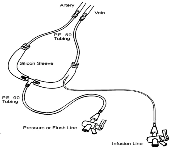
3.3. Pre-surgical protocol... 60
3.4. Surgical protocol... 60
3.5. Insulin sensitivity evaluation... 63
3.1.5.1. Glucose Analyser... 63
3.5.2. Determining a baseline glycaemia... 64
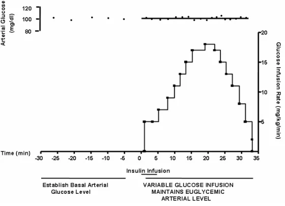
3.5.3. Rapid Insulin Sensitivity Test (RIST) ... 64
3.7. Data analysis... 68
3.8. Drugs and reagents... 69
4. RESULTS AND DISCUSSION... 71
4.1. Control of postprandial insulin sensitivity by the hepatic parasympathetic nerves in Wistar rats, and integration in the hepatic insulin sensitising substance (HISS) pathway... 71
4.1.1. Context and work hypothesis ... 71
4.1.2. Experimental protocols ... 72
4.1.3. Results... 73
4.1.3.1. HISS blockade by surgical manipulation... 73
4.1.3.2. HISS blockade by pharmacological manipulation ... 77
4.1.4. Discussion ... 79
4.2 Effect of age on postprandial insulin sensitisation... 85
4.2.1. General context and hypothesis... 85
4.2.2. Total insulin sensitivity in fasted versus fed male Wistar rats at 9 and 52 weeks of age ... 86
4.2.2.1 Experimental protocols ... 86
4.2.2.2. Results... 86
4.2.2.3. Discussion ... 87
4.2.3. HPN-dependent and HPN-independent components of insulin sensitivity in fed male Wistar rats at 6, 9, 16 and 52 weeks of age ... 88
4.2.3.1. Experimental protocols ... 88
4.2.3.2. Results... 89
4.2.3.3. Discussion ... 94
4.3. Gender comparison of postprandial insulin sensitivity with fed female Wistar rats at 9, 16 and 52 weeks of age. Further studies in 78 weeks old male and female Wistar rats.... 103
4.3.1. Context and work hypothesis ... 103
4.3.2. Experimental protocols ... 104
4.3.3. Results... 104
4.3.4. Discussion ... 112
4.4 Alterations in postprandial insulin sensitivity by high-sugar diets... 114
4.4.1. Context and general work hypothesis... 114
4.4.2 Effect of a high-sucrose diet on Wistar and SD rats ... 115
4.4.2.1. Experimental protocols ... 115
4.4.2.2. Results... 116
4.4.2.3. Discussion ... 122
4.4.3. Effect of the duration of exposure to a liquid high-sucrose diet in SD rats 128 4.4.3.1. Experimental protocols ... 128
4.4.3.2. Results... 128
4.4.4.1. Experimental protocols ... 131
4.4.4.2. Results... 131
4.4.4.3. Discussion ... 133
4.5. HISS mechanism status and role on insulin sensitivity in an animal model of essential hypertension (SHR)... 134
4.5.1. The hepatic parasympathetic nerves / hepatic nitric oxide branch of the HISS pathway in hypertension... 134
4.5.1.1. General context and work hypothesis ... 134
4.5.1.2. Experimental protocols ... 136
4.5.1.3. Results... 136
4.5.1.3.1. Assessment of the hypertensive condition ... 136
4.5.1.3.2. Other metabolic parameters... 136
4.5.1.3.3. Insulin sensitivity before and after parasympathetic blockade in hypertension ... 137
4.5.1.3.3. Role of hepatic nitric oxide on insulin sensitivity in essential hypertension ... 140
4.5.1.4. Discussion ... 141
4.5.2. The hepatic glutathione branch of the HISS pathway in essential hypertension... 148
4.5.2.1. General context and work hypothesis ... 148
4.5.2.2. Experimental protocols ... 149
4.5.2.3. Results... 149
4.5.2.4. Discussion ... 150
4.5.3. Postprandial increment of insulin sensitivity in SHR... 152
4.5.3.1. Context and work hypothesis ... 152
4.5.3.2. Experimental protocols ... 152
4.5.3.3. Results... 152
4.5.3.4. Discussion ... 153
4.6. Changes in insulin sensitivity in an animal model of essential hypertension determined by age... 154
4.6.1. General context and work hypothesis... 154
4.6.2. Experimental protocols ... 154
4.6.3. Results... 155
4.6.4. Discussion ... 156
5. OVERALL DISCUSSION... 159
5.1. Methodological considerations in the assessment of whole-body insulin sensitivity, and critical relevance of the prandial status... 159
5.2. Modulation of insulin sensitivity by gender and developmental stage... 164
5.5. The HISS pathway as a new paradigm in the cluster of hepatic disease,
autonomic dysfunction, insulin resistance, and type 2 diabetes... 171
5.6. Conclusion and implications... 175
LIST OF FIGURES
Figure 1- Current criteria to diagnose diabetes and prediabetes. ... 5
Figure 2 - Pathways of nutrient metabolism crosstalk... 19
Figure 3 - Autonomic nervous control of glucose homeostasis through central integration... 28
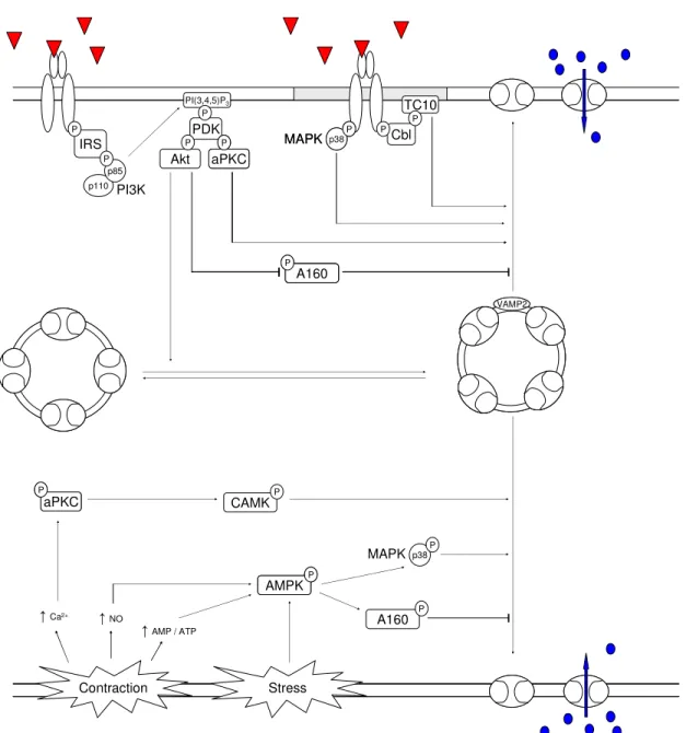
Figure 4 - Induction of GLUT4 translocation. ... 34
Figure 5 - Proposed hypothesis for the postprandial regulation of peripheral glucose uptake. ... 42
Figure 6 - Extracorporal arterial-venous circuit. ... 61
Figure 7 - Example of Rapid Insulin Sensitivity Test (RIST)... 65
Figure 8 – High-pressure liquid chromatography (HPLC) output example. ... 68
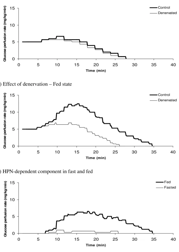
Figure 9 – Effect of denervation on RIST profiles of fasted and fed Wistar rats. ... 74
Figure 10 – Effect of denervation on RIST indexes of fasted and fed Wistar rats. ... 76
Figure 11 – Effect of pharmacological intervention on fed Wistar rats. ... 78
Figure 12 – Effect of age on RIST indexes of fasted and fed Wistar rats... 87
Figure 13 – Effect of age on RIST profiles of fed male Wistar rats... 90
Figure 14 – Effect of age on RIST indexes of fed male Wistar rats: HPN-dependent and –independent components. ... 93
Figure 15 – Effect of age on RIST profiles of fed female Wistar rats... 105
Figure 16 – Effect of age on RIST indexes of fed female Wistar rats: HPN-dependent and –independent components... 108
Figure 17 – RIST profiles of fed male and female Wistar rats at old age. ... 110
Figure 18 –RIST indexes of fed male and female Wistar rats at 78 weeks of age: HPN-dependent and –inHPN-dependent components... 112
Figure 21 – RIST indexes of standard and high-sucrose fed Sprague-Dawley rats. .... 121
Figure 22 – Effect of exposure time to high-sucrose diet in Sprague-Dawley rats... 129
Figure 23 – Effect of delivery form of high-sucrose diet in Sprague-Dawley rats. ... 132
Figure 24 – Effect of hypertension on postprandial glucose metabolism in comparison to two normotensive controls; before and after muscarinic antagonism. ... 137
Figure 25 – RIST profiles of spontaneously hypertensive rats (SHR), and their
normotensive controls Wistar and Wistar Kyoto; at 9 weeks of age... 138
Figure 26 – Effect of hypertension on postprandial glucose metabolism in comparison to two normotensive controls; before and after nitric oxide inhibition. ... 140
Figure 27 – Hepatic reduced and oxidised glutathione content in Wistar and
spontaneously hypertensive rats (SHR). ... 150
Figure 28 – Insulin sensitivity in the fasted and postprandial states, in an animal model of essential hypertension. ... 153
Figure 29 – Effect of maturation on spontaneously hypertensive rats (SHR) and
normotensive controls. ... 155
Figure 30 – Integrated view: Hepatic insulin sensitising substance (HISS) dysfunction and progression to type 2 diabetes... 176
LIST OF TABLES
Table I - Tissue distribution and function of facilitative glucose transporters... 32
Table II – Effect of denervation on dynamic curve main properties of fed and fasted Wistar rats... 75
Table III – Effect of age on dynamic curve main properties of fed male Wistar rats. ... 92
Table IV – Effect of age on dynamic curve main properties of fed female Wistar rats. ... 107
A160: Akt substrate of 160 kDa
Ach: acetylcholine
ACTH: adrenocorticotropic hormone
ADP: adenosine monophosphate
Akt: protein kinase B (also PKB)
APS: associated protein substrate
AMPK: 5’adenosine monophosphate-activated protein kinase
ANOVA: analysis of variance
aPKC: atypical protein kinase C
ATP: adenosine triphosphate
BMP-9: bone morphogenetic protein 9
CAMK: Ca2+/calmodulin protein kinase
cAMP: cyclic adenosine monophosphate
CAP: Cbl-associated protein
Cbl: Casitas b-lineage lymphoma protooncogene
CHB: vagus nerve common artery branch
C-peptide: connecting peptide
DNFB: 2,4-dinitro-1-fluorobenzene
ED50: effective dose
eNOS: endothelial nitric oxide synthase
ERK: extracellular signal-regulated kinase
GDP: guanosine diphosphate
GSH: reduced glutathione
GSH-Px: glutathione peroxidase
GSSG: oxidised glutathione
GSSG-R: oxidised glutathione redutase
GTP: guanosine triphosphate
GIP: glucose-dependent insulinotropic peptide
GKRP: glucokinase regulatory protein
GLP-1: glucagon-like peptide 1
HbA1c: glycosylated haemoglobin
HCl: hydrochloric acid
HIEC: hyperinsulinemic euglycaemic clamp
HISS: hepatic insulin sensitising substance
HNO: hepatic nitric oxide
HPLC: high-pressure chromatography
HPN: hepatic parasympathetic nerves
IDE: insulin degrading enzyme
IFG: impaired fasting glucose
IGF1: insulin-like growth factor 1
IGT: impaired glucose tolerance
iNOS: inducible nitric oxide synthase
ipv: intraportal
IR: insulin receptor
IRS: insulin receptor substrate
ITT: insulin tolerance test
iv: intravenous
KO: knock-out
L-NMMA: Ng-methyl-arginine
MAPK: p38 mitogen-activated protein kinase
mTOR: mammalian target of rapamycin protein
MTT: meal tolerance test
NEM: N-etilmaleimide
NO: nitric oxide
NOS: nitric oxide synthase
OGTT: oral glucose tolerance test
PDI: protein disulfide isomerase
PDK: 3-phosphoinositide-dependent kinase
PI(3,4,5)P3: phosphatidylinositol-3,4,5-triphosphate
PI(4,5)P2: phosphatidylinositol-4,5-biphosphate
PI3K: phosphoinositide-3-kinase
RSNO: S-nitrosothiols
SD: Sprague-Daley rat strain
SEM: standard error of the mean
SGLT: sodium-dependent glucose co-transporter
SH: thiol group
SHR: spontaneously hypertensive rat
SIN-1: 3-morpholinosydnonimine hydrochloride
SNARE: soluble SNF attachment protein
SNP: sodium nitroprusside
TCA: trichloroacetic acid
TRIS-HCl: Tris(hydroxymethyl)aminomethane hydrochloride
VAMP2: vesicle-associated membrane protein 2
WIS: Wistar rat strain
The postprandial state is characterised by an increment in whole-body insulin
sensitivity. This increased insulin-stimulated glucose disposal seen after a meal has been
proposed to be regulated by a mechanism to which the integrity of the hepatic parasympathetic
nerves (HPN) is crucial. The present thesis focus on the hypothesis that a dysfunction of the
HPN-dependent component of insulin sensitivity is involved in the insulin resistance seen in
various conditions related to the aetiology of type 2 diabetes.
The physiological impacts of age and gender were here studied in Wistar rats. In males,
age was correlated with a gradual decrease of total postprandial insulin sensitivity. However,
while the HPN-independent component of insulin action decreased early, between 6 and 9
weeks of age, and remained unchanged thereafter, the HPN-dependent component decreased
from 9 weeks of age throughout ageing. Females showed similar developmental changes,
although at different rates in some stages.
The repercussion of lifestyle on postprandial insulin sensitivity was evaluated by
providing Wistar and Sprague-Dawley rats with a high-sucrose supplement. The high-sucrose
diet induced a decrease in insulin sensitivity by affecting solely the HPN-dependent component.
Additionally, this development of postprandial insulin resistance was evident after only 2 weeks
of diet manipulation; prior to inducing weight gain and hyperglycaemia.
Studies in healthy animals argued in favour of the integration of the HPN on the hepatic
insulin sensitising substance (HISS) pathway. Blockade of the HPN through physical sectioning
or pharmacological muscarinic antagonism, or inhibition of the synthesis of hepatic nitric oxide,
another proposed step of the HISS pathway, were shown to similarly abrogate the postprandial
increment of insulin sensitivity; while having no effect on fasting insulin sensitivity.
The analysis of a rat model of essential hypertension (SHR) has shown an impairment
of the HISS-dependent component of insulin action, partially compensated by an increase of the
HISS-independent component; in relation to normotensive Wistar rats. That another
normotensive control strain for the SHR, the Wistar Kyoto, showed already a decreased in the
HISS-dependent component hints to a possible genetic interference on the HISS pathway;
particularly on parasympathetic function. Ageing studies further support these conclusions.
In summary, dysfunction of the HPN-determined branch of the HISS mechanism was
shown here to be involved in the development of insulin resistance related with ageing,
diet-induced metabolic deregulation, and hypertension. This was also shown to predate other risk
factors for the development of type 2 diabetes and related pathologies; and seems to constitute
an attractive target for behavioural and pharmacological interventions, especially those able to
O estado pós-prandial é caracterizado por um incremento da sensibilidade à insulina.
Este aumento da captação celular da glucose por acção da insulina, observado após a ingestão
de uma refeição, parece estar relacionado com um mecanismo dependente da integridade dos
nervos parassimpáticos hepáticos (HPN). A presente dissertação teve como base a hipótese de
que a disfunção desta componente dependente dos HPN está relacionada com a
insulino-resistência observada em várias condições envolvidas na etiologia da diabetes tipo 2.
O impacto da idade e do género foram aqui estudados em ratos Wistar. Em machos,
observou-se que a idade está relacionada com um decréscimo gradual da sensibilidade total à
insulina. No entanto, enquanto a componente independente dos HPN decresceu entre as 6 e 9
semanas de idade, mantendo-se depois inalterada, a componente dependente dos HPN decresceu
com o envelhecimento a partir das 9 semanas de idade. As fêmeas demonstraram alterações de
desenvolvimento semelhantes aos machos, apesar de algumas diferenças na taxa de decréscimo.
A influência do estilo de vida sobre a sensibilidade pós-prandial à insulina foi avaliada
fornecendo um suplemento de sacarose a ratos Wistar e Sprague-Dawley. A dieta rica em
sacarose induziu um decréscimo na sensibilidade à insulina, afectando apenas a componente
dependente dos HPN. Mais, o desenvolvimento de insulino-resistência pós-prandial foi obtido
após 2 semanas de manipulação; antes do surgimento de obesidade e hiperglicémia.
Estudos em animais saudáveis apoiam a integração dos HPN na via da substância
hepática sensibilizadora da insulina (HISS). O bloqueio dos HPN por intervenção cirúrgica ou
antagonismo muscarínico químico, assim como a inibição da síntese de óxido nítrico hepático,
outro passo proposto da via da HISS, provocaram a anulação total do incremento pós-prandial
de sensibilidade à insulina; não tendo tido, no entanto, qualquer efeito no estado de jejum.
A análise de um modelo animal de hipertensão essencial (SHR) revelou um decréscimo
da acção da insulina dependente da HISS, parcialmente compensada por um aumento da acção
da insulina independente da HISS; em relação aos ratos normotensos Wistar. O facto de outro
controlo normotenso dos SHR, o Wistar Kyoto, apresentar já uma diminuição da componente
dependente da HISS sugere uma possível determinação genética da via da HISS, provavelmente
actuando sobre a função parassimpática. Estudos com a idade fortalecem estas conclusões.
Em conclusão, a disfunção da componente dependente dos HPN na via da HISS foi aqui
demonstrada estar envolvida no desenvolvimento da insulino-resistência relacionada com o
envelhecimento, a desregulação induzida por factores nutricionais, e a hipertensão. Foi também
observado que essa disfunção antecipa outros factores de risco para o desenvolvimento de
diabetes tipo 2 e patologias relacionadas; o que parece constituir um alvo promissor para
1. INTRODUCTION
1.1. Thesis rationale
The basis of this doctoral thesis is related to the involvement of the hepatic
parasympathetic nerves (HPN) on the augmented whole-body insulin sensitivity seen in
the postprandial state; and the possible contribution of the disruption of HPN function to
the genesis of insulin resistance and type 2 diabetes.
Ingestion of a meal is the greatest challenge faced by glucose homeostasis. The
surge of nutrients has to be disposed off quickly, as their high concentrations in the
bloodstream may have pathophysiological effects; and properly, as misplaced reserves
may induce problems on the affected tissues. Thus, loss of the ability to adequately
dispose of ingested nutrients can be expected to lead to a deterioration of glucose
metabolism control, and favour the development of the aforementioned pathologies.
This dissertation regards studies designed to uncover the behaviour of metabolic
pathways determined by HPN function status, both in physiology and pathophysiology.
Likewise, the inquiry spread to address the impact of modern lifestyle, especially
feeding habits, on the HPN control of glucose metabolism. This was done bearing in
mind that the HPN may have a particular relevance during the postprandial state as a
determinant in the action of a hormone, the hepatic insulin sensitising substance (HISS),
which has been implicated in the peripheral allocation of the ingested glucose.
The term “HISS” is not yet a scientific household name. With it, as befits the scope
of a doctoral thesis, there are certainly many biological whys and hows to be addressed
in this “fairy tale”, as Marie Curie would call it1. For that, a case will be presented based both on the performed experiments and on the evidence found in other works, as to the
validity of this novel mechanism. Surely, when that is done, the scientific notions and
data provided throughout this work will have earned due consideration.
To begin with, the current dimension of diabetes will be alluded to, and the concepts
of diabetes and prediabetes will be clarified.
1 “I am among those who think that science has great beauty. A scientist in his laboratory is not only a
1.2. Diabetes as a modern epidemic
The Western World has seen in the last decades a sharp increase in the general
incidence of diabetes mellitus (Zimmet et al., 2001). More, the Developing Countries
have been witnessing ever more rapid rates of incidence and, similarly, an increasingly
lower age of disease onset (Tong and Cockram, 2003). This has made diabetes into one
of the most alarming worldwide health problems facing us presently, to the point of
being recently recognised by the United Nations as a full epidemic (United Nations
Resolution 61/225, 2007).
As if this scenario was not bad enough, it is expected to deteriorate further in the
near future. From the estimated 150 million people affected worldwide in 2000, the
incidence of diabetes is foreseen to rise to a total of 366 million people by 2030 (King et
al., 1998; Wild et al., 2004). Even worse if taken into account that these are
conservative estimates (Wild et al., 2004); the higher incidence of obesity and diabetes
in children, the rise in life expectancy enlarging the elderly strata of the population,
compound sedentary and stressful lifestyles, and urbanisation of developing countries,
can further exacerbate these extrapolations.
Diabetes mellitus has a considerable premature morbidity and mortality, especially
due to the accompanying panoply of complications and associated pathologies (Zimmet
et al., 2001). It is now considered to be globally the fifth leading cause of death (Roglic
et al., 2005). Additionally, it has a radical impact upon a growing portion of the world
population, in terms of life quality, and to society in a whole, in terms of an increased
expenditure of either private or public national systems of Health (Bruno et al., 2008;
Dall et al., 2009).
This is especially true for Portugal. With a number of documented cases of diabetes,
between the ages of 20 and 80, reaching around 0.5 million in 2003, predictions were
that the number of cases would rise by 2025 to 0.7 million (IDF, 2003; King et al.,
1998). Unfortunately, the recent disclosure of the preliminary report for the first
Portuguese epidemiologic study solely dedicated to diabetes (PREVADIAB-2009) has
surpassed the worst expectations (Pina e Brito, 2009). In it, it was found that more than
10% of individuals between ages 20 and 80 are already diabetic, which represents an
Portuguese (23%) were found to suffer from at least one kind of prediabetes2. Summarising, more than a third of the Portuguese population is either already diabetic,
or possesses a significant impairment of glucose metabolism that increases the risk of
developing diabetes in the near future. Considering that the rate of mortality from
diabetes in Portugal was more than double the mean in the European Union, and was
rising (WHO, 2003), this poses a dire prospect in terms of national Health.
Beyond finding ways to treat this hailing and financially cumbersome disease, it
becomes essential to understand the physiological mechanisms that are related to the
development of insulin resistance, the step that seems to predate and link diabetes and
its cluster of commonly associated pathologies (Gerich, 1999; Reaven, 1988).
To act in an early phase of this process would consequently provide the best
approach to a line of research capable of bearing answers into postponing the metabolic
degradation that eventually leads to type 2 diabetes. To contribute to this approach was
the main thought at the basis of this dissertation.
1.3. Concept, classification and diagnosis of diabetes and prediabetes
One of the oldest surviving accounts of the existence of diabetes is a basic
description recorded by Aretaus of Cappadocia in the 2nd century AD. Aretaus used the Greek word for ‘siphon’ to first coin the term ‘diabetes’, ‘because the fluid does not
remain in the body, but uses the man’s body as a channel whereby to leave it’
(Tattersall, 2003)3.
Diabetes mellitus is in our days more than simply a disease; it is now consensually
described as ‘a group of metabolic diseases characterised by hyperglycaemia resulting
from defects in insulin secretion, insulin action, or both. The chronic hyperglycaemia of
diabetes is associated with long-term damage, dysfunction, and failure of various
organs, especially the eyes, kidneys, nerves, heart, and blood vessels.’ (ADA, 1997).
2 The definition and characteristics of the prediabetes stage(s) will be further discussed in the next item. 3 This description is a clear allusion to the state of polyuria that sets later in the condition, often the
In our times, classification criteria aim mainly to reflect the aetiology and
pathogenesis of diabetes (ADA, 1997; Genuth et al., 2003). There are two main forms
of diabetes. Type 1 diabetes involves the autoimmune-mediated failure or destruction of
pancreatic –cell islets, eventually leading to absolute insulin deficiency (Dotta and
Eisenbarth, 1989; Muir et al., 1992). These patients, generally young people and
children (Taplin and Barker, 2008), rely on exogenous insulin for survival, and are
prone to ketoacidosis, coma and death. Type 2 diabetes is by far the most prevalent
form, representing 90% of diabetes cases. It is an heterogeneous and multifactorial
condition, probably determined by the crosstalk of several pathophysiological
mechanisms. Of both genetic and environmental origin (Beck-Nielsen and Groop,
1994), type 2 diabetes is characterized by defects in insulin action (insulin resistance)
and/or in insulin secretion ( -cell dysfunction) (Kashyap and Defronzo, 2007), either of
which may predominate (Gerich, 1999). Other forms of diabetes include gestational
diabetes mellitus, which is defined as any degree of inability to deal with a glucose load
(i.e. glucose intolerance) during pregnancy (independently of the onset of diabetes being
during or prior to that state), and several rarer forms induced by endocrinological, drug
or chemical, infectious, autoimmune, or genetic factors (ADA, 1997).
The epidemic of diabetes is thus more correctly referred to as an epidemic of type 2
diabetes. Indeed, even among young adults and children, where type 1 diabetes was the
most common chronic disease (Rosenbloom et al., 1999), the rising incidence of
physical inactivity, bad food habits, and obesity, is allowing a higher prevalence of type
2 diabetes (Cali and Caprio, 2008; Ten and Maclaren, 2004).
Diabetes is diagnosed when exaggerated blood glucose levels and/or abnormal
handling of a glucose load are detected (ADA, 2009). This diagnosis is based on
glycaemia magnitude in relation to established reference, or “cut-off”, values (Figure 1).
Hyperglycaemia is an important risk factor (Stratton et al., 2000); and the cut-off values
are chosen taking into account the prevalence thresholds for several diabetic
complications, especially retinopathy. From this stems that diagnosis can be made
already with or without the presence of known symptoms of diabetes4. With new developments in the clinical and research fields, the cut-off values have been
4 Common symptoms of already established diabetes include polyuria (excessive excretion of urine),
progressively adapted following current knowledge. It is presently considered that
diabetes is established if blood glucose level after an overnight fast is equal or over 126
mg/dl, and/or the blood glucose level obtained two-hours after an oral glucose tolerance
test (OGTT)5 is equal or over 200 mg/dl. Although the debate raged from early on about the best method for evaluating diabetes, fasting or 2h post-challenge plasma glucose,
mainly an effort was made to try to find cut-off points that represent similar biological
risk factors in the two approaches.
2h-post challenge blood glucose level (mg/dl)
< 140 140-199 200
< 100 Normal IGT
100-125 IFG IFG/IGT
Diabetes
(isolated
post-challenge
hyperglycaemia) Fasting
glucose level
(mg/dl) 126 Diabetes (isolated fasting
hyperglycaemia) Diabetes
Figure 1- Current criteria to diagnose diabetes and prediabetes. Glycaemic control evolves from
normal homeostasis to diabetes, with visible degradation either in the fasted state or in response to a glucose challenge, or both. Impaired glucose tolerance (IGT) and/or impaired fasting glucose (IFG) represent stages of prediabetes (adapted from (Tong and Cockram, 2003)).
A further distinction has been created, to describe an intermediate stage between
normal glucose homeostasis and diabetes; which has become known as ‘prediabetes’.
This stage can be characterized either by impaired fasting glucose (IFG) or impaired
glucose tolerance (IGT) (Figure 1). IFG is considered when the fasting glucose level is
higher than 100 mg/dl but lower than 126 mg/dl, and IGT is considered when blood
glucose assessed 2h after an OGTT is higher than 140 mg/dl but lower than 200 mg/dl
(Ryden et al., 2007). Less commonly, patients show both IFG and IGT. This last
condition presents a more extreme stage of prediabetes, sometimes able to develop
independently (Perreault et al., 2008), which usually progresses more rapidly towards
type 2 diabetes (de Vegt et al., 2001; Meigs et al., 2003; O'Rahilly et al., 1994).
5 Usually by ingestion of a glucose load containing 75g anhydrous glucose in water after an overnight
Prediabetes is estimated to affect presently almost 350 million people worldwide
(Garber et al., 2008). Its importance is highlighted by the increased medical costs
associated (Zhang et al., 2009), since a considerable proportion of prediabetics already
present microvascular and cardiovascular complications (Garber et al., 2008).
Some authors argue that, more than a risk factor for diabetes, prediabetes is in itself
a disease state that must be clinically addressed (Eldin et al., 2008). Others see the
constant lowering of cut-off points, especially those related to IFG, as a risk of losing
biological relevance and a danger of generalising treatments with possible side-effects
over groups that would never evolve type 2 diabetes or suffer from cardiovascular
complications (Rosenstock, 2007).
Anyway, IFG and IGT seem to be both related to the development of type 2 diabetes
through the presence of insulin resistance; even if possibly under slightly different
pathophysiological mechanisms (Meyer et al., 2006; Nathan et al., 2007; Perreault et al.,
2008; Weyer et al., 1999). IFG seems to be connected mainly to early defects in the
pancreas and liver; a loss of the first-phase of insulin secretion and a decreased capacity
of insulin to inhibit hepatic glucose output. Paradoxically, this seems to be accompanied
by normal skeletal muscle insulin sensitivity (Nathan et al., 2007). On the contrary, IGT
seems to only present hepatic insulin resistance at a later stage, or not at all, but
develops extensive peripheral insulin resistance by lack of ability to sensitise the
skeletal muscle6 (Crandall et al., 2008; Garber et al., 2008; Guerrero-Romero and Rodriguez-Moran, 2006; Stancakova et al., 2009). Eventually IGT may also show
alterations in both early- and late-stage insulin secretion (O'Rahilly et al., 1994).
Nonetheless, notwithstanding dynamic changes in insulin secretion, hyperinsulinemia is
usually a sustained feature in prediabetes (Weyer et al., 1999)7.
In real life these expressions of prediabetes show a considerable heterogeneity,
especially in individuals with isolated IFG (Kim and Reaven, 2008). This is not
surprising in terms of a natural history of development of type 2 diabetes. Traditionally,
6 Peripheral insulin resistance that leads to IGT development has been retraced even to the stage where
individuals are still within normal 2h-post challenge blood glucose levels (Stancakova et al, 2009).
7 The eventual failure of the ability of -cells to compensate for the rise on blood glycaemia by producing
type 2 diabetes has been seen as arising from early defects in peripheral insulin
sensitivity. The pancreas counterbalances the growing loss of tissue sensitivity by
secreting more insulin, leading to a maintained state of hyperinsulinemia. This process
continuously degrades, with both glycaemia and insulinemia rising, until -cell islets
reach a critical point and begin to fail. Other sites, as the liver and adipose tissue, begin
to also be affected. By then, usually diabetes sets with both full-blown hyperglycaemia
and possibly a decrease in insulinemia due to -cell exhaustion. Intuitive as this order of
events may seem, in reality these processes are so interrelated that evolution to type 2
diabetes may begin in any of those steps.
As in other biological processes, nature seldom finds only one way of doing and
undoing things, unlike what humans so often find comfortable to conceptualise.
However, it is true that some mechanisms may be more relevant than others. And in
pathophysiological issues this relevance may lie in those physiological pathways more
prone to be disrupted by environmental factors, since those are the ones that will
aggravate what can be the unavoidable disadvantages of the genetic background (Buren
and Eriksson, 2005). The natural history of progression from normal glucose
metabolism to type 2 diabetes, and logically the possibility of successful intervention to
delay it, may thus depend heavily on the correct identification of the aetiological defect.
It should also be critical to detect it at an early stage of the prediabetic range.
This rationale seems to be supported by epidemiological follow-up studies. In those
studies, people diagnosed with IFG and/or IGT are revaluated after several years of the
first diagnosis. Even taking into account all the circumstances that may induce
variability on the quantification given by the OGTT, which may (Ko et al., 1998;
Yudkin et al., 1990) or may not (de Vegt et al., 2001; Ko et al., 1998) make the OGTT a
lesser reproducible method than the measurements done on the fasting state (Balion et
al., 2007), reversion from IGT to normalcy seems to be more common than from IFG
(Lu et al., 2008). Considering that a first diagnosis of IGT or IFG is followed by an
increased awareness of each afflicted individual to the potential outcome of a
prediabetic condition, and that this may lead to immediate changes in lifestyle, this data
may hint to a higher sensitivity of IGT to environmental factors.
As mentioned before, IFG is diagnosed simply by assessing the blood glucose level
in the fasted state; and IGT is diagnosed through an OGTT. During an OGTT, after the
insulinaemia, are followed at regular intervals to assess the ability of the organism to
deal with the glucose challenge. Additional analytical quantifications and mathematical
models are able to provide more information on the dynamics of glucose
appearance/disappearance in the blood and the response of the pancreas in secreting
insulin and of the liver in insulin removal (Abdul-Ghani et al., 2007; Cobelli et al.,
2007).
The OGTT is usually reported as assessing glucose metabolism response in a
postprandial state, in opposition to the fasting state evaluation. However, this method
may not be the most suited for this purpose (Lefebvre and Luyckx, 1976). Indeed, the
comparison of the OGTT with a meal tolerance test (MTT)8 shows that a challenge of only glucose leads to a higher level of blood glycaemia and triggers insulin secretion in
a different dynamic curve than a challenge posed by a mixed meal (Berthiaume and
Zinker, 2002; Meier et al., 2008; Rijkelijkhuizen et al., 2009). This is explained by the
fact that glucose appearance in the blood is quicker when it comes from a diluted
solution than when the same quantity of glucose is part of a mixed meal, and by the
additive secretory response of the pancreas to nutrients other than carbohydrates (Bock
et al., 2007b), namely protein (Bock et al., 2007b; Krezowski et al., 1986). Additionally,
the arrival of solely glucose to the intestine was shown not to be enough to produce
some of the feeding signals involved in postprandial glucose homeostasis (Sadri et al.,
2006).
It becomes evident that a mixed meal challenge will constitute a more appropriate
test to fully assess the postprandial state. It would also be more accurate to evaluate
changes in real-life postprandial glucose metabolism, as has been already proposed
(Meier et al., 2008). Further, the previous considerations indicate that both clinically
advised measurements for evaluating diabetes and prediabetes, fasting and 2h
post-OGTT plasma glucose levels (ADA, 2009), may be unable to adequately detect the
impairment of real-life glucose metabolism responses in the postprandial state.
Now the question remains if those potential real-life alterations in postprandial
nutrient handling are relevant to the natural history of the development of type 2
diabetes; and if the additional effort in detecting them has any real clinical applicability
8 The MTT is initiated by the ingestion of a standardised mixed meal, but is in all the rest similar to the
(specifically, if they are able to predict development to type 2 diabetes; if they are
sensitive to strategies aimed at averting or postponing that development; and, in later
stages, if there is a potential for therapeutic pharmacological intervention).
However, before focusing on those points, the relevance of setting the cornerstone
of the present doctoral thesis on the study of alterations of insulin sensitivity can only be
ascertained after dealing with one other subject, envisioned today as one of the most
heated debates in the international field of diabetes clinical research: currently called
‘the metabolic syndrome’.
1.4.Clinical relevance of the metabolic syndrome, type 2 diabetes, and insulin
resistance
Type 2 diabetes is related to a multitude of complications, especially cardiovascular,
undoubtedly due to concomitant dysfunctions throughout several pathways involved in
energy management, as for example carbohydrate and lipid metabolism. Even
prediabetes can likewise be involved, since the risk of complications can already be
evident at this stage9 (Goldberg et al., 2009; Haffner et al., 1990). For this, prediabetes and type 2 diabetes can also be regarded as part of a broader picture.
In 1988, Gerald Reaven proposed (Reaven, 1988) that insulin resistance and
hyperinsulinemia are the main factors involved in the aetiology of conditions like type 2
diabetes, hypertension and coronary artery disease. This was termed “Syndrome X”. In
its initial proposition, Syndrome X was an informal cluster of risk factors that included
decreased sensitivity in insulin-stimulated glucose uptake, glucose intolerance,
hyperinsulinemia, dyslipidemia (increased very-low-density lipoprotein triglycerides
and decreased high-density lipoprotein cholesterol), and hypertension. Although it was
named “syndrome”, this was more a recognition of a set of whole-body metabolic and
hemodynamic alterations induced by the sustained hyperinsulinemia that compensated
for insulin resistance and glucose intolerance than a suggestion for a real new disease
(Kim and Reaven, 2004). In a later paper, the same author expanded the number of
cardiovascular conditions possibly related to the Syndrome X (Reaven, 1993).
9 In the same way, the reversion from prediabetes, specifically IGT, to normal glucose tolerance, achieved
Soon after, the initial proposal was taken by others; not always with the same focus.
From 1988 to 1991, the Syndrome X was given a multitude of other incarnations
(‘deadly quartet’, ‘insulin resistance syndrome’, ‘visceral fat syndrome’, ‘atherogenic
metabolic triad’, among others) (Oda, 2008). While some preserved insulin resistance
and hyperinsulinemia at the core of this cluster (Balkau and Charles, 1999; DeFronzo
and Ferrannini, 1991; Ferrannini, 2006; Ferrannini and Balkau, 2002), others argued
that the central aetiological role should be given to obesity (Oda, 2008; Samaras et al.,
2006; Yudkin, 2007). From 1991 onwards, several organisations established clinical
guidelines for this supposed entity, by then mostly named “metabolic syndrome”
(Grundy, 2006a). These guidelines also reflected the variability of aetiological points
and diseases considered by previous descriptions of the syndrome (Kim and Reaven,
2004; Oda, 2008); with some of them even excluding insulin resistance as a parameter
worth evaluating, either due to technical or conceptual issues (Alberti et al., 2006; Kim
and Reaven, 2004)10. Not surprisingly, a comparison between the applicability of several of the most used definitions for the metabolic syndrome wielded the result of
less than 30% of people surveyed being eligible for treatment simultaneously by all
three of them (Day, 2007).
Not surprisingly with a high prevalence worldwide (Grundy, 2008), by now the
proper clinical relevance, and even desirability, of the existence of the “metabolic
syndrome” category has been put in question (Alberti and Zimmet, 2008; Gale, 2008;
Kahn et al., 2005). While supporters of the metabolic syndrome argue that it is not an
attempt to label a new disease, but to identify a risk state, like prediabetes (Alberti and
Zimmet, 2008; Grundy, 2006b), detractors highlight the confusion arising from
considering the same conditions as both risk factors and outcomes, and the redundancy
of the diagnosis of the metabolic syndrome in comparison to the diagnosis and
treatment of each of its components (Gale, 2008; Kim and Reaven, 2008; Sundstrom et
al., 2006).
It has been shown that the prevalence of the individual components of the metabolic
syndrome increases with decreasing insulin sensitivity (Lee et al., 2007). However, that
a common aetiological feature has yet to be established, even among well known
10 It is also relevant to point out that even those criteria guidelines that took into account measurements of
associations of included pathologies, further complicates the analysis of these matters
(Gale, 2008).
Besides those objections, the simultaneous occurrence of obesity may be more a
complication than an aetiological factor for this cluster. There is no doubt that obesity is
related to insulin resistance. Indeed, obesity may aggravate insulin resistance and other
features of the metabolic profile by fat mass deposition (Alberti et al., 2006) and an
higher release of free fatty acids (Opie, 2007), or/and a rise in secretion of inflammatory
cytokines from the adipose tissue (Oda, 2008; Yudkin, 2007). However, the other
features of the metabolic syndrome seem to develop independently of these
obesity-induced factors (Petersen et al., 2007; Reaven, 1993), when already in the presence of
peripheral insulin resistance (Petersen et al., 2007).
To highlight this fact, it has been observed that a minority of primarily obese people
develop diabetes (Perreault et al., 2008); while lean prediabetics generally progress to
diabetes and obesity (Perreault et al., 2008). Consistent with this, obesity has been
described has presenting, in the absence of the other defects, a minor risk factor
(Kaplan, 1998). Thus, obesity may be related to the metabolic syndrome not by a direct
effect but by increasing the chance of an individual to become less sensitive to
insulin-stimulated glucose uptake (Matthews, 1999; Reaven, 2006) and/or by accelerating the
metabolic degradation that follows insulin resistance.
Elevated blood pressure is another common feature of the metabolic syndrome with
ties with insulin resistance, obesity, and cardiovascular complications (Antic et al.,
2003; Reaven, 2006). And although only half of patients with essential hypertension
show insulin resistance and hyperinsulinemia (Reaven et al., 1996; Zavaroni et al.,
1992), those are precisely the ones that develop the other features of the metabolic
syndrome (Reaven, 2006; Zavaroni et al., 1999).
In healthy individuals, insulin has a net vasodilator effect (Anderson et al., 1991);
which reflects the balance between the vasoconstrictor and vasodilator effects of insulin.
These effects are originated both through the central nervous system and directly at the
peripheral vasculature (Cabou et al., 2007; Sartori et al., 2005); and are integrated in the
overall control of blood pressure. Several mechanisms have been proposed to contribute
to the link between insulin resistance and essential hypertension (Antic et al., 2003;
nitric oxide, a potent vasodilator11, from endothelial cells (Claxton and Brands, 2003; Cook et al., 2003; Macedo and Lautt, 1996; Mather et al., 2001; Montagnani et al.,
2002; Sartori et al., 2005; Steinberg et al., 1994; Turini et al., 2007), the increased
reabsorption of sodium and water due to hyperinsulinemia (DeFronzo et al., 1975),
and/or an exaggerated overactivity of the sympathetic nervous system in response to
insulin (Kopp, 2005; Lembo et al., 1992). Again, insulin resistance seems to lie at the
genesis of a pathology included in the metabolic syndrome.
Ageing has also been related with features of the metabolic syndrome; such as
obesity, hypertension, and diabetes (Denys et al., 2009). The normal process of ageing
is known to gradually present insulin resistance (Scheen, 2005), which has been
proposed as a possible common origin to this cluster (Muller et al., 1996). The
development of these other would also determine the acceleration of the age-determined
defects (Morley, 2008).
If the criteria for metabolic syndrome diagnosis was created to find individuals who
have an increased risk of developing diabetes and cardiovascular disease (Oda, 2008),
then it seems more profitable to centre this question in insulin resistance instead; as was
initially proposed (Reaven, 1988). Furthermore, excessive dependence upon the
diagnosis of the metabolic syndrome may provide a sense of false security for those
with abnormalities not yet serious enough to be classified as suffering from the
metabolic syndrome (Kim and Reaven, 2004); which falls paradoxically short from the
declared objective of creating a social weapon for public awareness about the epidemic
of type 2 diabetes and associated pathologies, like obesity and hypertension.
Even considering the aforementioned relevance, insulin resistance estimates are
badly regarded in the clinical field; either when derived from fasting or post-challenge
insulin blood level measurements (Chevenne et al., 1999; Kahn et al., 2005; Samaras et
al., 2006). Also, prediabetes, with no organ damage per se yet established, is regarded
solely as an indicator of potential future complications (Genuth et al., 2003). In spite of
this, the alternative (Samaras et al., 2006), nearly comes up to waiting for the ‘metabolic
11 This dysfunction can be due either to an inability of insulin action or to a defect on nitric oxide
syndrome’ to become visible. This may sum to an aggravation of metabolic and
hemodynamic complications, leading, in the near future, to a harder-to-maintain quality
of life.
On the other hand, the cautions of clinical intervention seem well founded. Mainly,
it is ruled by finding the best way to foresee and prevent life-threatening conditions or
those that lead to gross deterioration of general health; making a diagnosis through the
most practical and cost-effective method of analysis. This implies a difficult balance
between the ability of a risk factor to predict a real future progression of a disease and
the possible negative side-effects that a pharmacological intervention may carry.
The present situation seems to hinge in a dispute between the relevance of insulin
resistance to the aetiology and early prediction of future metabolic degradation and the
necessity of recognising an immediate negative impact upon a patient to make a medical
intervention worthwhile in terms of cost effectiveness and patient safety. Browsing
through the literature, one could become convinced that a stalemate about this question
has been reached. If so, perhaps exploring the possibly distinct defects inherent to each
type of prediabetes (i.e., determined either in fasting or post-ingestion) may shed more
light into the relevance of the loss of insulin sensitivity to the development of
prediabetes and diabetes; and to the features associated with the metabolic syndrome.
1.5.Tracking the influence of the prandial state to prediabetes and diabetes
Circulating glucose is able to glycosylate valine residues of erythrocyte
haemoglobin, in a process that evolves linearly with blood glucose concentration (Little
and Sacks, 2009). Due to the long half-life of erythrocytes, glycosylated haemoglobin
(HbA1c) is used to estimate the degree of glycemic control in the last 2 to 3 months. A
higher HbA1c is indicative of exposure to longer or more extreme hyperglycaemia12 (ADA, 2010; Fonseca et al., 2009; Genuth et al., 2003; Kilpatrick et al., 2009; TIEC,
12 Glycosylated haemoglobin (HbA1c) has been pondered as a potential tool in the diagnosis of diabetes.
2009). The current recommendation is that HbA1c should be lower than 6.5%, to reduce
the risk of microvascular complications (ADA, 2010; Ceriello and Colagiuri, 2008).
Prospective studies have shown clearly that intensive glycemic control can lead to a
lowering of HbA1c, and a decrease of the risk of microvascular complications with type
2 diabetes (Matthews, 1999). This was achieved solely by intervening on fasting
glucose levels; which correlate well with HbA1c (Wettre et al., 1993), but produced a
much smaller impact on cardiovascular disease risk reduction than expected and, on
follow-up studies, soon shown an inability to staunch the progression of glycemic
deregulation (Del Prato, 2002). Indeed, the relative importance of either augmented
fasting glucose levels or postprandial glucose excursions to the formation of HbA1c has
been subject to debate (Avignon et al., 1997; Bonora et al., 2001). Nonetheless, it now
seems that, in individuals with declared diabetes, postprandial hyperglycaemia is the
primer determinant factor in subjects with moderate glycemic control (ie, with moderate
values of HbA1c) (Monnier et al., 2003), while fasting hyperglycaemia is more
determinant in stages of higher values of HbA1c (Bonora et al., 2001; Monnier et al.,
2003). This raises the hypothesis that reaching fasting hyperglycaemia may represent a
state of worsened metabolic profile; and that at this stage the importance of fasting
hyperglycaemia masks the greater relevance of postprandial glucose excursions to
HbA1c on earlier glucose metabolism deregulation (Monnier et al., 2003). The rise of
HbA1c with worsening diabetes would thus indicate a transition between an earlier
phase of metabolic compromise dictated by postprandial hyperglycaemia and a later
phase of diabetes deterioration determined by the deleterious effect of fasting
hyperglycaemia (Monnier et al., 2003). This has been shown in type 2 diabetics, where
the primary loss was of postprandial glycemic control, mainly at breakfast, followed by
deterioration of the pre-meal periods, and only after that was possible to observe a
maintained hyperglycaemia during the night period where fasting is more pronounced
(Monnier et al., 2007).
The above argument also hints to the hypothesis that the distinct prediabetic states
mentioned earlier, IFG and IGT, influence differently the formation of HbA1c, the
development towards diabetes, and cardiovascular complications.
IGT and IFG are both predictors for type 2 diabetes, showing a higher risk than
normal subjects in prospective or retrospective studies; but IGT seems to provide a
more wide recognition of individuals that progress to this disease (DECODE, 1999;
of future cardiovascular complications, morbidity and mortality (Heine et al., 2004);
independently of further development of diabetes (Qiao et al., 2003a). Several studies
showed that the risk of cardiovascular disease and mortality of people with IGT was
almost as high as on subjects already diabetic, and that IFG showed a similarly lower
risk as individuals with normal fasting glycaemia (DECODE, 2001; IDF, 2003; Qiao et
al., 2002; Tominaga et al., 1999). Furthermore, most individuals with HbA1c between
6.0 and 7.0% have normal fasting glucose but already abnormal 2h post-OGTT glucose
levels (Woerle et al., 2004).
This makes the isolated IGT prediabetics the most promising group for intervention
to prevent the development of type 2 diabetes and metabolic complications (DECODE,
1999). Interestingly, controlling postprandial glycaemia in patients that have already
IFG was shown not to prevent the development of type 2 diabetes, probably because by
then they had developed -cell dysfunction (Kirkman et al., 2006). Furthermore, HbA1c
may not reflect all the relevant influence of hyperglycaemia. Indeed, the same HbA1c
level may be related to different glycemic control profiles, depending on glucose level
fluctuations (Del Prato, 2002). Since fluctuations are expected to be of a higher
magnitude precisely during postprandial excursions, this provides an additional
contribution of postprandial glucose levels to the development of type 2 diabetes and
other complications, even if these hyperglycaemias are sporadic and otherwise glycemic
control is still maintained (Bonora, 2002; Wettre et al., 1993).
The prior mentioned results where obtained by using the OGTT, but the same is
expected to happen in response to a meal (Del Prato, 2002). Certainly, glucose
excursions measured by the OGTT are more pronounced than when the MTT is used
(Berthiaume and Zinker, 2002; Meier et al., 2008); and individuals characterised as IGT
by an OGTT can show a normal glucose level in response to a MTT, due to an adequate
hyperinsulinemia brought by the sum of glucidic and non-glucidic driven insulin
secretion (Bock et al., 2006), and to other factors that influence insulin sensitivity and
are only present in response to a mixed meal (Sadri et al., 2006). But this does not
necessarily indicate that the MTT is a less adequate test in detecting defects in glucose
metabolism. Indeed, the MTT is a more physiological test, as people usually consume
mixed meals and not pure glucose loads. Furthermore, after the MTT, the rise in other
nutrients, as lipids and aminoacids13, may independently uncover further risks for
complications (Del Prato, 2002; Heine et al., 2004; Henkel et al., 2002; Kim et al.,
2000; Rebolledo and Actis Dato, 2005)14.
Since subjects with postprandial glucose intolerance have been described to present
similar endogenous glucose production after feeding as normal individuals, the site of
defect of postprandial glucose disposal in IGT must lie at the peripheral tissues (Weyer
et al., 1999). This stage corresponds to an hypersecretion of insulin; which may partially
offset peripheral insulin resistance, but is not enough to avoid higher-than-normal
postprandial glycemia. Later, with -cell exhaustion, IGT is expected to finally evolve
to type 2 diabetes.
Interestingly, a lower insulin-stimulated glucose uptake at the peripheral tissues may
also be happening in the early stages of IFG. In the fasted state, these individuals
present an higher hepatic glucose output, and lower skeletal muscle glucose disposal,
than normal subjects; leading to fasting hyperglycemia. On the other hand, in the
postprandial state, the rise in circulating insulin is able to induce an adequate shutdown
of the hepatic glucose output. However, not even this insulin level seems able to
compensate for the skeletal muscle insulin resistance (Bock et al., 2007a); albeit this
defect is not yet severe enough to provoke postprandial hyperglycemia.
Skeletal muscle, the major peripheral insulin-sensitive tissue, seems to be affected
by insulin resistance before the liver or the adipose tissue (Petersen et al., 2007). As
reported, peripheral insulin resistance is the underlying defect for IGT subjects, and may
be even present in early IFG ones. In fact, it may even be acquired as a hereditary trait,
since normoglycemic first-degree relatives of type 2 diabetic patients can already show
similar skeletal muscle insulin resistance to IGT subjects (Abdul-Ghani et al., 2006).
Furthermore, the mentioned studies support the notion that these defects are expressed
earlier, and in a more insidious way, in the postprandial state. This, taken together with
the fact that microvascular and macrovascular complications begin several years before
overt diabetes, provides the clinical relevance desired to warrant a closer look into the
contribution of postprandial insulin resistance in the development of type 2 diabetes and
other related pathologies.
14 However, it must be referred that glucose intolerance was shown to predate lipid intolerance (Henkel et