O
h
r
c
i
r
g
a
in
e
a
s
l
R
e
Semra Soylu Güngör1, Ferah Sönmez2, Dilek Yılmaz2 1Pediatri ABD, 2Pediatrik Nefroloji BD, Pediatri ABD, Adnan Menderes Üniversitesi Tıp Fakültesi, Aydın, Türkiye The Efect of Corticosteroids
The Efect of Corticosteroids on
Urinary Calcium Excretion. A Pilot Study
Kortikosteroidlerin İdrar
Kalsiyum Atılımına Etkileri. Pilot Çalışma
DOI: 10.4328/JCAM.4457 Received: 02.03.2016 Accepted: 22.03.2016 Printed: 01.07.2016 J Clin Anal Med 2016;7(4): 524-8
Corresponding Author: Dilek Yılmaz, Pediatrik Nefroloji BD, Pediatri ABD, Aydın (09110), Türkiye. T.: +90 2564441256 GSM: +905055954369 F.: +90 2562144086 E-Mail: [email protected] Özet
Amaç: Kortikosteroidler, nefrotik sendrom ve bronşiyal hiperreaktivite teda-visindeki temel ilaçlar olup, yüksek dozda ve uzun süre kullanılmaları gereke-bilir. Sistemik kortikosteroidlerin hiperkalsiüriye neden olduğu, klinik ve de-neysel çalışmalarda gösterilmiştir. Bu çalışmada, nefrotik sendrom ve bron-şiyal hiperreaktivite tanılı çocuklarda, kortikosteroidlerin idrar kalsiyum atılı-mı üzerindeki etkisini ve ilişkili parametreleri araştırmayı amaçladık. Gereç ve Yöntem: Nefrotik sendrom ve bronşiyal hiperreaktivite tanılı, 1-15 yaş arası 39 çocuk ile aynı yaş ve cinsiyetteki 15 sağlıklı kontrol grubu çocuk çalışmaya dahil edildi. Nefrotik sendrom tanılı 2 mg/kg/g oral prednizolon alan 19 hasta grup 1 olarak ve inhale 2x200 µgr budesonid tedavisi alan bronşiyal hiperre-aktivite tanılı 20 hasta grup 2 olarak çalışmaya alındı. Tüm çocukların kemik yapım (serum osteokalsin ve alkalen fosfataz), kemik yıkım (idrar deoksipiri-dinolin/kreatinin) ve kemik metabolizma belirteçleri (paratiroid hormon, kal-siyum ve fosfat) çalışıldı. Bulgular: Nefrotik sendrom grubunda, tedavi sonra-sı idrar kalsiyum/kreatinin oranında artış saptandı. Serum osteokalsin sevi-yesi, oral prednizolon tedavisi alan nefrotik sendrom grubunda azalmış sap-tandı. Fakat idrar deoksipridinolin crosslink/kreatinin oranı, beklenenin aksi-ne, hem nefrotik sendrom hem de bronşiyal hiperreaktivite tanılı hastalar-da düşük saptandı. Tartışma: Bu çalışmahastalar-da, oral prednizolon tehastalar-davisi kulla-nımının renal kalsiyum atılımını arttırdığını fakat inhale budesonid kullanımı-nın arttırmadığını gördük. Oral prednizolon tedavisinin kemik yapımını baskı-ladığını düşünüyoruz. Düşük bulunan idrar deoksipridinolin crosslink/kreatinin oranıyla ilgili, vaka sayısı çok olan daha fazla prospektif çalışma gereklidir.
Anahtar Kelimeler
Bronşiyal Hiperreaktivite; Kalsiyum; Kortikosteroid; Nefrotik Sendrom
Abstract
Aim: Corticosteroids are the main drugs in the treatment of nephrotic syn-drome and bronchial hyperreactivity and can be used for long periods in high doses. From clinical use and experiments, systemic corticosteroids are known to cause hypercalciuria. In this study we aim to determine the efect of cor-ticosteroids on urinary calcium excretion and to assess related parameters in children with chronic disease. Material and Method: Thirty-nine children with nephrotic syndrome and bronchial hyperreactivity from ages 1-15 and 15 same-aged healthy controls of the same sex are included in the study. Nineteen patients with nephrotic syndrome using 2 mg/kg/day oral predniso-lone are included in group 1, and 20 patients with bronchial hyperreactivity who use inhaled 2x200 µgr budesonide are included in group 2. All children’s bone formation (serum osteocalcin and alkaline phosphatase), resorption (urine deoxypyridinoline crosslinks/creatinine), and metabolism markers (parathyroid hormone, calcium, and phosphate) were analyzed. Results: Post-treatment urinary calcium/creatinine ratio was increased in the nephrotic syndrome group. Osteocalcine levels were found decreased in nephrotic syndrome patients who take oral prednisolone treatment. Urine deoxypyr-idinoline crosslinks/creatinine ratio levels were found low in both nephrotic syndrome and bronchial hyperreactivity patients, contrary to expectations. Discussion: In this study we found that oral prednisolone usage increased re-nal calcium excretion while inhaled budesonide did not increase rere-nal calcium excretion. We believe that oral prednisolone repressed the bone formation. To further investigate low urine deoxypyridinoline crosslinks/creatinine ratio, more prospective studies with a greater number of participants are required.
Keywords
The Efect of Corticosteroids
Introduction
Corticosteroids are one of the main drugs commonly used in the treatment of chronic diseases to alleviate anti-inlamma-tory, antiallergic, and immunosuppressive efects of the dis-eases [1]. In children, nephrotic syndrome (NS) and bronchial hyperreactivity (BH) are the diseases for which steroids are the irst choice treatment [2]. Corticosteroids show important side efects that inluence many systems, with several hypotheses proposed regarding one of the rare efects, viz., hypercalciuria (HC) [3]. Only a limited number of studies on the hypercalciuric efect of corticosteroids use (Cs) in children were available in the literature.
Turkey is a country where pediatric urinary tract stone disease is endemically prevalent [4]. The global incidence of HC in chil-dren was reported to be 0.6-12.7%; 2.8-12.5% occur in Turkey and 9.6% in Aydın city [5-7]. HC can cause hematuria, recurrent urinary tract infections, abdominal pain, and urinary inconti-nence [5-6]. Therefore, the diagnosis and treatment of HC is crucial, especially to explain the causes and to avoid unneces-sary and detailed tests.
Systemic Cs were reported to cause HC by inhibiting the osteo-blastic activity and increasing the osteoclastic activity in the bone, as well as by increasing urinary calcium (Ca) excretion from the kidney, according to some studies [7]. Serum alkaline phosphatase (ALP) and osteocalcin levels show osteoblastic tivity in the bone. To clarify the aspect of bone osteoclastic ac-tivity, the urinary deoxypyridinoline/creatinin ratio can be used. Deoxypyridinoline plays a role in the ixation and strengthening of type I collagen in the bone and its excretion via urine in the case of collagen destruction [3].
In this study we aim to evaluate the role of the Cs, used both in the oral and inhalation forms, on the urinary excretion of Ca and the factors afecting it.
Material and Method
Thirty-nine children with NS and BH from 1 to 15 years of age, and 15 healthy controls of the same ages and sex were includ-ed in the study. Written informinclud-ed consent was receivinclud-ed from the parents prior to treatment, and the study protocol was ap-proved by the local ethics committee of Adnan Menderes Uni-versity. Patients who had used oral, parenteral, or inhaled Cs, Ca, and vitamin D within three months of the study, and those with chronic diseases (such as cystic ibrosis, chronic pulmo-nary disease, and bronchiolitis obliterans) were excluded from the study. Patients and their irst-degree relatives were investi-gated for asthma, eczema, allergic rhinitis, recurrent wheezing, urticaria, and nephrolithiasis. Only patients experiencing their irst attack were included in the study. The non-control patients were divided into two groups. Group 1 included 19 patients tak-ing Cs to treat NS, by 2 mg/kg/day oral prednisolone treatment for one month. Group 2 included 20 patients taking Cs to treat BH, using inhaled 2x200 µgr budesonide for three months. The NS cases were followed up in the clinic and categorized as those with onset or in remission. Patients with edema, severe proteinuria (three consecutive days of >40 mg/m2/hour or pro-tein/creatinine >2g/g and +3-4 protein on the urine strip), and hypoalbuminemia <2.5 g/dl) were deined as having an “onset.” Those who had experienced three days of protein release in
the urine < 4 mg/m2/hour and 0 or trace amount of protein or protein/creatinine < 0.2 g/g on the urine strip were deined as being in remission [2].
According to the criteria for the global initiative for asthma (GINA), one of the indicators in the clinic, the BH cases were deined as having moderate persistent asthma (using the B2 agonist every day, having daily asthma symptoms, and experi-encing more than three symptomatic nights in a week). Asth-matic exacerbations are termed episodes characterized by a progressive increase in the symptoms of shortness of breath, cough, wheezing, or chest tightness and progressive decrease in lung function [8].
Blood and urine samples of the patients in the NS and BH groups were collected at the beginning of the attack and at remission (four weeks post treatment) and stored at -80οC. Urine deoxypyridinoline crosslinks/creatinine (uDPC/Cr) were considered bone resorption markers, whereas the sOsteocalcin and ALP values were deined as bone formation markers, and PTH, Ca, and P were considered bone metabolism markers. Se-rum calcium (sCa), phosphate, sodium, potassium, creatinine, and ALP were analyzed using the standard methods. Because the children in the nephrotic syndrome group had serum albu-min values of 2.5 mg/dl, corrected Ca was used. Intact PTH and osteocalcin tests were measured with the hormone analyzer. Urinary prostaglandin E2 (uPGE2) measurement was performed with the ELISA method. The urinary N-acetyl-B-D glucosamini-dase (uNAG) level was studied employing the spectrophotomet-ric method using the Diazyme N-acetyl-B-D glucosaminidase commercial kit (catalog no: DZ062 A). The urine deoxypyridino-line crosslinks (DPC) test was measured utilizing the competi-tive enzyme immunoassay method.
The Ca (mg/dl), Creatinine (Cr) (mg/dl) ratio (uCa/Cr) was mea-sured in the morning fasting urine to obtain the calcium excre-tion. In our study, the HC limits were deined as above the 0.21 value of the uCa/Cr ratio, according to the HC study done earlier in Aydın involving 2500 children [9]. Because diet is one of the main factors afecting calcium excretion, dietary calcium intake accounted for all of the cases in the three days prior to sam-pling. Normal glomerular iltration rate (>90 ml/min/1,73 m2) was estimated using the Schwartz Formula [10].
Compliance with the normal distribution of quantitative vari-ables was analyzed with the Kolmogorov-Smirnov test. Paired t-test or one-way ANOVA was used to compare data in accor-dance with the normal distribution between the groups. De-scriptive statistics were shown as average standard deviation. Wilcoxon signed rank test for paired data was used. Kruskal-Wallis test was used for between-group comparisons of data not suitable for normal distribution. Descriptive statistics are shown as median (25-75 percentile); p < 0.05 was considered as statistically signiicant.
Results
The Efect of Corticosteroids
sufered from Henoch-Schönlein purpura (HSP). In the bronchial hyperreactivity group, 80% of the cases had asthma while 20% of the cases showed asthma+gastroesophageal relux. No dif-ferences were observed in terms of sex, body weight, height, and dietary Ca intake among the groups (p>0.05). Although a family history of stone disease was identiied more in group BH than in the others, it showed no statistical signiicance (p=0.064). Family history of atopy was signiicantly higher in the BH group than in the NS group (p<0.001). Also, higher blood pressure prior to treatment was recorded in the NS group than in the other groups (p< 0.05).
In the assessment of the pre-treatment values among the groups, the urinary Ca/Cr ratio was signiicantly lower in the NS group than in the BH group (p=0.025) (Fig. 1). The urinary NAG/ Cr ratio was higher in the NS group than in the BH and control groups (p<0.001) and the urinary PGE2/Cr ratio was higher in the NS group than in the control (p=0.038) (Table 2). In the con-trol group, a positive correlation was found between sOsteocal-cin, ALP (p=0.006, r=0.673), and NAG/Cr (p=0.019, r=0.595) and between PGE2/Cr and DPC/Cr (p=0.001, r=0.746).
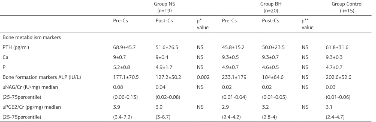
In the post-treatment assessment, the sOsteocalcin values were found to be signiicantly lower in the NS group than the control (p=0.027) (Fig.2) and the ALP values were found to be signiicantly lower in the NS than in the other two groups (p=0.001). No diference was observed in the post-treatment sCa levels among the groups (p=0.132). The urinary DPC/Cr ra-tio was found to be lower in the NS and BH groups than in the control (p=0.001) (Fig. 3). No diference was observed in any of the other serum and urine tests among the groups (p>0.05) (Table 2). The post-treatment uCa/Cr and DPC/Cr values were found to be signiicantly correlated (p=0.001, r=0.706) in the nephrotic syndrome group.
In the nephrotic syndrome group, the post-treatment uCa/Cr ratio was found to be higher than the pre-treatment values of uCa/Cr (p= 0.002) (Fig. 1). The post-treatment serum ALP and urinary DPC/Cr levels were found to be lower than those of the pre-treatment (p=0.002, p<0.001, respectively). The serum os-teocalcin levels were insigniicantly lower post treatment. No diference was recorded between the pre- and post-treatment results for the other values (p>0.05) (Table 2).
In the bronchial hyperreactivity group, the post-treatment uDPC/Cr ratio was found to be lower than the pre-treatment
uDPC/Cr ratio (p=0.001) (Fig. 3). The pre- and post-treatment values of osteocalcin and the other parameters were not found to show signiicant diference (p>0.05).
Table 1. Clinical and demographic data of the study groups Group NS
(n=19) Group BH (n=20) Group Control (n=15)
Age(year) 7.07±3.07 7.92±3.01 8,7±3.49
Body weight (kg) 25±1.4 26.3±1.7 27.4±0.8
Height (cm) 120±10 115±12 127±14
Sex
female (%) 36.8 50 46.7
male (%) 63.2 50 53.3
Blood pressure (mmHg) 120/80 95/63 90/60
Daily dietary calcium intake (mg/day)
577.6± 286.6 435.5±178.5 496.2±226.2
Stone disease in the family (%)
15.8 50 0
Family history of atopy (%) 5.3 85 0
Figure 1. Pre-Cs and post-Cs treatment uCa/Cr values of groups. (Descriptive statistics was used median and non-outlier range)
Figure 2. Pre and posttreatment sOsteocalcine values of groups. (Descriptive statistics was used median and non-outlier range)
The Efect of Corticosteroids
Discussion
In this study, we found that the usage of oral prednisolone increased renal Ca excretion, whereas the usage of inhaled budesonide caused no adverse efect. In the NS group, the os-teocalcin levels as an indicator of bone formation were found to decrease. Also, the uDPD/Cr level, as an indicator of bone resorption, was lower in both the NS and BH patients, contrary to expectation.
Osteocalcin is a speciic indicator for osteoblastic activity [7]. In some studies a drop in the sOsteocalcin levels with high-dose inhaled corticosteroid usage was reported [7,11], whereas in some other studies, no such diference was reported in sOsteo-calcin levels with budesonide usage [12]. In children diagnosed with congenital adrenal hyperplasia and on oral steroid treat-ment for more than two years, the bone mineral density was found to be normal, the sOsteocalcin level was found to be low, while the uDPD/Cr level was high [13]. Inhaled budesonide was reported to exert low systemic efects and bioavailability. In our study, the sOsteocalcin levels, one of the bone formation mark-ers, was found to have decreased in the NS patients, although it showed no change in the BH patients using 400µgr inhaled budesonide for three months. In conclusion, the type, dosage, and period of usage were believed to be efective in maintain-ing the sOsteocalcin level.
According to Wetzson RJ et al. [14], bone speciic ALP was found to be low in children with steroid sensitive-NS compared with the control group. According to Aceto [15] and Bak et al. [16], no diferences were observed during the diferent stages of treat-ment for calcium, phosphate, alkaline phosphatase, and 25-OH vitamin D. Vitamin D-binding globulin and 25-hdroxy D3 were low in the nephrotic urine [16]. Serum Ca and the most active vitamin D metabolites, 1.25-dyhidroxy D3 were normal or low, and serum parathyroid hormone was normal or high in those with nephrotic syndrome [16]. However, the serum vitamin D levels could not be assessed in this study. Therefore, we are un-able to comment on vitamin D. In our study, no diferences were obtained in any of the bone metabolism markers: Ca, P, and PTH. In the NS group the ALP level dropped signiicantly ater treatment. These results show a decrease in the osteoblastic activity in the bone with the use of oral Cs. In the BH group, we
found no diference at all with this dosage and periods of use of inhaled Cs. This demonstrated that inhaled Cs did not exert systemic efects.
Measurements of the type I collagen metabolites (the pre-dominant collagen in bone) such as DPC have been reported to be useful for monitoring the bone turnover in many difer-ent disorders and drug usages [17]. The urine deoxypyridinoline level, which is a bone turnover marker, is expected to increase with the use of Cs. To our knowledge, a few studies are avail-able on the efect of Cs therapy on the urine deoxypyridino-line crosslink level, and Rao et al. [18] reported an increase in the post-treatment uDPC/Cr levels in children on a 20-month inhaled budesonide or luticasone therapy. But Ton et al. [11] also recorded a decrease in the post-treatment uDPC/Cr level in children on Cs. They associated this with the adaptation of the bone to the prednisolone treatment. Koşan et al. [19] dem-onstrated an increase in the urinary DPC/Cr excretion at the 4th and 12th weeks of treatment in children with nephrotic syn-drome. However, in our study, diferent post Cs treatment levels of DPC/Cr were found in the NS and BH patients, which were low compared with the values of the pretreatment levels and control group levels. The indings of the present study revealed that the etiology was unknown for the lower post-treatment DPD/Cr levels in the NS and BH participants compared with the pre-treatment DPD/Cr levels and those of the control.
Many articles stated that the inhaled Cs neither inluenced nor decreased the excretion of urinary Ca in children [8, 20-21]. Bootsma et al. [20] reported that a nine-week treatment of 750 µg/day luticasone and 1500 µg/day beclomethasone showed no efect on the spot urinary Ca excretion. Akil et al. [8] also re-ported lower spot uCa/Cr levels in children diagnosed with asth-ma taking approxiasth-mately 400-600 µg/day inhaled budesonide as treatment for a year, compared with the healthy children. However, in the study by Bentur et al., [22] in 16% of children taking 400 µg/gün inhaled budesonide for two months, the post treatment uCa/Cr level was found to signiicantly increase. Not many publications related to the hypercalciuric efects of oral Cs in children with nephrotic syndrome are available. Düzen et al. [23] reported an increase in the levels of urinary calcium excretion in patients on 10 mg/day prednisolone treat-Table 2. Comparison of pre-Cs and post-Cs treatment values of groups NS and BH
Group NS
(n=19) Group BH (n=20) Group Control (n=15)
Pre-Cs Post-Cs p*
value
Pre-Cs Post-Cs p**
value Bone metabolism markers
PTH (pg/ml) 68.9±45.7 51.6±26.5 NS 45.8±15.2 50.0±23.5 NS 61.8±31.6
Ca 9±0.7 9±0.4 NS 9.3±0.5 9.3±0.7 NS 9.3±0.3
P 5.2±0.8 4.9±1.7 NS 4.9±0.7 4.6±0.5 NS 4.7±0.7
Bone formation markers ALP (IU/L) 177.1±70.5 127.2±50.2 0.002 233.1±179 184±64.6 NS 202.6±52.6
uNAG/Cr (IU/mg) median 0.08 0.04 NS 0.02 0.02 NS 0.03
(25-75percentile) (0.06-0.13) (0.02-0.08) (0.01-0.04) (0.01-0.05) (0.01-0.06)
uPGE2/Cr (pg/mg) median 3.9 3.9 NS 2.9 3.2 NS 3.1
(25-75percentile) (3.4-7.2) (3-6.7) (2.4-4.2) (2.8-4) (2.4-4.7)
The Efect of Corticosteroids
ment for one month. Similarly, Koşan et al. [19] reported an increase in the levels of urinary calcium excretion during the 4th and 12th weeks of steroid treatment in children with NS. Our results showed that in the NS group, the uCa/Cr ratio which had increased post treatment was found to be highly positively correlated to the post-treatment DPC/Cr. We also observed a decrease in the osteocalcin levels post steroid treatment. From these indings, we concluded that a one-month usage of oral corticosteroids resulted in an increase in the bone resorption, as well as a decrease in bone formation by way of systemic ef-fects. However, the BH group revealed no change in the levels of urinary Ca excretion even ater utilizing 400 mcg of inhaled budesonide for two months.
The NAG/Cr ratio was seen to increase in idiopathic HC due to the primary tubular disorder [24]. The increased NAG/Cr ratio that had been recorded as high, prior to the treatment in the NS group, is believed to be related to the tubular inlamma-tion due to the disease itself. Also, the post-treatment NAG/ Cr levels were not found to signiicantly difer from the values prior to treatment in both the NS and BH groups. These results demonstrated that oral and inhaled Cs usage has no efect on the proximal tubules.
Prostaglandin E2 is well recognized for its role in HK by increas-ing the 1.25(OH)2 D synthesis [25]. In our study the uPGE2/ Cr levels were found to be higher in the NS group than in the control group (p=0.038). Also, in the NS and BH post-treatment values, the uPGE2/Cr, which is highly and positively correlated with the DPC/Cr, was assumed to afect the bone resorption. This study has limitations because the number of cases studied was small and vitamin D levels could not be investigated.
Conclusion
The short-term usage of moderate and low-dose inhaled ste-roids was found to have no efect on the osteoblastic and os-teoclastic activities or on renal Ca excretion. However, it was observed that one-month of oral steroid usage signiicantly increased the levels of urinary Ca excretion whereas it sup-pressed the osteoblastic activity in the bone.
Acknowledgement: We thank Dr.Ayşe Yenigün, Dr.Çiğdem Yenisey, and Dr.İmran Kurt Ömürlü for their contributions.
Competing interests
The authors declare that they have no competing interests.
References
1. Cooper MS, Seibel MJ, Zhou H. Glucocorticoids, bone and energy metabolism. Bone 2015;pii: S8756-3282(15)00219-7. doi: 10.1016/j.bone.2015.05.038. 2. Gbadegesin R, Smoyer WE. Nephrotic syndrome. In: Geary DF, Schaefer F, eds. Comprehensive Pediatric Nephrology, 1st ed. Philadelphia, PA: Mosby Elsevier; 2008.p.205–18.
3. Leonard MB. Glucocorticoid-induced osteoporosis in children: impact of the underlying disease. Pediatrics. 2007;119 Suppl 2:S166-74.
4. Yılmaz D, Sönmez F, Yenisey C, et al. The role of active vitamin D on stone for-mation and hypercalciuria. Nobel Med 2013;9:88-91.
5. Milliner DS. Urolithiasis. In: Avner ED, Harmon WE, Niaudet P (eds). Pediatric Nephrology (5th ed). Baltimore: Lippincott Williams and Wilkins. 2004:1409-17. 6. Hoppe B, Leumann E, Milliner DS. Urolithiasis and nephrocalcinosi in childhood. In: Geary DF, Schaefer F (eds). Compherensive Pediatric Nephrology. New York; Elsevier/WB Saunders. 2008.p.499-523.
7. Akil İ, Yüksel H, Ürk V. Bronş astımlı çocıklarda inhale steroidin kemik metabolizmasının biyokimyasal göstergeleri ve renal kalsiyum atılım hızına etkisi. Astım Allerji Immünoloji 2003;1:5-10.
8. Reddel HK, Levy ML. Global Initiative for Asthma Scientiic Committee and Dissemination and Implementation Committee. The GINA asthma strategy
re-port: what’s new for primary care? NPJ Prim Care Respir Med 2015;25:15050. doi: 10.1038/npjpcrm.2015.50.
9. Sönmez F, Akçanal B, Altıncık A. Urinary calcium excretion in healthy Turkish children. Int Urol Nephrol 2007;39:917-22.
10. Schwartz GJ, Haycock GB, Edelmann CM Jr, et al. A simple estimate of glo-merular iltration rate in children derived from body length and plasma creatinine. Pediatrics 1976;58 :259-63.
11. Ton FN, Gunawardene SC, Lee H, et al. Efects of low-dose prednisone on bone metabolism. J Bone Miner Res 2005 ;20:464-70.
12. Birkebaek NH, Esberg G, Andersen K, et al. Bone and collagen turnover during treatment with inhaled dry powder budesonide and beclomethasone dipropionate. Arch Dis Child 1995;73:524–7.
13. Abd El Dayem SM, El-Shehaby AM, Abd El Gafar A, et al. Bone density, body composition, and markers of bone remodeling in type 1 diabetic patients. Scand J Clin Lab Invest 2011;71(5):387-393.
14. Wetzsteon RJ, Shults J, Zemel BS, et al. Divergent efects of glucocorticoids on cortical and trabecular compartment BMD in childhood nephrotic syndrome. J Bone Miner Res 2009;24(3):503-13.
15. Aceto G, D’Addato O, Messina G, et al. Bone health in children and adolescents with steroid-sensitive nephrotic syndromeassessed by DXA and QUS. Pediatr Nephrol 2014 ;29(11):2147-2155.
16. Bak M, Serdaroglu E, Guclu R. Prophylactic calcium and vitamin D treat-ments in steroid-treated children with nephrotic syndrome. Pediatr Nephrol 2006;21:350-4.
17. Benarba B, Meddah B, Tir Touil A. Response of Bone Resorption Markers to Aristolochia longa Intake by Algerian Breast Cancer Postmenopausal Women. Adv Pharmacol Sci 2014;2014:820589.
18. Rao R, Gregson RK, Jones AC. Systemic efects of inhaled corticosteroids on growth and bone turnover in childhood asthma. Eur Respr J 1999;13:87-94. 19. Koşan C, Ayar G, Orbak Z. Efects of steroid treatment on bone mineralme-tabolism in children with glucocorticoid-sensitive nephrotic syndrome. WestIndian Med J 2012;61:627-30.
20. Bootsma P, Dekhuijzen RD, Festen J. Fluticasone propionate does not inluence bone metabolism in contrast to beclametasone dipropionate. Am J Respr Crit Care 1996;153:924-30.
21. Altıntaş U, Bingol G, Can S. The efcts of long term use of corticosteroids on linear growth, adrenal function and bone mineral density in children. Allergol Et Immunopatol 2005;33:204-9.
22. Bentur L, Jarrous T, Bentur Y. The efect of inhaled corticosteroids on the urinary calcium to creatinine ratio in childhood asthma. Therapie 2003;58:313-6. 23. Duzen O, Erkoc R, Begenik H, et alN. The course of hypercalciuria and related markers of bone metabolism parameters associated with corticosteroid treat-ment. Ren Fail 2012;34(3):338-42.
24. Sikora P, Glatz S, Beck Bodo. Urinary NAG in children with urolithiasis, nephro-calcinosis or risk of urolithiasis. Pediatr Nephrol 2003;18:996-9.
25. Srivastava T, Alon US. Pathophysiology of hypercalciuria in children. Pediatr Nephrol 2007;22:1659-73.
How to cite this article: