DO:
D 10.1590/S1516-14392011005000073
*e-mail: [email protected]
Incorporation of Strontium up to 5 Mol. (%) to Hydroxyapatite
did not Affect its Cytocompatibility
Débora dos Santos Tavaresa*, Cristiane Xavier Resendea, Maíra Paiva Quitana,
Letícia de Oliveira Castrob, José Mauro Granjeirob,c, Gloria de Almeida Soaresa
a
Materials Engineering Department, COPPE, Rio de Janeiro Federal University – UFRJ,
CP 68505, CEP 21941-972, Rio de Janeiro, RJ, Brazil
b
Cell and Molecular Biology Department and Cell Therapy Center UPC-HUAP,
Fluminense Federal University – UFF, Outeiro de S. J. Batista, s/n, CP 24150, Niterói, RJ, Brazil
c
Dipro/Bioengineering, INMETRO, Av. Nossa Senhora das Graças, nº 50,
CEP 25250-020, Xerém, Duque de Caxias, RJ, Brazil
Received: November 9, 2010; Revised: July 29, 2011
The aim of this work was to produce hydroxyapatite (HA) granules containing 0, 0.5, 1 and 5 mol. (%) of strontium (Sr), evaluate the physico-chemical properties and also the cytotoxicity by three different parameters of cell viability (OSD 10993-5, 10993-12). The physico-chemical characterization was carried out by using X-ray
diffraction (XRD), Fourier-transform infrared spectroscopy (FTIR) and X-ray luorescence (XRF). The XRD proile presented the main peaks of HA (JCPDS 860740) and the absorption bands of HA were identiied by
FTOR. The XRF results showed that the strontium concentration was close to the theoretical value. Regarding the cytotoxicity assays, the incorporation of strontium up to 5 mol. (%) to the HA did not affected dehydrogenase
activity (XTT, 2,3-bis[2-methyloxy-4-nitro-5-sulfophenyl]-2H-tetrazolium-5-carboxanilide), membrane integrity
(neutral red uptake) or DNA contend (incorporation of crystal violet), in relation to HA alone. On conclusion, hydroxyapatite containing from 0.5 to 5 mol. (%) of Sr was successfully produced and presented no cytotoxicity.
Keywords: strontium, hydroxyapatite, cytotoxicity
1. Introduction
Bone loss due to chronic diseases or trauma can result in large defects, which challenge clinicians, and most of the time bone grafts are needed. Research is being conducted in order to improve bone formation through the implantation of biomaterials with a superior
bioactivity. Calcium phosphate ceramics, such as hydroxypapatite
(HA), are widely used as bone grafts because of their similarity to the mineral phase of the bone. Moreover, they seem to induce the formation of a biological apatite layer, and are thus considered bioactive materials1,2. Biological apatite is a calcium-deicient apatite
with several substitutions in its crystal lattice, such as F–, Cl–, Na+,
K+, Fe2 +, Zn2+, Sr2+, Mg2+, and CO 3
–2[3,4,5]. Granular material is often
used for bone illing in order to avoid phagocytosis, since it has been shown that particle size inluences the induction of bone resorption6.
Strontium (Sr) is a bone-seeking trace element and 98% of its
total content is in the skeleton7. Ot was reported that low dosages
can improve bone formation while high dosages induce defective bone mineralization8,9. Recent in vitro studies showed that strontium ranelate, a novel agent used in osteoporosis (characterized by low bone mass and susceptibility to fractures) treatment, inhibits bone resorption via the osteoclasts and promotes osteoblast replication and bone formation10. On vivo tests with strontium-substituted hydroxyapatite (Sr/Ca = 0.10) cement were performed using a rabbit
hip replacement model and an apatite layer was found six months later between the cement and the cancellous bone11. As reported,
strontium-containing hydroxyapatite (Sr-HA) appears to be an interesting bone substitute material. Because of the difference in the atomic radius between strontium and calcium, the crystal lattice becomes distorted and therefore the Sr-HA stability decreases, facilitating its replacement by new bone12.
On vitro studies of Sr-HA containing 0, 1, 5, 10 and 100 mol. (%) Sr presented good biocompatibility, but the cytotoxicity increased as the strontium concentration increased13. Moreover, osteoblast-like MG63 cells cultured on Sr-HA nanocrystals containing 0, 1, 3, 7 atom. (%) exhibited good proliferation and increased values of
the differentiation parameters14. Differences in the responses of
biomaterials have been reported depending on the cytotoxicity test used. Therefore, it is important to evaluate this response by using a combination of more than one parameter related to cell functions in order to increase the reliability of results15.
Previous works indicated that even Sr-HA with 100 mol. (%) of
strontium has a good biocompatibility, but which also showed that cytotoxicity increased proportionally to the strontium content. Due to the ambiguous results in the literature, the aim of this study was to produce dense HA granules containing 0, 0.5, 1 and 5 mol. (%) of strontium (which is closer to the biological content in HA), evaluate the physico-chemical properties and also the cytotoxicity of produced biomaterials by three different parameters of cell viability, according to OSD 10993-5[16] and 10993-12[17] standards.
2. Materials and Methods
2.1. Synthesis of HA and processing the dense granules
Strontium-substituted hydroxyapatites were synthesized by a wet chemical route. A KH2PO4 solution (MERCK, 99.5 wt. (%) pure)
was prepared and dropped into the Ca(NO3)2 (VETEC, 99 wt. (%) pure) and Sr(ND3)2 (MERCK, 99 wt. (%) pure) solution over a period
pH of the solution was adjusted to 11 using an aqueous solution of
ammonium 50% (v/v), with a inal concentration of 12.5%. The
reaction product was kept in suspension by constant stirring and
heating to 40 °C for 2 hours. The precipitate was centrifuged and washed several times with ultra-pure water until pH 7 was reached. Finally, the precipitate was dried for 24 hours at 37 °C. The dried
powder was crushed and sieved using a 125 µm sieve to obtain the
ine fractions. All materials were uniaxially pressed into cylindrical tablets under a compressive strength of 216 MPa, followed by sintering at 1150 °C for 4 hours in air (2.8 °C/min heating rate). The
tablets were crushed and sieved to select dense granules in the range
of 300-600 µm.
The Ca + Sr/P molar ratio was equal to 1.5 according to the apatite chemical formula of Ca9-xSrx(PO4)6(DH)2 in order to produce non-stoichiometric apatites, which are usually more soluble than the stoichiometric ones. Table 1 gives the concentrations used for each apatite composition.
2.2. Physico-chemical characterization
The calcium phosphate phase of the granular material was
determined using an X-ray diffractometer (SHIMADZU XRD 6000) with monochromatized Cu kα radiation and an operational tube with
a voltage and current of 40 kV and 30 mA, respectively. The angle of diffraction was from 10 to 60° and the granular material was crushed.
The resulting trace was analyzed and compared with the standard library of known diffraction patterns. The granules (mixed with KBr) were also examined by FTOR in order to identify the vibrational
modes of the molecules (Perkin Elmer, Spectrum 100) in the range of 500-4000 cm–1. At least 16 scans were conducted and the average
of the results was reported. The chemical composition of the samples
was determined by X-ray luorescence (XRF) on RIGAKU RIX 3100 equipment with a rhodium tube (4 kW). Therefore, the strontium content and also the Ca+Sr/P ratio could be veriied.
2.3. Cytotoxicity test
Samples were extracted in a culture medium (100 mg.mL–1, apatite granules/DMEM free of bovine fetal serum) at 37 °C for 24 hours and the extracts were collected for the cytotoxicity assay
according to OSD 10993-5[16] and 10993-12[17]. As control were used
1% phenol solution (positive) and titanium powder (100 mg.mL–1, as a negative control). MC3T3 osteoblasts (CRL 2594 – ATCC) were seeded into a 96-well cell culture plate (1 × 10⁴ . well–1) and cultured in DMEM containing NaHCO3 (1.2 g.L–1), ampicillin (0.025 g.L–1), and
streptomycin (0.1 g.L–1) supplemented with 10% bovine fetal serum for 24 hours at 37 °C under 5% CO2/95% air conditions. Afterwards, the medium was replaced by each extract and supplemented with
10% bovine fetal serum (BFS) and maintained at 37 °C under 5% CO2/95% air conditions.
After 24 hours of cell exposure to each extract media, cytotoxicity was evaluated using a commercial kit (Cytotox, Xenometrix,
Germany) which allows the use of three different parameters of cell survival and integrity on the same sample:
2,3-bis[2-methyloxy-4-nitro-5-sulfophenyl]-2H-tetrazolium-5-carboxanilide (XTT), neutral red (NR) and crystal violet dye elution (CVDE). The XTT
cell proliferation assay is based on the ability of mitochondrial dehydrogenase enzymes to convert the yellow water-soluble tetrazolium salt XTT into orange colored soluble compounds of
formazan, measured by their absorbance at 480 nm18. Neutral red is a
survival/viability test based on the ability of living cells to incorporate the neutral red dye in their lysosomes, where it accumulates on membrane-intact cells19; the quantity of dye incorporated can be measured at 540 nm. The CVDE assay evaluates cell density by staining DNA; the absorbance at 540 nm is proportional to the amount
of cells in each well20. The absorbance data were obtained with a microplate UV/Vis spectrophotometer (PowerWave MS2, BioTek
Onstruments, USA).
All of the tests were performed in quintuplicate. Mean values and standard deviations were submitted to one-way analysis of variance
(ANOVA) and Tukey’s post-test considering signiicant differences
if p < 0.05.
3. Results and Discussion
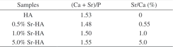
The chemical compositions of the powders obtained from the
X-ray luorescence are shown in Table 2. Regarding the X-ray luorescence results, the strontium concentration incorporated in
the HA was very close to the theoretical value (Table 1). Moreover,
the (Ca+Sr)/P in all of the substituted HA was close to 1.5, and these results are comparable to the Ca/P ratio in pure HA, 1.53, corresponding to a calcium-deicient apatite.
Figure 1 shows the XRD patterns of hydroxyapatite with the
different Sr/Ca molar ratios after sintering. All of the patterns show the mean peaks of hydroxyapatite at 25.8, 31.8, 32.3 and 32.9o (JCPDS 860740), compatible with a Sr-HA solid solution, since
the crystalline structure of HA was maintained and no other phase containing strontium was formed after the calcium partial substitution by strontium21, according to XRD data showed in Figure 1. The XRD
of 0, 0.5, and 1 mol. (%) Sr-HA granules exhibited a peak at 31.2o,
which is typical of β-TCP and a decreased in its intensity was noted
with the augmentation of strontium content. The presence of β-TCP is, probably, a consequence of the transformation of HA to TCP that occurs in calcium-deicient apatites. A previous report had already described the presence of TCP in Sr-HA, and its formation was
attributed to the non-stoichiometry of the apatite structure causing structural and compositional changes in elevate temperatures22. Structural changes to TCP upon calcining calcium deicient HA at temperatures between 500 and 1100 °C (up to 1450 °C) were also
reported earlier23. Furthermore, other undesirable phases can be
found as reported by Dagang et al.24 during the synthesis of Sr-HA. Dne of the most critical parameters in determining properties and
thermal stabilities of HA powder is the Ca/P ratio25. Accordingly, the
β-TCP peak was not observed in 5% Sr-HA samples probably due to its Ca + Sr/P molar ratio, which was higher when compared to the
Table 1. Molar concentrations used for the powder synthesis of Ca9-xSrx(PO4)6(DH)2, where x = 0.0, 0.5, 1.0 and 5.0.
Sr (x) KH2PO4 Ca(NO)2 Sr(ND3)2
0.0 0.80 0.91 0.000
0.5 0.80 0.90 0.009
1.0 0.80 0.90 0.018
5.0 0.80 0.86 0.090
Table 2. XRF analysis1 of granular material showing (Ca + Sr)/P and Sr/Ca
ratios.
Samples (Ca + Sr)/P Sr/Ca (%)
HA 1.53 0
0.5% Sr-HA 1.48 0.55
1.0% Sr-HA 1.50 1.0
5.0% Sr-HA 1.55 5.0
other apatites synthesized herein (Table 2). On addition, the partial
replacement of Ca2+ by Sr2+ increases the solubility of HA. This phenomenon was studied by Christoffersen et al.26, who reported that
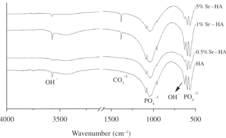
the solubility of apatite increases with an increasing strontium content. The FTOR spectra of HA and Sr-doped HA are shown in Figure 2 and different modes of molecular vibration characteristics of the apatites can be observed. The bands at 1091 (ν3), 1049 (ν3),
967 (ν3), 598 (ν4) and 568 cm–1 (ν4) are typical of phosphate (PO 4–3)
molecular vibration and were present in all of the spectra from the
synthetic apatites. The sharp band at 3572 and 632 cm–1 corresponds
to the DH– stretching mode, characteristic of HA. A well-deined band of carbonate, at 1385 cm–1, was detected when the strontium
concentration was equal or superior to 1% Sr. The band present in this region suggests that phosphate ions are partially substituted by carbonate. This carbonate substitution may be due to the reactive absorption of atmospheric carbon dioxide by the alkaline solution during the slurry preparation and reaction. A similar carbonate
absorption band in the region of 1367-1527 cm–1 was previously
reported on Sr-HA27, which increased of intensity with strontium
content. Another study showed that carbonate incorporation decreases the crystallinity of the apatites and enhances solubility28.
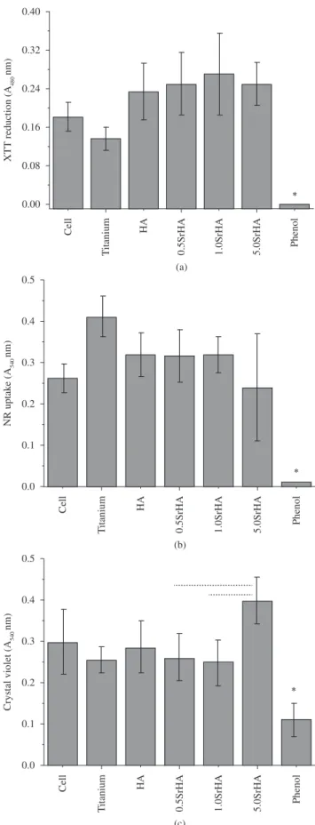
Cytotoxicity assays provide a measure of cell death caused by
contact with materials or their extracts, and only one test is not capable of detecting the cytotoxicity of biomaterials29,30. Hence, membrane integrity (NR), metabolic activity (XTT) and cell density (CVDE)
were evaluated. Figure 3 shows the in vitro cytotoxicity measured
by XTT, NR, and CVDE after 24 hours in culture with the apatite
(partially substituted with Sr and pure HA) extracts.
The XTT analysis showed no signiicant difference among the
Sr-HA or HA extracts or cells exposed only to DMEM plus BFS. Metabolic activity was around 1.5 fold greater in the Sr-HA groups, irrespective to Sr concentration, in relation to cells in DMEM alone
(p < 0.05). As expected, the positive control (1% phenol) signiicantly
decreased cell viability.
A similar trend was presented by Fu et al.13 when the strontium concentration was up to 5%, using the same assay. Capuccini
et al.31 showed that the presence of strontium signiicantly improved
osteoblast adhesion in early culture phases. Moreover, cell adhesion increased with increasing strontium content (1, 5, and 10 Sr at.%). The cytotoxicity of Sr-HA containing 1, 5, 10, and 100 mol. (%) was also evaluated by Yuan-Fei et al.32 via methyltetrazolium (MTT) assay and
showed that all of the bioceramics studied had good biocompatibility.
Furthermore, osteoblast-like MG63 cells cultured on the Sr-HA nanocrystals containing 0, 1, 3, and 7 atom. (%) displayed good
proliferation and increased values of the differentiation parameters14.
Dsteoprecursor cell behavior was examined through MTT reduction
at 3, 7, and 14 days of incubation by culturing them onto 10 mol %
Sr-HA surface. The authors concluded that Sr-HA induced better cell attachment and proliferation when compared to HA33.
Regarding the NR assays it is possible to observe that no effect on the membrane integrity occurred by the exposition to extracts, except in the phenol group (almost 100% reduction), as already reported4,34 (Figure 3b).
The crystal violet intercalate to DNA and correlates to the cell
number. Despite the slightly more CV intensity for 5% Sr-HA group (p < 0.05, 30%), no other signiicant difference was observed. In
resume, this data indicates no effect of Sr-HA extracts on cell viability.
Also, phenol reduced 70% the number of adhered cells (p < 0.05).
Ot is well known that different HA can be synthesized by substituting calcium, phosphate or hydroxyl groups resulting in biomaterials with different biological and chemical properties. Lima et al.4 assessed the number of viable Balb/c 3T3 ibroblasts (through
an hemocytometer) after exposure to several metal modiied apatites
extracts for 24 hours as follows: Ca9.5M0.5(PO4)6(DH)2 (M=Fe, Zn,
Cu, Co, Sr, Pb) and Ca10(VD4)6(DH)2. Sr-HA did not present any
toxicity while phenol killed 82% of the cells. It was observed an increase of 70% and 20% in the number of cells exposed to the
extracts of Fe-HA and Sr-HA, respectively. This study showed that cells response is dependent on the metal that substitutes calcium or phosphate ions in the crystal lattice of HA, and that 0.5% Sr-HA did not affect cell viability.
On general, the assays mainly used to evaluate cell responses on bioceramics are the XTT (reduced in soluble formazan compounds) and the MTT (reduced in insoluble formazan compounds), which are based on similar principles. Therefore, to the best of our knowledge,
the literature is poor regarding NR uptake and CVDE assays to compare with the results of the Sr-modiied apatites synthesized
herein.
Cytotoxicity is a challenging process to analyze, as there are
several ways to activate cellular disruption. On addition, all of these assays have their disadvantages that must be taken into consideration when analyzing the obtained data29,35. For instance, metabolic
impairment assays such as MTT and XTT can be biased by occasional
Figure 1. XRD patterns of pure and strontium-doped hydroxyapatites (
•
-β-TCP and ♦-HA).Figure 3. Cytotoxicity assay for pure HA and strontium-doped hydroxyapatites at different concentrations on mouse osteoblasts: a) XTT reduction; b) neutral red uptake; and c) crystal violet dye elution. The bars in the igure represent the standard deviation of means. Phenol group was statistically different from all samples tested in the three assays (*, p < 0.05). (- - -) indicate a statistical difference between the samples (p < 0.05).
bacterial contamination, which can overestimate cell viability36; in contrast, some reducing agents and respiratory chain inhibitors of mitochondrial MTT reduction could underestimate cell viablity29. An
increase in NR uptake by lysosomal swelling agents such as weakly basic substances37 could lead to an underestimation of cytotoxicity
and CV dye can also stain dead cells due to its non-speciicity in
binding to DNA29. Concerning the CV test result, a considerable cell
number was observed in the phenol group. However, those cells are
in a death process since no metabolic activity was detected and CV
also dyes dead cells that are still attached to the plate. This reinforces the necessity to evaluate more than one aspect of cell survival and integrity before attesting a biomaterial cytocompatibility. Likewise, biomaterials may affect diverse aspects points of cell viability.
Taken together, our results showed that incorporation of strontium in the hydroxyapatite, despite the potential increase in the Sr-HA solubility, did not affect the viability of murine osteoblasts, since
no signiicant effect was observed in the metabolic activity, cell
membrane integrity and DNA contend.
4. Conclusion
Granular hydroxyapatite containing from 0 to 5 mol. (%) of Sr was successfully produced and presented no cytotoxicity. The strontium incorporated in the HA crystal resulted in a solid solution with a minor phase (β-TCP) identiied by XRD, except for Sr = 5 mol. (%). Further studies are required to evaluate the potential beneits of Sr-HA as a
bone substitute biomaterial and its effects in osteoporosis treatment.
Acknowledgements
We wish to thank FAPERJ, FINEP, MS/DECIT, CAPES and CNPq for inancial support and NUCAT/UFRJ for XRF analysis.
References
1. Dorozhkin SV. Calcium orthophosphates. Journal of Materials Science: Materials in Medicine. 2007; 42(4):1061-1095.
2. Vallet-Regi M and Gonzalez-Calbet JM. Calcium phosphates as substitution of bone tissues. Progress in Solid State Chemistry. 2004; 32(1-2):1-31. http://dx.doi.org/10.1016/j.progsolidstchem.2004.07.001 3. Calasans-Maia MD, Rossi AM, Dias EP, Santos SRA, Áscoli F and
Granjeiro J.M. Stimulatory effect on osseous repair of zinc-substituted hydroxyapatite: histological study in rabbit’s tibia. Key Engineering Materials. 2008; 20(2):1269-1272. http://dx.doi.org/10.4028/www. scientiic.net/KEM.361-363.1269
4. Lima OR, Santos SRA, Santiago DL, Rossi AM and Granjeiro JM. Cytotoxicity analysis of apatites modified with divalent metals. Key Engineering Materials. 2008; 20(2):1135-1138. http://dx.doi. org/10.4028/www.scientiic.net/KEM.361-363.1135
5. Wopenka B and Pasteris JD. A mineral perspective on the apatite in bone. Materials Science and Engineering C. 2005; 25(2):131-143. http://dx.doi. org/10.1016/j.msec.2005.01.008
6. Ongham E and Fisher J. The role of macrophages in osteolysis of total joint replacement. Biomaterials. 2005; 26(11):1271-1286. PMid:15475057. http://dx.doi.org/10.1016/j.biomaterials.2004.04.035
7. Verberckmoes SC, Behets GJ, Oste L, Bervoets AR, Lamberts LV, Drakopoulos M et al. Effects of strontium on the physicochemical characteristics of hydroxyapatite. Calcified Tissue International. 2004; 75(5):405-415. PMid:15592797. http://dx.doi.org/10.1007/s00223-004-0260-4
8. Marie PJ, Garba MT, Hott M and Miravet L. Effect of low doses of stable strontium on bone metabolism in rats. Mineral Electrolyte Metabolism. 1985; 11(1):5-13. PMid:3974537.
10. Marie PJ. Strontium as therapy for osteoporosis. Current Opinion in Pharmacology. 2005; 5:633-636. PMid:16183330. http://dx.doi. org/10.1016/j.coph.2005.05.005
11. Ni GX, Lu WW, Xu B, Chiu KY, Yang C, Li ZY et al. Interfacial behaviour of strontium-containing hydroxyapatite cement with cancellous and cortical bone. Biomaterials. 2006; 27(29):5127-5133. PMid:16781769. http://dx.doi.org/10.1016/j.biomaterials.2006.05.030
12. Xue-Yang L and De-Min C. Effects of strontium substituted hydroxyapatite of different porosities on osteoblasts in vitro. Journal of Oral Tissue Engineering. 2005; 2(2):66-75.
13. Fu YF, Chen DM and Zhang JZ. In vitro study on the cytotoxicity of strontium substituted hydroxyapatite. Shanghai Kou Qiang Yi Xue. 2002; 11(3):229-232. PMid:14983257.
14. Capuccini C, Torricelli P, Boanini E, Gazzano M, Giardino R and Bigi A. Onteraction of Sr-doped hydroxyapatite nanocrystals with osteoclast and osteoblast-like cells. Journal of Biomedical Materials Research Part A. 2009; 89(3):594-600 PMid:18437694. http://dx.doi.org/10.1002/ jbm.a.31975
15. Weyermann J, Lochmann D and Zimmer A. A practical note on the use of cytotoxicity assays. International Journal of Pharmaceutics. 2005; 288:369-376. PMid:15620877. http://dx.doi.org/10.1016/j. ijpharm.2004.09.018
16. Onternational Drganization for Standardization - OSD. ISO 10993-5: Biological evaluation of medical devices. Geneva: OSD; 1999. part 5: Tests for cytotoxicity: On vitro methods.
17. Onternational Drganization for Standardization - OSD. ISO 10993-12: Biological evaluation of medical devices. Geneva: ISO; 1996. Part 12: Sample preparation and reference materials.
18. Scudiero DA, Shoemaker RH, Paull KD, Monks A, Tierney S, Nofziger TH et al. Evaluation of a soluble tetrazolium/formazan assay for cell growth and drug sensitivity in culture using human and other tumor cell lines. Cancer Research. 1988; 48(17):4827-4833. PMid:3409223. 19. Winckler J. Vital staining of lysossomes and other cell organelles of
the rat with neutral red. Progress in Histochemistry and Cytochemistry. 1974; 6(3):1-91.
20. Kueng W, Silber E and Eppenberger U. Quantiication of cells cultured on 96-well plates. Analytical Biochemistry. 1989; 182(1):16-19. http://dx.doi.org/10.1016/0003-2697(89)90710-0
21. Bigi A, Boanini E, Capuccini C and Gazzano M. Strontium-substituted hydroxyapatite nanocrystals. Inorganica Chimica Acta. 2007; 360(3):1009-1016. http://dx.doi.org/10.1016/j.ica.2006.07.074 22. Kim HW, Koh YH, Kong YM, Kang JG and Kim HE. Strontium
substituted calcium phosphate biphasic ceramics obtained by a powder precipitation method. Journal of Materials Science: Materials in Medicine. 2004; 15(10):1129-1134. PMid:15516874. http://dx.doi. org/10.1023/B:JMSM.0000046395.76435.60
23. Gibson IR, Rehman I, Best SM and Bonield W. Characterization of the transformation from calcium-deicient apatite to β-tricalcium phosphate. Journal of Materials Science: Materials in Medicine. 2000; 11(12):799-804. PMid:15348063. http://dx.doi.org/10.1023/A:1008905613182 24. Dagang G, Kewei X, Xiaoyun Z and Yong H. Development of a
strontium-containing hydroxyapatite bone cement. Biomaterials.
2005; 26 (19) 4073-4083. PMid:15664634. http://dx.doi.org/10.1016/j. biomaterials.2004.10.032
25. Fanovich MA and Porto Lopez JM. Inluence of temperature and additives on the microstructure and sintering behaviour of hydroxyapatites with different Ca/P ratios. Journal of Materials Science: Materials in Medicine. 1998; 9(1):53-60. PMid:15348702. http://dx.doi.org/10.1023/A:1008834712212 26. Christoffersen J, Christoffersen MR, Kolthoff N and Barenholdt O.
Effects of strontium Oons on Growth and Dissolution of Hydroxyapatite and on Bone Mineral Detection. Bone. 1997; 20(1):47-54. http://dx.doi. org/10.1016/S8756-3282(96)00316-X
27. Zhang W, Shen Y, Pan H, Lin K, Liu X, Darvell BW et al. Effects of strontium in modiied biomaterials. Acta Biomaterialia. 2011; 7:800-808. PMid:20826233. http://dx.doi.org/10.1016/j.actbio.2010.08.031 28. Landi E, Tampieri A, Celotti G, Vichi and Sandri M. Inluence of synthesis
and sintering parameters on the characteristics of carbonate apatite. Biomaterials. 2004; 25(10):1763-1770. PMid:14738839. http://dx.doi. org/10.1016/j.biomaterials.2003.08.026
29. Chiba K, Kawakami K and Tohyama K. Simultaneous evaluation of cell viability by neutral red, MTT and crystal violet staining assays of the same cells. Toxicology in Vitro. 1998; 12(3):251-258. http://dx.doi. org/10.1016/S0887-2333(97)00107-0
30. De-Deus G, Canabarro A, Alves G, Linhares A, Senne MI and Granjeiro JM. Dptimal cytocompatibility of a bioceramic nanoparticulate cement in primary human mesenchymal cells. Journal of Endodontics. 2009; 35(10):1387-1390. PMid:19801236. http://dx.doi.org/10.1016/j.joen.2009.06.022
31. Capuccini C, Torricelli P, Sima F, Boanini E, Ristoscu C, Bracci B et al. Strontium-substituted hydroxyapatite coatings synthesized by pulsed-laser deposition: On vitro osteoblast and osteoclast response. Acta Biomaterialia. 2008; 4(6):1885-1893. PMid:18554996. http://dx.doi. org/10.1016/j.actbio.2008.05.005
32. Yuan-Fei F and De-Min C. Inluence of Sr2+ on strontium substituted
hydroxyapatite’s (Sr-HA) cytotoxicity. Journal of Oral Tissue Engineering. 2005; 2:76-80.
33. Weichang X, Moore JL, Hosick HL, Bose S, Bandyopadhyay A, Lu WW et al. Dsteoprecursor cell response to strontium-containing hydroxyapatite ceramics. Journal of Biomedical Materials Research Part A. 2006; 7(4):804-814.
34. Takamori ER, Figueira EA, Taga R, Sogayar MC and Granjeiro JM. Evaluation of the Cytocompatibility of Mixed Bovine Bone. Brazilian Dental Journal. 2007; 18(3):179-184. PMid:18176706. http://dx.doi. org/10.1590/S0103-64402007000300001
35. Murray PE, Godoy CG and Godoy FG. How is the biocompatibility of dental biomaterials evaluated? Medicina Oral, Patología Oral y Cirurgía Bucal. 2007; 12(E2):258-266.
36. Ciapetti G, Granchi D, Verri E, Savarino L, Cavedagna D and Pizzoferrato, A. Application of a combination of neutral red and amino Black staining for rapid, reliable cytotoxicity testing of biomaterials. Biomaterials. 1996; 17(13):1259-1264. http://dx.doi.org/10.1016/0142-9612(96)88670-4 37. Ohkuma S and Poole B. Cytoplasmic vacuolation of mouse peritoneal