Single Nucleotide Polymorphism and Susceptibility
towards Cancer: Evidence from a Meta-Analysis
Sushil Kumar Sahu
1, Tathagata Choudhuri
1,2*1 Division of Infectious Disease Biology, Institute of Life Sciences, Bhubaneswar, India, 2 Department of Biotechnology, Siksha Vhabana, Visva Bharati,
Santiniketan, Bolpur, India
Abstract
Background: The Bcl-β-associated X protein (Bax) is a proapoptotic member of the Bcl-β family known to be activated and upregulated during apoptosis. Single nucleotide polymorphisms (SNPs) in Bax promoter may participate in the process of carcinogenesis by altering its own expression and the cancer related genes. Bax-β48G>A polymorphism has been implicated to alter the risk of cancer, but the listed results are inconsistent and inconclusive. In the present study, we performed a meta-analysis to systematically summarize the possible association of this polymorphism with the risk of cancer.
Methodology: We conducted a search of case-control studies on the associations of Bax-β48G>A polymorphism with susceptibility to cancer in Pub Med, Science Direct, Wiley Online Library and hand search. Data from all eligible studies based on some key search terms, inclusion and exclusion criteria were extracted for this meta-analysis. Hardy-Weinberg equilibrium (HWE) in controls, power calculation, heterogeneity analysis, Begg’s funnel plot, Egger’s linear regression test, forest plot and sensitivity analysis were performed in the present study.
Results: Cancer risk associated with Bax-β48G>A polymorphism was estimated by pooled odds ratios (ORs) and 95% confidence intervals (95% CIs). The pooled ORs were calculated in allele contrast, homozygous comparison, heterozygous comparison, dominant and recessive model. Statistical significance was checked through Z and p-value in forest plot. A total of seven independent studies including 177β cases and 1708 controls were included in our meta-analysis. Our results showed that neither allele frequency nor genotype distributions of this polymorphism were associated with risk for cancer in any of the genetic model. Furthermore, Egger’s test did not show any substantial evidence of publication bias.
Conclusions/Significance: This meta-analysis suggests that the Bax-β48G>A polymorphism is not an important cancer risk factor. Nevertheless, additional well-designed studies with larger sample size focusing on different ethnicities and cancer types are required to further validate the results.
Citation: Sahu SK, Choudhuri T (β01γ) Lack of Association between Bax Promoter (-β48G>A) Single Nucleotide Polymorphism and Susceptibility towards Cancer: Evidence from a Meta-Analysis. PLoS ONE 8(10): e775γ4. doi:10.1γ71/journal.pone.00775γ4
Editor: Bhaskar Saha, National Center for Cell Science, India
Received April 16, β01γ; Accepted September 4, β01γ; Published October 17, β01γ
Copyright: © β01γ Sahu, Choudhuri. This is an open-access article distributed under the terms of the Creative Commons Attribution License, which permits unrestricted use, distribution, and reproduction in any medium, provided the original author and source are credited.
Funding: SKS was supported by Council of Scientific and Industrial Research, Government of India. This work was supported by an intramural grant of Institute of Life Sciences, Department of Biotechnology, Government of India. The funders had no role in study design, data collection and analysis, decision to publish, or preparation of the manuscript.
Competing interests: The authors have declared that no competing interests exist. * E-mail: [email protected]
Introduction
The specific causes of cancer are not yet known. However, epidemiological studies contribute to suggest the relationship of genetic polymorphism to cancer susceptibility. Genetic association studies with single nucleotide polymorphisms (SNPs) targeting cancer are emerging area of research. SNP can be defined as a genomic locus where two or more alternative bases occur with substantial frequency of greater than 1% [1]. A total of around 10 million SNPs has been
Bax gene is a well-studied tumor suppressor gene in various cell types. Bax is encoded by six exons and expresses a complex pattern of alternative RNA splicing that forms a β1kd membrane (α) and two forms of cytosolic protein ( and ). It belongs to a proapoptotic member of the Bcl-β family and has been implicated in the induction of apoptosis [8]. It remains inactive in cytosol of non-apoptotic cells during homeostatic conditions. Upon a cell death signal, Bax undergoes a conformational change that leads to their insertion, oligomerization and formation of large pores through the outer mitochondrial membrane [9]. Many apoptogenic factors like cytochrome c [10,11], Smack/Diablo [1β], Omi/HtrAβ [1γ], endonuclease G [14] and apoptosis inducing factor [15] are released through this pore and recruit various molecules of apoptotic pathways. Numerous studies have demonstrated that alternation of Bax expression plays an important role in pathogenesis of cancer [16–β1]. Genetic change of the Bax gene may have distinguished significance in cancer initiation and progression since it has a series of target genes including many oncogenes and tumor suppressor genes [ββ–β6]. Studies on the association between SNPs in Bax and human cancer have provided new insights into the molecular mechanisms of cancer development. To date, at least 111 Bax SNPs have been reported in Enterzdatabase. In case of gastrointestinal cancer missense mutations of the Bax gene in codon 169 (Thr > Ala or Thr > Met) causes inhibition of the proapoptotic activity of the protein and enhance the development of cancer [β7]. A guanine substituting adenosine at position 1β5 (G1β5A) in the Bax promoter is associated with higher stages of chronic lymphocytic leukemia (CLL) and failure to treatment response [β8]. A silent point mutation in Bax codon 184 (TCG > TCA) has been reported in lung cancer patient [β9]. Although Bax gene polymorphism has been suggesting its potential involvement in cancer development, current knowledge of the SNP located within the 5’-untranslated region of the promoter of the Bax gene, β48G>A, in cancer is still dispersive and limited. Bax-β48GA polymorphism has been associated with down-regulation of Bax gene expression, advanced disease stage, lower treatment response and decreased survival rate in CLL and lung cancer. Also, there are contradictory reports showing lack of association between this polymorphism with risk of CLL and breast cancer. Thus, some group of researchers reported Bax-β48GA polymorphism could be the bio-marker of susceptibility or protectibility to cancer [γ0–γβ], while other [γγ–γ7] shows a lack of association. However, with relatively small sample sizes, these former studies provided limited information and could not draw a convincible conclusion. Therefore, we performed a meta-analysis with all available studies (Checklist S1. PRISMA checklist.) to provide a more reliable conclusion about the relationship between Bax-β48GA polymorphism and cancer risk.
Materials and Methods
Publication Search
An extensive search was performed on Pub Med, Science Direct, Wiley Online Library and hand search with a
combination of following search terms: Bax, polymorphism, SNP, mutation and/or cancer up to March, β01γ. We evaluated all associated publications to identify the most eligible literature. The selection procedure of this study has been shown in Figure 1. The results were limited to papers published in English. Finally, seven studies were identified as eligible articles in this meta-analysis for Bax-β48GA polymorphism, including four studies on CLL (81β cases and 46β controls), one study on squamous cell carcinoma (814 cases and 9γ4 controls), one study on lung cancer (9γ cases βγ0 controls) and one study on breast cancer (5γ cases 8β controls).
Inclusion and Exclusion Criteria
The following criteria were used to select studies for this meta-analysis (a) published in peer reviewed journals, (b) articles about Bax-β48GA polymorphism and risk of cancers and (c) articles containing useful allele and genotype frequency. The exclusion criteria were (a) not case-control studies, (b) control population including with cases of cancers, and (c) duplication of a previous publication.
Data Extraction
Two investigators (SKS and TC) reviewed and extracted the information from all eligible publications independently from each other according to the inclusion and exclusion criteria listed above. The following characteristics were collected from each study: first author’s name, year of publication, country of origin, cancer type, genotyping method for the assessment of Bax-β48GA polymorphism, total number of controls and cases with G/G, G/A and A/A genotypes.
Statistical Analysis
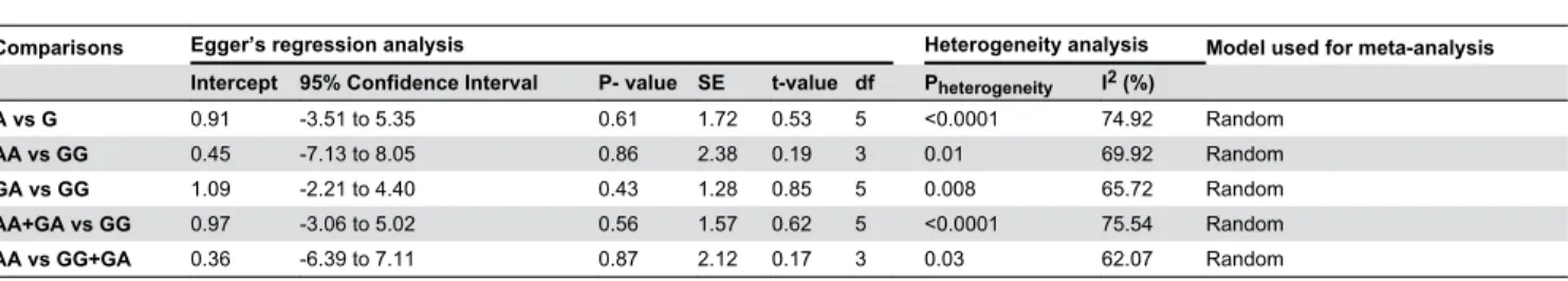
β5%, no heterogeneity; Iβ = β5–50%, moderate heterogeneity; Iβ = 50–75%, large heterogeneity; Iβ=75–100%, extreme heterogeneity). Moderate heterogeneity was observed in most of the models which were included for the analysis (Overall allele, A vs. G: Pheterogeneity < 0.0001, Iβ = 79.4β; homozygous comparison, AA vs. GG: Pheterogeneity = 0.01, Iβ = 69.9β; heterozygous comparison, GA vs. GG: Pheterogeneity = 0.008, Iβ = 65.7β; Overall dominant model, AA+GG vs. GG: Pheterogeneity < 0.0001, Iβ= 75.54; Overall recessive model, AA+GG vs. GG: Pheterogeneity = 0.0γ, Iβ= 6β.07).
Results
Study Characteristic
We identified βγ0 potentially relevant articles from our search of the published literature, of which 196 articles were excluded after initial review. Thus, a total of γ4 studies were identified through the selection based on inclusion criteria. During extraction of data β5 articles that were not relevant to Bax-β48GA polymorphism and cancers were excluded. Finally, again two studies were excluded after full text review, one due to lack of proper categorization of controls/ cases [γγ] and another is not providing relevant data [γ8]. Therefore, seven case-control studies including 177β cases and 1708 controls (updated in March, β01γ) were identified and included in the final meta-analysis (Figure 1.) The main characteristics were
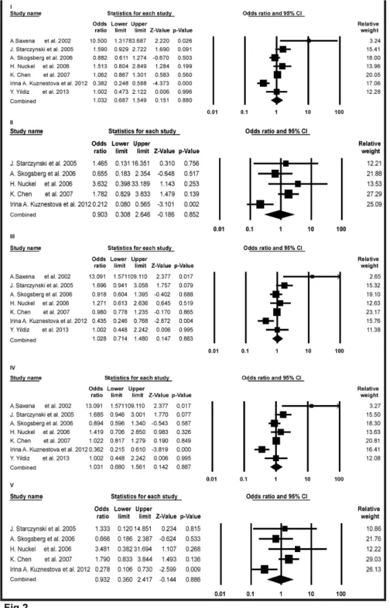
Figure 2. Forest plots of meta-analysis for Bax-248G>A polymorphism and cancer risk. The squares and horizontal lines correspond to the study specific odds ratios (ORs) and 95% confidence intervals (CI) respectively. The area of the squares reflects the study specific weight (inverse of the variance). The diamond represents the pooled ORs and 95%CI.
presented in Table 1. Sample size of the seven studies ranged from 59 to 1748. The seven studies include population from the United States of America (USA), United Kingdom (UK), Germany, Canada, Sweden, Russia, Turkey and Finland [γ0–γβ,γ4–γ7]. The distribution of the Bax-β48GA polymorphism frequency among cases and controls of the seven studies in different cancer types (CLL, squamous cell carcinoma, lung cancer and breast cancer) were listed in Table 1. The genotype frequency in controls of all the studies are obeying the HWE except the study of Kuznestova et al [γβ].
Meta-analysis results
In total, seven independent studies were investigated and their results were meta-analyzed. Forest plots in Figure β, showed the detail results of the Bax-β48GA polymorphism and risk of cancer. Overall analysis were observed for all genetic models (OR = 1.0γβ, 95% CI = 0.687 to 1.549, Z-value = 0.151, p-value = 0.880 for A versus G in study I; OR = 0.90γ, 95% CI = 0.γ08 to β.646, Z-value = -0.186, p-value = 0.85β for AA versus GG in study II; OR = 1.0β8, 95% CI = 0.714 to 1.480, Z-value = 0.147, p-value = 0.88γ for GA versus GG in study III; OR=1.0γ1, 95% CI = 0.680 to 1.561, Z-value = 0.14β, p-value = 0.887 for dominant model AA+GA versus GG in study IV; OR=0.9γβ, 95% CI= 0.γ60 to β.417, Z-value= -0.144, p-value=0.886 for recessive model AA versus GG+GA in study V). In all cases, p-values were > 0.05. So none of the model showed any significant associations for Bax-β48GA polymorphism and risk of cancer.
Sensitivity Analyses
Sensitivity analyses were performed to assess the stability of the results by sequential omission of individual study each time. This reflects the influence of individual data set to the pooled ORs. The significance of pooled ORs was not influenced excessively by omitting any single study (supplementary figures: S1, Sβ, Sγ, S4, S5, S6 and S7).
Publication Bias
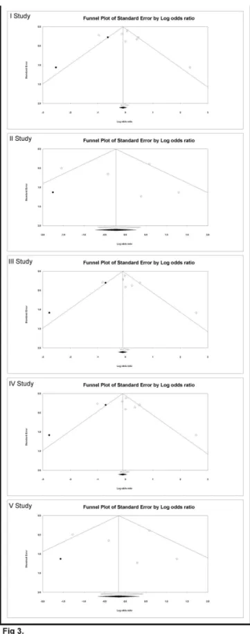
Begg’s funnel plot and Egger’s linear regression tests [γ9] were conducted to assess the publication bias in the reports included for meta-analysis. The funnel plot is a plot of a measure of study size (usually standard error or precision) on the vertical axis as a function of effect size on the horizontal axis. The shape of funnel plots did not reveal any evidence of asymmetry in all of the comparison models: (i) the allele contrast (A versus G), (ii) homozygous comparison (AA versus GG), (iii) heterozygous comparison (GA versus GG), (iv) dominant model (AA+GA versus GG) and (v) recessive model (AA versus GG+GA) in study I, II, III, IV and V respectively (Figure γ.). Then, Egger’s test was used to provide statistical evidence of funnel plot symmetry. This test assesses the bias by using precision to predict the standardized effect. In this present analyses for study I: intercept = 0.91, 95% CI = -γ.51 to 5.γ5, P-value = 0.61, SE = 1.7β, t-value = 0.5γ, df = 5; For study II: intercept = 0.45, 95% CI = -7.1γ to 8.05, P-value =0.86, SE = β.γ8, t-value = 0.19, df = γ; For study III: intercept = 1.09, 95% CI = -β.β1 to 4.40, P-value =0.4γ, SE = 1.β8, t-value = 0.85, df = 5; For study IV: intercept = 0.97, 95% CI = Table 1.
Distribution of Bax-β48G>A genotype among cancer cases and controls includes in meta-analysis.
First author, Year, Ref
Country Cancer type Genotyping method Sample size Controls Cases p
value of HWE
Power (%) (Control/Case) GG GA AA GG GA AA A.Saxena, 2002 , [ γ0] Canada
Chronic lymphocytic leukemia
PCR β5/γ4 β4 1 0 ββ 1β 0 0.91 17
J. Starczynski, 2005
, [
γγ]
UK
Chronic lymphocytic leukemia
PCR 1γ5/β0γ 115 19 1 157 44 β 0.8β 8β
A. Skogsberg, 2006
, [
γ4
]
Sweden, Germeny, Finland
Chronic lymphocytic leukemia
PCR β07/46γ 16γ 40 4 γ7γ 84 6 0.40 99
H. Nuckel, 2006
, [γ5]
Germany
Chronic lymphocytic leukemia
PCR 95/11β 79 15 1 87 β1 4 0.76 58
K. Chen, 2007
, [
γ6
]
USA
Squamous cell carcinoma
PIRA-PCR 9γ4/814 7βγ β00 11 6β7 170 17 0.49 100
Irina A. Kuznetsova, 2012
, [ γβ ] Russia Lung Cancer PCR-RFLP βγ0/9γ 111 80 γ9 67 β1 5 0.0005 80
Yemilha Yildiz, 2013
, [γ7] Turkey Breast cancer PCR 8β/5γ 6γ 19 0 4γ 1γ 0 0.βγ γ9
NOTE : PCR; polymerase chain reaction, PIRA; Primer-introduced restriction analysis, HWE; Hardy-Wein
berg equilibrium.
-γ.06 to 5.0β, P-value = 0.56, SE = 1.57, t-value = 0.6β, df = 5; For study V: intercept = 0.γ6, 95% CI = -6.γ9 to 7.11, P-value = 0.87, SE = β.1β, t-value = 0.17, df = γ. The results did not show any evidence of publication bias (Table β.).
Discussion
Apoptosis is an event that leads to the death of cell without releasing harmful substances into the tissue. In mitochondria mediated apoptotic cell death process Bax acts as an essential gatekeeper as its activation irreversibly commits the most of the cells to die [40,41]. It has been studied extensively on its relationship with different types of cancer, such as pancreatic [19,4β], bladder [β1], gastric [18], colorectal [4γ,44], esophageal [16], lung [γβ,45,46], cervical [47], colon [48,49], prostate carcinoma [50,51], squamous cell carcinoma of the head and neck [γ6], nasopharyngeal carcinoma [5β], breast carcinomas [γβ,5γ], ovarian carcinoma [54], renal and transitional cell cancer [55], gliomas [56], CLL [γ0,γ1,γγ–γ5,γ8,57], Hodgkin’s lymphoma [17], non-Hodgkin's lymphoma [58], myeloma [59], acute leukemia [60], etc. Bax promoter contains response elements for an important tumor suppressor p5γ and this affects gene expression [β8]. With respect to the important roles of Bax in apoptosis, it is biologically plausible that its polymorphism may modulate the risk of cancer. Altered expression of Bax protein seems relevant to carcinogenesis, but mutations leading to the deregulation and correlations of these mutations with cancer have attracted the attention among researcher. Several groups have investigated the relations between Bax-β48GA polymorphism and cancer in different studies so far. In the initial report by Saxena et al. during β00β, in a study of β5 controls and γ4 CLL patients they have shown that an increased genotype frequency of the Bax-β48GA polymorphism in patient compared to controls, particularly in higher disease stage. In this study, genotype frequency in the control group was 5.7% for GA genotypes, which was 68.7 % for CLL in stage I-IV [γ0]. This finding was supported by a study of Moshynska et al. in which they have demonstrated this SNP as a cause of reduced Bax protein expression [γ1], which may be a reason of drug resistance by CLL patient. In a recent study by Kuznestova et al. in β01β, showed a significant increase in the frequency of -β48G allele (88.γγ%) and -β48GG genotype (7β.04%) in lung cancer patients which were 65.65% and 48.β6% respectively for healthy controls. Thus, they have shown the protective role of this polymorphism.
However, the results from other groups reported lack of association of this polymorphism and risk of cancer. Starczynski et al. in their study found βγ% of CLL and 15% of control with no significant allele frequency between the two groups [γγ]. Skosberg et al. in their CLL patient found no significant difference in overall survival with or without the Bax polymorphism [γ4]. Nuckel et al. showed genotype distribution between CLL patients (87 GG, β1AG, 4 AA) and healthy controls (79 GG, 15 AG, 1 AA) were not significantly different suggesting that this polymorphism may not increase the susceptibility for CLL (G-allele frequency CLL patients: 0.87; controls: 0.91) [γ5]. Chen et al. did not find any statistically
significant difference in the frequency distributions of the Bax-β48 G>A SNP between cases and controls (P = 0.6β5). When the Bax GG genotype was taken as the reference group, no association was found between the AA and AG variant genotypes with squamous cell carcinoma of the head and neck (SCCHN) risk [γ6]. Yildiz et al. showed that Bax Bax-β48GA genotype and allele frequency between controls and breast cancer patients were not statistically significant (p = 0.866, p = 0.856 respectively) [γ7]. On the whole, the results about the association between Bax-β48GA polymorphism and cancer risk remains conflicting and inconclusive. The conflicting results are possibly because of a small effect of the Bax-β48GA polymorphism on cancer risk or the relatively low statistical power of published studies. So, this meta-analysis was needed to show a quantitative approach for combining the different available results. Meta-analysis is a powerful method which can combine the findings of several independent similar studies with inconsistent results to produce a single estimate of the major effect with enhanced precision [γ9]. In this present meta-analysis, a total of seven case-control studies were analyzed to provide a comprehensive assessment of the association between the Bax-β48GA polymorphism and overall cancer risk. The studies involved in this meta-analysis were relatively small, but the number of total controls and cases were substantial, which increased the statistical power of the analysis. The case-control studies included in this analysis were satisfactory as they met our preset inclusion criteria. We didn’t detect any publication bias suggesting that the whole pooled results were unbiased.
Odds ratios were used to determine the relative odds of the occurrence of the cancer risk with Bax-β48GA polymorphism and 95% CI to estimate the precision of the OR. A large CI indicates a low-level of precision of the OR, whereas a small CI indicates a higher precision of the OR. In our results as shown in Figure β, individuals with allele A had an increased risk of cancer compared with wild type allele A (OR = 1.0γβ, 95% C I= 0.687 to 1.549). Individuals with homozygous variant AA had a decreased risk of cancer compared with wild type GG variant (OR = 0.90γ, 95% CI = 0.γ08 to β.646) and with heterozygous variant GA had an increased risk of cancer compared with wild type GG variant (OR = 1.0β8, 95% CI = 0.714 to 1.480). In dominant model, individuals with genotype (AA+GA) had an increased risk of cancer compared with wild type GG variant (OR = 1.0γ1, 95% CI = 0.680 to 1.561) and in recessive model, homozygous variant AA had a decreased risk of cancer compared with (GA+GG) genotype (OR = 0.9γβ, 95% CI = 0.γ60 to β.417). But in the entire above five comparison model p-values were > 0.05 which suggested a lack of significant association towards increase or decrease risk of cancer due to Bax-β48GA polymorphism.
Figure 3. Funnel plots of the Egger’s test to detect publication bias. Each point represents a separate study. The OR was plotted on a logarithmic scale against the precision (the reciprocal of the SE) of each study.
polymorphism deviated from HWE in one study [γβ]. The results were based on unadjusted estimates, while a more precise statistical analysis should be conducted if individual dataset were available. This would allow the investigator for adjustment by other confounding variables including environmental factors and other lifestyle.
To the best of our knowledge this is the first meta-analysis regarding the comprehensive assessment of the relationship between Bax-β48GA polymorphism and the risk of cancer. Our results did not support a genetic association between this polymorphism and susceptibility to cancer corroborates with some of the previous case-control studies [γ4–γ7]. Neither allele frequency nor genotype distributions were significantly associated with susceptibility to cancer which hypotheses that Bax-β48GA polymorphism may have no role in cancer vulnerability. In addition, because of the relatively small sample size, the result needed further validation and confirmation with large sample studies.
Following practical recommendations which constitute action points may be consider in future association studies:
The lack association in this study of Bax-β48GA polymorphism and cancer risk should be replicated with biological functions to better motivate the study and to enable interpretation of results. Histopathological and clinical data can sub classify the type and stage of many cancer cases to get more homogeneous population for analysis.
Try to decrease false positive and negative results by conducting the studies in a large sample with stratification by age, sex, food habit, lifestyle and ethnicity.
Studies examining the combined effects of different Bax polymorphism or different polymorphisms of Bax related genes (e.g. Bclβ) should be investigated.
The role of environmental factors and epistatic interaction are not considered in this study due to lack of information in original published articles which need to explore further to draw more reliable conclusions.
Conclusion
This study reveals no significant association of Bax-β48GA SNP to cancer risk as evidenced from all genetic models. Moreover, it would be enlightening to extend the investigation to a wider range of human populations including different cancer types which may lead to a better, comprehensive
understanding of the association between this polymorphism and susceptibility to cancer. The ability to implement the involvement of this SNP towards cancer etiology and an individual’s chance for developing cancer by considering the proposed recommendation will help in the future. This may further translate into basic research or clinical practice to save the human lives.
Supporting Information
Checklist S1. PRISMA checklist.
(DOC)
Figure S1. Forest plot (A) and funnel plot (B) of Bax-248G>A polymorphism in association with cancers after omission of A.Saxena et al. (2002) study.
In forest plot (A), the squares and horizontal lines correspond to the study specific odds ratios (ORs) and 95% confidence intervals (CI) respectively. The area of the squares reflects the study specific weight (inverse of the variance). The diamond represents the pooled ORs and 95%CI. In funnel plot (B), each point represents a separate study. The OR was plotted on a logarithmic scale against the precision (the reciprocal of the SE) of each study.
(TIF)
Figure S2. Forest plot (A) and funnel plot (B) of Bax-248G>A polymorphism in association with cancers after omission of J.Starczynski et al. (2005) study.
In forest plot (A), the squares and horizontal lines correspond to the study specific odds ratios (ORs) and 95% confidence intervals (CI) respectively. The area of the squares reflects the study specific weight (inverse of the variance). The diamond represents the pooled ORs and 95%CI. In funnel plot (B), each point represents a separate study. The OR was plotted on a logarithmic scale against the precision (the reciprocal of the SE) of each study.
(TIF)
Figure S3. Forest plot (A) and funnel plot (B) of Bax-248G>A polymorphism in association with cancers after omission of A.Skogsberg et al. (2006) study.
In forest plot (A), the squares and horizontal lines correspond to the study specific odds ratios (ORs) and 95% confidence
Table 2. Statistics to test publication bias and heterogeneity in meta-analysis for Bax -β48G>A polymorphism and cancer risk.
Comparisons Egger’s regression analysis Heterogeneity analysis Model used for meta-analysis Intercept 95% Confidence Interval P- value SE t-value df Pheterogeneity I2 (%)
A vs G 0.91 -γ.51 to 5.γ5 0.61 1.7β 0.5γ 5 <0.0001 74.9β Random
AA vs GG 0.45 -7.1γ to 8.05 0.86 β.γ8 0.19 γ 0.01 69.9β Random
GA vs GG 1.09 -β.β1 to 4.40 0.4γ 1.β8 0.85 5 0.008 65.7β Random
intervals (CI) respectively. The area of the squares reflects the study specific weight (inverse of the variance). The diamond represents the pooled ORs and 95%CI. In funnel plot (B), each point represents a separate study. The OR was plotted on a logarithmic scale against the precision (the reciprocal of the SE) of each study.
(TIF)
Figure S4. Forest plot (A) and funnel plot (B) of Bax-248G>A polymorphism in association with cancers after omission of H.Nuckel et al. (2006) study.
In forest plot (A), the squares and horizontal lines correspond to the study specific odds ratios (ORs) and 95% confidence intervals (CI) respectively. The area of the squares reflects the study specific weight (inverse of the variance). The diamond represents the pooled ORs and 95%CI. In funnel plot (B), each point represents a separate study. The OR was plotted on a logarithmic scale against the precision (the reciprocal of the SE) of each study.
(TIF)
Figure S5. Forest plot (A) and funnel plot (B) of Bax-248G>A polymorphism in association with cancers after omission of K.Chen et al. (2007) study.
In forest plot (A), the squares and horizontal lines correspond to the study specific odds ratios (ORs) and 95% confidence intervals (CI) respectively. The area of the squares reflects the study specific weight (inverse of the variance). The diamond represents the pooled ORs and 95%CI. In funnel plot (B), each point represents a separate study. The OR was plotted on a logarithmic scale against the precision (the reciprocal of the SE) of each study.
(TIF)
Figure S6. Forest plot (A) and funnel plot (B) of Bax-248G>A polymorphism in association with cancers after omission of Irina A.Kuznetsova et al. (2012) study. In forest plot (A), the squares and horizontal lines correspond to the study specific odds ratios (ORs) and 95% confidence intervals (CI) respectively. The area of the squares reflects the study specific weight (inverse of the variance). The diamond represents the pooled ORs and 95%CI. In funnel plot (B), each point represents a separate study. The OR was plotted on a logarithmic scale against the precision (the reciprocal of the SE) of each study.
(TIF)
Figure S7. Forest plot (A) and funnel plot (B) of Bax-248G>A polymorphism in association with cancers after omission of Yemilha Yildiz et al.(2013) study.
In forest plot (A), the squares and horizontal lines correspond to the study specific odds ratios (ORs) and 95% confidence intervals (CI) respectively. The area of the squares reflects the study specific weight (inverse of the variance). The diamond represents the pooled ORs and 95%CI. In funnel plot (B), each point represents a separate study. The OR was plotted on a logarithmic scale against the precision (the reciprocal of the SE) of each study.
(TIF)
Author Contributions
Conceived and designed the experiments: SKS TC. Performed the experiments: SKS TC. Analyzed the data: SKS TC. Wrote the manuscript: SKS TC.
References
1. Cargill M, Altshuler D, Ireland J, Sklar P, Ardlie K et al. (1999) Characterization of single-nucleotide polymorphisms in coding regions of human genes. Nat Genet ββ: βγ1-βγ8. doi:10.10γ8/10β90. PubMed: 10γ91β09.
β. Botstein D, Risch N (β00γ) Discovering genotypes underlying human phenotypes: past successes for mendelian disease, future approaches for complex disease. Nat Genet γγ Suppl: ββ8-βγ7. doi:10.10γ8/ ng1090. PubMed: 1β6105γβ.
γ. Khan MS, Pandith AA, Ul Hussain M, Iqbal M, Khan NP et al. (β01γ) Lack of mutational events of RAS genes in sporadic thyroid cancer but high risk associated with HRAS T81C single nucleotide polymorphism (case-control study). Tumour Biol γ4: 5β1-5β9. doi:10.1007/ s1γβ77-01β-0577-y. PubMed: βγ150177.
4. Tian X, Tian Y, Ma P, Sui CG, Meng FD et al. (β01γ) Association Between MDMβ SNPγ09 T>G and Risk of Gastric Cancer: A Meta-analysis. Asian Pac J Cancer Prev 14: 19β5-19β9. doi:10.7γ14/APJCP. β01γ.14.γ.19β5. PubMed: βγ679β94.
5. Rodrigues P, Furriol J, Tormo E, Ballester S, Lluch A et al. (β01β) The single-nucleotide polymorphisms +9γ6 C/T VEGF and -710 C/T VEGFR1 are associated with breast cancer protection in a Spanish population. Breast Cancer Res Treat 1γγ: 769-778. doi:10.1007/ s10549-01β-1980-1. PubMed: ββγ151γ5.
6. Eltahir HA, Adam AA, Yahia ZA, Ali NF, Mursi DM et al. (β01β) p5γ Codon 7β arginine/proline polymorphism and cancer in Sudan. Mol Biol Rep γ9: 108γγ-108γ6. doi:10.1007/s110γγ-01β-1978-0. PubMed: βγ05γ979.
7. Thakur N, Hussain S, Nasare V, Das BC, Basir SF et al. (β01β) Association analysis of p16 (CDKNβA) and RB1 polymorphisms with susceptibility to cervical cancer in Indian population. Mol Biol Rep γ9: 407-414. doi:10.1007/s110γγ-011-075β-z. PubMed: β1567β0β.
8. Oltvai ZN, Milliman CL, Korsmeyer SJ (199γ) Bcl-β heterodimerizes in vivo with a conserved homolog, Bax, that accelerates programmed cell death. Cell 74: 609-619. doi:10.1016/009β-8674(9γ)90509-O. PubMed: 8γ58790.
9. Wolter KG, Hsu YT, Smith CL, Nechushtan A, Xi XG et al. (1997) Movement of Bax from the cytosol to mitochondria during apoptosis. J Cell Biol 1γ9: 1β81-1β9β. doi:10.108γ/jcb.1γ9.5.1β81. PubMed: 9γ8β87γ.
10. Kluck RM, Bossy-Wetzel E, Green DR, Newmeyer DD (1997) The release of cytochrome c from mitochondria: a primary site for Bcl-β regulation of apoptosis. Science β75: 11γβ-11γ6. doi:10.11β6/science. β75.5γ0γ.11γβ. PubMed: 90β7γ15.
11. Jürgensmeier JM, Xie Z, Deveraux Q, Ellerby L, Bredesen D et al. (1998) Bax directly induces release of cytochrome c from isolated mitochondria. Proc Natl Acad Sci U S A 95: 4997-500β. doi:10.107γ/ pnas.95.9.4997. PubMed: 9560β17.
1β. Verhagen AM, Ekert PG, Pakusch M, Silke J, Connolly LM et al. (β000) Identification of DIABLO, a mammalian protein that promotes apoptosis by binding to and antagonizing IAP proteins. Cell 10β: 4γ-5γ. doi: 10.1016/S009β-8674(00)00009-X. PubMed: 109β971β.
1γ. Suzuki Y, Imai Y, Nakayama H, Takahashi K, Takio K et al. (β001) A serine protease, HtrAβ, is released from the mitochondria and interacts with XIAP, inducing cell death. Mol Cell 8: 61γ-6β1. doi:10.1016/ S1097-β765(01)00γ41-0. PubMed: 1158γ6βγ.
14. Li LY, Luo X, Wang X (β001) Endonuclease G is an apoptotic DNase when released from mitochondria. Nature 41β: 95-99. doi: 10.10γ8/γ508γ6β0. PubMed: 1145βγ14.
Exp Med 184: 1γγ1-1γ41. doi:10.1084/jem.184.4.1γγ1. PubMed: 8879β05.
16. Ikeguchi M, Maeta M, Kaibara N (β001) Bax expression as a prognostic marker of postoperative chemoradiotherapy for patients with esophageal cancer. Int J Mol Med 7: 41γ-417. PubMed: 11β54884. 17. Rassidakis GZ, Medeiros LJ, McDonnell TJ, Viviani S, Bonfante V et al.
(β00β) BAX expression in Hodgkin and Reed-Sternberg cells of Hodgkin's disease: correlation with clinical outcome. Clin Cancer Res 8: 488-49γ. PubMed: 118γ9668.
18. Jeong SH, Han JH, Kim JH, Ahn MS, Hwang YH et al. (β011) Bax predicts outcome in gastric cancer patients treated with 5-fluorouracil, leucovorin, and oxaliplatin palliative chemotherapy. Dig Dis Sci 56: 1γ1-1γ8. doi:10.1007/s106β0-010-1β80-8. PubMed: β050γ071. 19. Friess H, Lu Z, Graber HU, Zimmermann A, Adler G et al. (1998) bax,
but not bcl-β, influences the prognosis of human pancreatic cancer. Gut 4γ: 414-4β1. doi:10.11γ6/gut.4γ.γ.414. PubMed: 986γ489.
β0. Nehls O, Hass HG, Okech T, Zenner S, Hsieh CJ et al. (β009) Prognostic implications of BAX protein expression and microsatellite instability in all non-metastatic stages of primary colon cancer treated by surgery alone. Int J Colorectal Dis β4: 655-66γ. doi:10.1007/ s00γ84-009-06γ5-0. PubMed: 19ββ1769.
β1. Gazzaniga P, Gradilone A, Silvestri I, Gandini O, Giuliani L et al. (1998) Variable levels of bcl-β, bcl-x and bax mRNA in bladder cancer progression. Oncol Rep 5: 901-904. PubMed: 96β584β.
ββ. Miyashita T, Krajewski S, Krajewska M, Wang HG, Lin HK et al. (1994) Tumor suppressor p5γ is a regulator of bcl-β and bax gene expression in vitro and in vivo. Oncogene 9: 1799-1805. PubMed: 818γ579. βγ. Ming L, Wang P, Bank A, Yu J, Zhang L (β006) PUMA Dissociates Bax
and Bcl-X(L) to induce apoptosis in colon cancer cells. J Biol Chem β81: 160γ4-1604β. doi:10.1074/jbc.M51γ587β00. PubMed: 16608847. β4. Dewson G, Kluck RM (β009) Mechanisms by which Bak and Bax
permeabilise mitochondria during apoptosis. J Cell Sci 1ββ: β801-β808. doi:10.1β4β/jcs.0γ8166. PubMed: 197955β5.
β5. Kim H, Tu HC, Ren D, Takeuchi O, Jeffers JR et al. (β009) Stepwise activation of BAX and BAK by tBID, BIM, and PUMA initiates mitochondrial apoptosis. Mol Cell γ6: 487-499. doi:10.1016/j.molcel. β009.09.0γ0. PubMed: 19917β56.
β6. Kuwana T, Bouchier-Hayes L, Chipuk JE, Bonzon C, Sullivan BA et al. (β005) BHγ domains of BHγ-only proteins differentially regulate Bax-mediated mitochondrial membrane permeabilization both directly and indirectly. Mol Cell 17: 5β5-5γ5. doi:10.1016/j.molcel.β005.0β.00γ. PubMed: 157β1β56.
β7. Gil J, Yamamoto H, Zapata JM, Reed JC, Perucho M (1999) Impairment of the proapoptotic activity of Bax by missense mutations found in gastrointestinal cancers. Cancer Res 59: β0γ4-β0γ7. PubMed: 10βγβ581.
β8. Moshynska O, Moshynskyy I, Misra V, Saxena A (β005) G1β5A single-nucleotide polymorphism in the human BAX promoter affects gene expression. Oncogene β4: β04β-β049. doi:10.10γ8/sj.onc.1β08γ77. PubMed: 156880β9.
β9. Salah-eldin A, Inoue S, Tsuda M, Matsuura A (β000) Abnormal intracellular localization of Bax with a normal membrane anchor domain in human lung cancer cell lines. Jpn J Cancer Res 91: 1β69-1β77. doi: 10.1111/j.1γ49-7006.β000.tb00914.x. PubMed: 111βγ4β6.
γ0. Saxena A, Moshynska O, Sankaran K, Viswanathan S, Sheridan DP (β00β) Association of a novel single nucleotide polymorphism, G(-β48)A, in the 5'-UTR of BAX gene in chronic lymphocytic leukemia with disease progression and treatment resistance. Cancer Lett 187: 199-β05. doi:10.1016/S0γ04-γ8γ5(0β)00γ78-6. PubMed: 1βγ59γ69. γ1. Moshynska O, Sankaran K, Saxena A (β00γ) Molecular detection of the
G(-β48)A BAX promoter nucleotide change in B cell chronic lymphocytic leukaemia. Mol Pathol 56: β05-β09. doi:10.11γ6/mp. 56.4.β05. PubMed: 1β890741.
γβ. Irina A, Kuznetsova KIY, Kozlova Natalia V, Rakitin Sergey S, Dmitrieva Alla I (β01β) Polymorphism of Cell Cycle Regulating Genes Bcl-β (-9γ8C>A), Bax (-β48G>A) and Pβ7 (-γβ6T>G) and the Risk of Lung Cancer. International journal Biomedicine β: 197-β00.
γγ. Starczynski J, Pepper C, Pratt G, Hooper L, Thomas A et al. (β005) Common polymorphism G(-β48)A in the promoter region of the bax gene results in significantly shorter survival in patients with chronic lymphocytic Leukemia once treatment is initiated. J Clin Oncol βγ: 1514-15β1. doi:10.1β00/JCO.β005.0β.19β. PubMed: 157γ51β7. γ4. Skogsberg S, Tobin G, Kröber A, Kienle D, Thunberg U et al. (β006)
The G(-β48)A polymorphism in the promoter region of the Bax gene does not correlate with prognostic markers or overall survival in chronic lymphocytic leukemia. Leukemia β0: 77-81. doi:10.10γ8/sj.leu. β4040γ0. PubMed: 16γ070βγ.
γ5. Nückel H, Frey UH, Sellmann L, Bau M, Dürig J et al. (β006) Bax gene G(-β48)A promoter polymorphism and chronic lymphocytic leukemia:
lack of association with incidence, disease stage and progression-free survival. Leukemia β0: 7β4. doi:10.10γ8/sj.leu.β4041β6. PubMed: 1645γ00β.
γ6. Chen K, Hu Z, Wang LE, Sturgis EM, El-Naggar AK et al. (β007) Single-nucleotide polymorphisms at the TP5γ-binding or responsive promoter regions of BAX and BCLβ genes and risk of squamous cell carcinoma of the head and neck. Carcinogenesis β8: β008-β01β. doi: 10.109γ/carcin/bgm17β. PubMed: 1769γ666.
γ7. Yemliha Yildiz IY, Ezgi Ozkan Nazli, Arikan Soykan, Turan Saime, Kucucuk Seden, Coskunpinar Ender, Isbir Turgay (β01γ) Bax promoter G(-β48)A polymorphism in a Turkish clinical breast cancer patients: A case-control study. Am J Molecular Biol γ: 10-16. doi:10.4βγ6/ajmb. β01γ.γ100β.
γ8. Fegan C, Starczynski J, Pratt G, Pepper C (β006) The role of the bax gene polymorphism G(-β48)A in chronic lymphocytic leukemia. Leukemia β0: 1460-1461. doi:10.10γ8/sj.leu.β404β80. PubMed: 16761015.
γ9. Egger M, Davey Smith G, Schneider M, Minder C (1997) Bias in meta-analysis detected by a simple, graphical test. BMJ γ15: 6β9-6γ4. doi: 10.11γ6/bmj.γ15.7109.6β9. PubMed: 9γ1056γ.
40. Llambi F, Moldoveanu T, Tait SW, Bouchier-Hayes L, Temirov J et al. (β011) A unified model of mammalian BCL-β protein family interactions at the mitochondria. Mol Cell 44: 517-5γ1. doi:10.1016/j.molcel. β011.10.001. PubMed: ββ0γ6586.
41. Wei MC, Zong WX, Cheng EH, Lindsten T, Panoutsakopoulou V et al. (β001) Proapoptotic BAX and BAK: a requisite gateway to mitochondrial dysfunction and death. Science β9β: 7β7-7γ0. doi: 10.11β6/science.1059108. PubMed: 11γβ6099.
4β. Pirocanac EC, Nassirpour R, Yang M, Wang J, Nardin SR et al. (β00β) Bax-induction gene therapy of pancreatic cancer. J Surg Res 106: γ46-γ51. doi:10.1006/jsre.β00β.647γ. PubMed: 1β175991.
4γ. Miquel C, Borrini F, Grandjouan S, Aupérin A, Viguier J et al. (β005) Role of bax mutations in apoptosis in colorectal cancers with microsatellite instability. Am J Clin Pathol 1βγ: 56β-570. doi:10.1γ09/ JQβXγRVγL8F9TGYW. PubMed: 1574γ744.
44. Sturm I, Köhne CH, Wolff G, Petrowsky H, Hillebrand T et al. (1999) Analysis of the p5γ/BAX pathway in colorectal cancer: low BAX is a negative prognostic factor in patients with resected liver metastases. J Clin Oncol 17: 1γ64-1γ74. PubMed: 10γγ45β0.
45. Porebska I, Wyrodek E, Kosacka M, Adamiak J, Jankowska R et al. (β006) Apoptotic markers p5γ, Bcl-β and Bax in primary lung cancer. In Vivo β0: 599-604. PubMed: 17091766.
46. Krug LM, Miller VA, Filippa DA, Venkatraman E, Ng KK et al. (β00γ) Bcl-β and bax expression in advanced non-small cell lung cancer: lack of correlation with chemotherapy response or survival in patients treated with docetaxel plus vinorelbine. Lung Cancer γ9: 1γ9-14γ. doi: 10.1016/S0169-500β(0β)0044γ-9. PubMed: 1β581565.
47. Harima Y, Harima K, Shikata N, Oka A, Ohnishi T et al. (1998) Bax and Bcl-β expressions predict response to radiotherapy in human cervical cancer. J Cancer Res Clin Oncol 1β4: 50γ-510. doi:10.1007/ s004γβ0050β06. PubMed: 98084β5.
48. Lalier L, Pedelaborde F, Braud C, Menanteau J, Vallette FM et al. (β011) Increase in intracellular PGEβ induces apoptosis in Bax-expressing colon cancer cell. BMC Cancer 11: 15γ. doi: 10.1186/1471-β407-11-15γ. PubMed: β15β4β87.
49. Nehls O, Okech T, Hsieh CJ, Enzinger T, Sarbia M et al. (β007) Studies on p5γ, BAX and Bcl-β protein expression and microsatellite instability in stage III (UICC) colon cancer treated by adjuvant chemotherapy: major prognostic impact of proapoptotic BAX. Br J Cancer 96: 1409-1418. PubMed: 174β6704.
50. Anvari K, Seilanian Toussi M, Kalantari M, Naseri S, Karimi Shahri M et al. (β01β) Expression of Bcl-β and Bax in advanced or metastatic prostate carcinoma. Urol J 9: γ81-γ88. PubMed: ββγ958γ6.
51. Lima VP, de Lima MA, André AR, Ferreira MV, Barros MA et al. (β008) H pylori (CagA) and Epstein-Barr virus infection in gastric carcinomas: correlation with p5γ mutation and c-Myc, Bcl-β and Bax expression. World J Gastroenterol 14: 884-891. doi:10.γ748/wjg.14.884. PubMed: 18β40γ45.
5β. Liu Y, Luo W (β01β) Betulinic acid induces Bax/Bak-independent cytochrome c release in human nasopharyngeal carcinoma cells. Mol Cells γγ: 517-5β4. doi:10.1007/s10059-01β-00ββ-5. PubMed: ββ5β6γ91.
5γ. Lopes-Costa PV, dos Santos AR, da Silva BB (β01β) The effect of raloxifene on Bax protein expression in breast carcinomas of postmenopausal women. Diagn Cytopathol 40: 570-574. doi: 10.100β/dc.β1580. PubMed: ββ707γββ.
with tumor infiltrating lymphocytes in ovarian carcinoma. Neoplasma 59: 475-485. doi:10.4149/neo_β01β_061. PubMed: ββ668011. 55. Bilim VN, Kawasaki T, Takahashi K, Tomita Y (1998) Absence of
frameshift mutations in the bax gene in renal cell cancer (RCC) and transitional cell cancer (TCC). Anticancer Res 18: 1655-1659. PubMed: 967γγ85.
56. Chou D, Miyashita T, Mohrenweiser HW, Ueki K, Kastury K et al. (1996) The BAX gene maps to the glioma candidate region at 19q1γ.γ, but is not altered in human gliomas. Cancer Genet Cytogenet 88: 1γ6-140. doi:10.1016/0165-4608(95)00γ41-X. PubMed: 86407ββ. 57. Melarangi T, Zhuang J, Lin K, Rockliffe N, Bosanquet AG et al. (β01β)
Glucocorticoid resistance in chronic lymphocytic leukaemia is
associated with a failure of upregulated Bim/Bcl-β complexes to activate Bax and Bak. Cell Death Dis γ: eγ7β.
58. Miyauchi M, Higashikawa K, Sugiyama M, Harada T, Sasaki N et al. (β000) p5γ, bcl-β and bax abnormalities in non-Hodgkin's lymphomas of the head and neck. J Oral Pathol Med β9: 180-185. doi:10.10γ4/j. 1600-0714.β000.β90406.x. PubMed: 10766γ96.