Polymorphism and Cancer Risk: A Meta-Analysis
Mei-Ling Zhu
1,2,3., Mengyun Wang
2,3., Zhi-Gang Cao
2,4., Jing He
1,2,3, Ting-Yan Shi
1,2,3, Kai-Qin Xia
1,2,3,
Li-Xin Qiu
1,2,3*
, Qing-Yi Wei
3,5*
1Department of Oncology, Fudan University Shanghai Cancer Center, Shanghai, China,2Department of Oncology, Shanghai Medical College, Fudan University, Shanghai, China,3Cancer Research Laboratory, Fudan University Shanghai Cancer Center, Shanghai, China,4Department of Breast Surgery, Cancer Center and Cancer Institute, Fudan University, Shanghai, China,5Department of Epidemiology, The University of Texas MD Anderson Cancer Center, Houston, Texas, United States of America
Abstract
Background:
Excision repair cross complementing group 5 (
ERCC5
or
XPG
) plays an important role in regulating DNA
excision repair, removal of bulky lesions caused by environmental chemicals or UV light. Mutations in this gene cause a rare
autosomal recessive syndrome, and its functional single nucleotide polymorphisms (SNPs) may alter DNA repair capacity
phenotype and cancer risk. However, a series of epidemiological studies on the association between the
ERCC5
Asp1104His
polymorphism (rs17655, G
.
C) and cancer susceptibility generated conflicting results.
Methodology/Principal Findings:
To derive a more precise estimation of the association between the
ERCC5
Asp1104His
polymorphism and overall cancer risk, we performed a meta-analysis of 44 published case-control studies, in which a total of
23,490 cases and 27,168 controls were included. To provide additional biological plausibility, we also assessed the
genotype-gene expression correlation from the HapMap phase II release 23 data with 270 individuals from 4 ethnic
populations. When all studies were pooled, we found no statistical evidence for a significantly increased cancer risk in the
recessive genetic models (His/His vs. Asp/Asp: OR = 0.99, 95% CI: 0.92–1.06,
P
= 0.242 for heterogeneity or His/His vs. Asp/His
+
Asp/Asp: OR = 0.98, 95% CI: 0.93–1.03,
P
= 0.260 for heterogeneity), nor in further stratified analyses by cancer type,
ethnicity, source of controls and sample size. In the genotype-phenotype correlation analysis from 270 individuals, we
consistently found no significant correlation of the Asp1104His polymorphism with
ERCC5
mRNA expression.
Conclusions/Significance:
This meta-analysis suggests that it is unlikely that the
ERCC5
Asp1104His polymorphism may
contribute to individual susceptibility to cancer risk.
Citation:Zhu M-L, Wang M, Cao Z-G, He J, Shi T-Y, et al. (2012) Association between theERCC5Asp1104His Polymorphism and Cancer Risk: A Meta-Analysis. PLoS ONE 7(7): e36293. doi:10.1371/journal.pone.0036293
Editor:Rui Medeiros, IPO, Inst Port Oncology, Portugal
ReceivedJanuary 31, 2012;AcceptedMarch 29, 2012;PublishedJuly 18, 2012
Copyright:ß2012 Zhu et al. This is an open-access article distributed under the terms of the Creative Commons Attribution License, which permits unrestricted use, distribution, and reproduction in any medium, provided the original author and source are credited.
Funding:This study was supported by a grant from ‘‘China’s Thousand Talents Program’’ Recruitment at Fudan University, a grant from the Ministry of Health (201002007) and the National Natural Science Foundation of China (81101808). The funders had no role in study design, data collection and analysis, decision to publish, or preparation of the manuscript.
Competing Interests:The authors have declared that no competing interests exist.
* E-mail: [email protected] (L-XQ); [email protected] (Q-YW)
.These authors contributed equally to this work.
Introduction
Exposure to environmental carcinogens can cause different
types of DNA damage that subsequently lead to carcinogenesis of
different tissues, if left unrepaired. During the evolution, humans
have developed a versatile DNA repair machinery to ensure
genome integrity in response to the insults of cancer-causing
agents. DNA repair is a complex biological process consisting of
several distinct pathways. To date, more than 150 human DNA
repair genes have been identified in five major pathways:
nucleotide excision repair (NER), base excision repair (BER),
mismatch repair (MMR), double-strand break repair (DSBR), and
transcription coupled repair (TCR). Of those pathways, NER is
the most studied DNA repair mechanism responsible for various
types of DNA damage, including thymidine dimers, oxidative
DNA damage, bulky adducts cross-links, and alkylating damage
[1]. At least eight core genes (i.e.,
ERCC1, XPA, XPB/ERCC3,
XPC, XPD/ERCC2, XPE/DDB1, XPF/ERCC4,
and
XPG/ERCC5
)
in the NER pathway play vital roles in repairing DNA damage
and maintain genome integrity [2,3].
PCNA could be involved in triggering the 3
9
incision in NER.
ERCC5 cleaves the 5
9
flap, splayed arm and a variety of bubble
substrates at ss/dsDNA junctions with the 5
9
overhang and makes
the 3
9
incision in NER [14] (
Figure 1
).
As a structure-specific endonuclease and also a 5
9
-3
9
exo-nuclease, the ERCC5 protein is required for two sub-pathways in
NER. One is TCR, which preferentially removes DNA damage in
the transcribed DNA strand of active genes; the other is global
genomic repair (GGR), which removes lesions throughout the
genome [15,16]. Additionally, ERCC5 also possesses some
secondary functions independent of its cleavage activity in
supporting a role of TFIIH in receptor-mediated transcription
[17,18]. Furthermore, data from S. cerevisiae studies demonstrate
a role for Rad2 (the ERCC5 homolog) in RNA polymerase
II-mediated transcription [19]. In addition, ERCC5 is thought to
have a possible role in the removal of oxidative damage by BER
and possibly other pathways [20,21]. Numerous studies using
various tumor cell lines or tissues indicates that
ERCC5
plays a key
role in carcinogenesis and that its deficiency leads to DNA repair
defects, genomic instability, failure of modulation of gene
transcription [22–26]. Genetic disorders resulting from mutations
in the
ERCC5
gene, such as xeroderma pigmentosum (XP),
Cockayne syndrome (CS), and tri-chothiodystrophy (TTD),
un-derscore the biological importance of this gene [14], and most of
these syndromes follow a recessive genetic model, in which
heterozygotes are unaffected, but mutant homozygotes manifest
the disease [27].
The
ERCC5
gene is located on chromosome 13q22-q33, consists
of 15 exons that range from 61 to 1074 bp and 14 introns that range
from 250 to 5763 bp, and spans 32 kb [28]. To date, at least 73
non-synonymous SNPs (nsSNPs) in the
ERCC5
coding region have been
identified (http://www.ncbi.nlm.nih.gov/SNP/), and 24 SNPs in
the gene region have been studied for their association with cancer
risk (
Table S1
), of which only four were nsSNPs (
Figure S1
). For
example, the Asp1104His polymorphism (rs17655 G
.
C) is
common [minor allele frequency (MAF)
.
0.05] and regarded as
a tagger, which was most frequently investigated for its association
with cancer risk. Interestingly, relatively few nsSNPs are present in
the eight NER core genes, suggesting the conservativeness of these
genes for their biological importance.
In a published meta-analysis, a total of 12 SNPs of the eight
NER core genes, including 6 nsSNPs, have been investigated for
the associations with cancer risks [29–44], among which 5 nsSNPs
were found to be associated either with risk of a specific cancer or
risk of the overall cancer risks mostly under recessive genetic
models (
Table S2
), but no published meta-analysis has
summa-rized all reported studies of the Asp1104His polymorphism in
association with risk of all cancer types. It is biologically plausible
that the Asp1104His polymorphism, causing a change from
aspartate to histidine at codon 1104 in
ERCC5
protein, may result
in an alteration of the gene function, thus likely altering risk of
developing cancers, possibly following a recessive genetic model.
To date, although a number of studies have been performed to
investigate the association between the
ERCC5
Asp1104His
polymorphism and cancer risk, the evidence regarding the role
of SNPs of the
ERCC5
gene as a genetic marker for cancer risk is
conflicting, partially because of the possible lack of a main effect of
the SNP on risk of any single type
o
f cancer, a possibly low
penetrance or weak effect, or the relatively small sample size in
each of published studies. Therefore, we performed a
meta-analysis to identify statistical evidence for an association between
the
ERCC5
Asp1104His polymorphism and cancer risk using all
published data to date.
Materials and Methods
Identification and Eligibility of Relevant Studies
We searched two electronic databases (MEDLINE and
EMBASE) for all relevant articles with the following terms:
‘‘
ERCC5
’’ or ‘‘
XPG
’’, ‘‘DNA repair’’, ‘‘polymorphism’’ or
‘‘vari-Figure 1. Structural characteristics and function of
ERCC5
protein modified from the picture in the reference[14].(A) Human ERCC5 protein harbors the N- and I-nuclease domains (blue) and 600 amino acid spacer region (Orange). Conserved residues (Glu77, Glu791 and Asp812) located in the active site (red). Interaction regions with TFIIH, RPA, and PCNA (PIP) and the ubiquitin-binding domain (UBM) are indicated. (B) ERCC5 cleaves 59flap, splayed-arm and bubble substrates at ss/dsDNA junctions and makes the 39incision in NER. (C) ERCC5 protein plays versatile roles in cellular processes including DNA repair, genomic integrity maintenance and modulation of gene transcription.ant’’, ‘‘case-control’’, ‘‘risk’’, ‘‘association’’, and ‘‘cancer’’ or
‘‘carcinoma’’ or ‘‘neoplasm’’ or ‘‘malignance’’ (last search was
updated on Sept 1, 2011). References of the retrieved articles or
reviews on this topic were also manually screened for additional
relevant eligible studies.
We defined inclusion criteria as follows: written in English or
Chinese; case-control design; sufficient information for estimating
odds ratio (OR) and their 95% confidence interval (CI); observed
genotype frequencies in the controls in agreement with
Hardy-Weinberg equilibrium (HWE). Abstracts and unpublished reports
were not considered. Investigations in subjects with family history
or cancer-prone disposition were also excluded. Meanwhile, if
studies had overlapping subjects, we selected the most recent study
that included the largest number of individuals in the publications.
Additionally, we also checked for minor allelic frequency (MAF)
among studies by different genotype frequencies in ethnic groups
based on Hapmap or dbSNP frequencies reported for the different
ethnic groups, and the datasets were excluded if they had a very
high probability of inaccurate reported.
Data Extraction
Two investigators (Zhu ML and Wang MY) independently
extracted the following information from each study: the first
author, year of publication, country of origin, ethnicity, cancer
type, source of controls (population-based, hospital-based,
family-based and mixed controls), genotyping method, number of
genotyped cases and controls, numbers of genotypes for
ERCC5
Asp1104His (rs17655) in cases and controls, and main findings.
For studies including subjects of different ethnic groups, we
extracted data separately for each ethnic group and categorized as
Caucasian, Asian, African and others. When a study did not state
what ethic groups were included or if it was impossible to separate
participants according to the data presented, we termed the
sample as ‘others’.
Correlation Analysis of
ERCC5
mRNA Expression
We provide biological plausibility of the studied SNP, we
downloaded the Asp1104His genotyping data from the HapMap
phase II release 23 data set consisting of 3.96 million SNP
genotypes from 270 individuals from four populations (CEU: 90
Utah residents from northern and western Europe; CHB: 45
unrelated Han Chinese in Beijing; JPT: 45 unrelated Japanese in
Tokyo; YRI: 90 Yoruba in Ibadan, Nigeria) (http://www.sanger.
ac.uk/humgen/hapmap3) [45]. The data on
ERCC5
mRNA
expression levels from EBV-transformed B lymphoblastoid cell
lines from the same 270 HapMap individuals were available online
(http://app3.titan.uio.no/biotools/tool.php?app = snpexp) as well
[46,47]. Then, we conducted linear regression model-trend test for
assessing the correlation between Asp1104His and
ERCC5
mRNA
expression for different populations.
Statistical Methods
We assessed the association between the
ERCC5
Asp1104His
polymorphism and cancer risk by crude ORs and 95% CIs. Then,
we calculated the pooled ORs and 95% CIs under an assumption
of a recessive genetic model (His/His vs. Asp/Asp or His/His vs.
Asp/His
+
Asp/Asp). In addition, we performed stratification
analyses by cancer type (if one cancer type contained less than
three studies, it was merged into the ‘other cancers’ group),
ethnicity, source of controls, study design and sample size (
,
500,
500–1000, and
.
1000).
We evaluated the between-study heterogeneity by using the Chi
square-based Q-test, which was considered significant if
P
,
0.10.
Values from single studies were combined using models of both
random effects (DerSimonian and Laird method 1986) [48] and
fixed effects (Mantel–Haenszel method) [49]. When
P
value of the
heterogeneity test was
.
0.10, the fixed-effects model was used,
which indicates no significant heterogeneity of the effect size across
all studies; otherwise, the random-effects model was more
appropriate, which tends to provide wider CIs, when the results
of the constituent studies differ among themselves. To evaluate the
effect of individual studies on overall risk of cancers, we conducted
sensitivity analyses by excluding each study individually and
recalculating the ORs and 95% CI. We also used the inverted
funnel plot and the Egger’s test to examine the potential influence
of publication bias (linear regression analysis) [50]. HWE among
controls for each study was examined by the Pearson’s
goodness-of-fit chi-square test. The boxplot presentation and trend tests
were performed with Statistical Analysis System software (v.9.1
SAS Institute, Cary, NC) All other statistical analyses were carried
out with STATA software, version 11.0 (Stata Corporation,
College Station, TX). All
P
values were two-sided with a
signifi-cance level of 0.05, unless specified otherwise.
Results
Study Characteristics
We identified a total of 74 relevant publications after initial
screening. Among these, 62 publications had met the inclusion
criteria and were subjected to further examination. We excluded 8
publications because they did not present detailed genotyping
information [51–58]. We also excluded 3 publications because
they included the overlapped data with those included in the
analysis [59,60,61]. Furthermore, we removed 7 publications
because their genotype distributions among the controls deviated
from HWE [62–68]. Therefore, our final data pooling consisted of
44 publications [69–112] with a total of 23490 cancer cases and
27168 controls, of which there were actual 49 case-control studies,
because 5 publications provided more than one individual study
(Figure S2).
These 49 studies included 9 breast cancer studies, 8
skin cancer studies, 5 lung cancer studies, 5 bladder cancer studies,
3 head and neck cancer studies, 3 colorectal cancer studies, 3
non-Hodgkin lymphoma studies, and 13 studies of other cancers. Of
these, there were 27 hospital-based studies, 20 population-based
studies, 1 family-based study, and 1 study with mixed controls. In
addition, 29 of 49 studies were conducted in Caucasians, 4 were
conducted in African-Americans, 6 were conducted in Asians, and
the remaining 10 were conducted in other ethnic groups. Several
genotyping methods were used, including the polymerase chain
reaction–restriction fragment length polymorphism (PCR-RFLP),
which was the most frequently used method, TaqMan,
sequenc-ing, Illumina, SNaPshot, SNPlex and Mass spectrometry, but two
publications did not provide information about genotyping
methods. Additionally, all studies were in keep with HapMap or
dbSNP frequencies reported for the different ethnic groups
(
Table S3
).
Quantitative Synthesis
When all eligible studies were pooled into one dataset for the
meta-analysis, we found no statistical association between the
ERCC5
Asp1104His polymorphism and overall cancer risk under
the recessive genetic models: His/His vs. Asp/Asp: OR = 0.99,
95% CI: 0.92–1.06 or His/His vs. Asp/His
+
Asp/Asp:
OR = 0.98, 95% CI: 0.93–1.03.
stratified analyses by tumor type, source of controls, and sample
size in cases (
Table 1, Figure 2
).
Heterogeneity and Sensitivity Analyses
There were no between-study heterogeneity among overall
studies of the
ERCC5
Asp1104His polymorphism in the recessive
genetic models (
x
2= 54.45,
P
= 0.242 for heterogeneity test and
x
2= 53.86,
P
= 0.260 for heterogeneity test for His/His vs. Asp/
Asp and His/His vs. Asp/His
+
Asp/Asp, respectively). In the
sensitivity analyses, the influence of each study on the pooled OR
was checked by repeating the meta-analysis while omitting each
study, one at a time. This procedure validated the stability of our
results. Furthermore, the inclusion of 7 studies, whose genotype
distributions among the controls deviated from HWE, affected
between-study heterogeneity for His/His vs. Asp/Asp (
x
2= 72.21,
P = 0.060), but did not influence the result of the meta-analysis
significantly: His/His vs. Asp/Asp: OR = 1.00, 95% CI: 0.93–
1.09. His/His vs. Asp/His
+
Asp/Asp: OR = 0.98, 95% CI: 0.93–
1.03.
Publication Bias
We conducted Begg’s funnel plot and Egger’s test to access the
publication bias of all included studies. The shape of the funnel
plot seemed symmetrical (
Figure S3
), suggesting that there was no
obvious publication bias. Furthermore, the Egger’s test further
provided statistical evidence that there was no significant
publication bias in this meta-analysis (the Egger’s test: His/His
vs. Asp/Asp:
P
= 0.897, His/His vs. Asp/His
+
Asp/Asp:
P
= 0.749).
Correlation Analysis for
ERCC5
mRNA Expression and
Asp1104His Genotypes
In the genotype-phenotype correlation analysis using the
lymphoblastoid cell lines derived from peripheral lymphocytes
from 270 people, we found no trend for the allele effect on
ERCC5
mRNA expression for Europeans (
P
trend= 0.308), Asians (
P
trend
= 0.091) and Africans (
P
trend= 0.308) (
Figure 3
).
Discussion
On the basis of eligible 49 case-control studies with a total of
23490 cancer cases and 27168 controls, our meta-analysis
comprehensively evaluated the association between the
ERCC5
Asp1104 His polymorphism and risk of different types of cancers,
and we did not find statistical evidence for such an association in
the recessive genetic models as shown in the XP syndrome.
Similarly, in subgroup analyses, we consistently showed no
statistical association between the polymorphism and cancer risk
Table 1.
Meta-analysis of the association between the
ERCC5
Asp1104His polymorphism and cancer risk under the XP recessive
genetic model for 49 studies.
Variables
No. of studies
No. of subjects
Cases/controls His/His vs. Asp/Asp His/His vs. Asp/His+Asp/Asp
OR (95%CI)c P
ORa Phetb OR (95%CI)c PORa Phetb
All 49 23490/27168 0.99 (0.92–1.06) 0.707 0.242 0.98 (0.93–1.03) 0.393 0.260
Cancer type
Breast 9 4915/5277 0.99 (0.85–1.15) 0.882 0.362 0.95 (0.83–1.09) 0.497 0.420 Skin 8 3909/4263 0.95 (0.78–1.16) 0.622 0.586 0.98 (0.86–1.13) 0.809 0.782
Lung 5 4702/5654 0.99 (0.86–1.15) 0.937 0.032 0.98 (0.90–1.06) 0.566 0.049
Bladder 5 2304/2253 0.94 (0.74–1.20) 0.621 0.112 0.95 (0.75–1.21) 0.686 0.083 Head and neck 3 1429/1954 0.90 (0.67–1.22) 0.510 0.194 0.88 (0.67–1.16) 0.364 0.240
Colorectal 3 743/879 0.97 (0.59–1.58) 0.900 0.372 0.92 (0.57–1.48) 0.720 0.262
NHL 3 2105/1957 1.07 (0.83–1.38) 0.594 0.238 1.03 (0.80–1.32) 0.839 0.345 Others 13 3383/4931 1.02 (0.86–1.21) 0.850 0.384 1.03 (0.89–1.19) 0.696 0.244
Ethnicity
Caucasian 29 13316/15586 0.99 (0.89–1.10) 0.814 0.287 0.98 (0.90–1.08) 0.709 0.452 African-American 4 1318/1330 1.11 (0.89–1.38) 0.365 0.020 1.03 (0.86–1.24) 0.747 0.016
Asian 6 2314/2443 0.90 (0.76–1.06) 0.216 0.292 0.93 (0.82–1.06) 0.262 0.091
Others 10 6542/7809 1.00 (0.88–1.15) 0.982 0.878 0.98 (0.90–1.07) 0.679 0.852
Source of controls
Hospital 27 9787/11586 0.95 (0.85–1.05) 0.298 0.186 0.97 (0.89–1.05) 0.426 0.129
Population 20 10333/11150 1.02 (0.91–1.14) 0.732 0.325 0.99 (0.89–1.09) 0.795 0.427 Others 2 3370/4432 1.02 (0.85–1.23) 0.810 0.853 0.98 (0.90–1.08) 0.706 0.795
Sample size
,500 33 7469/10388 0.98 (0.87–1.11) 0.755 0.055 0.96 (0.86–1.06) 0.396 0.067
500–1000 11 8170/7890 1.02 (0.90–1.15) 0.781 0.704 1.01 (0.91–1.11) 0.923 0.649
.1000 5 7851/8890 0.96 (0.84–1.09) 0.516 0.893 0.97 (0.90–1.05) 0.474 0.916
aPvalue of the Z-test for odds ration test. bPvalue of the Q-test for heterogeneity test. cFixed effects model.
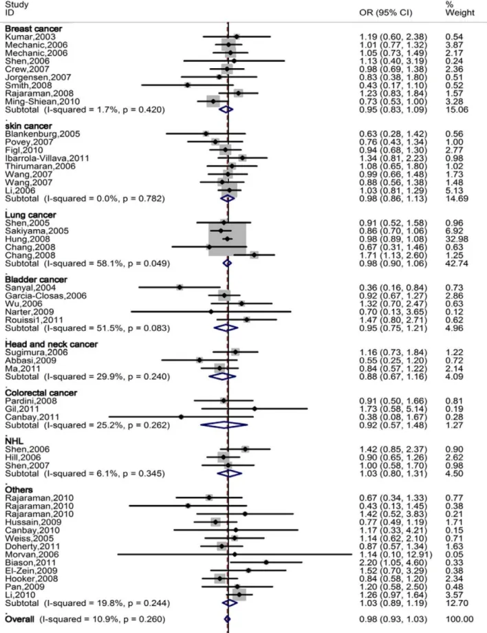
Figure 2. Forest plot of overall cancer risk of different cancer types associated with
ERCC5
Asp1104His polymorphism (His/His vs. Asp/His+Asp/Asp) by the fixed effects for each of the 44 published studies.For each study, the estimates of OR and its 95% CI were plotted with a box and a horizontal line. The symbol filled diamond indicates pooled OR and its 95% CI.in any of the subgroups. Furthermore, the observed null
associations were supported by the data that the variant genotypes
of the SNP were not associated with mRNA expression levels of
ERCC5
in the lymphoblastoid cell lines.
DNA repair deregulation is a crucial factor in the multistep
process of carcinogenesis, and the
ERCC5
gene is a vital
component of the DNA repair machinery. It has been observed
that deficiency of
ERCC5
may result in severe autosomal recessive
diseases including XP, CS and TTD [14] characterized by solar
hypersensitivity of the skin, high predisposition for developing
cancers (mainly epithelial and melanoma) on areas exposed to
sunlight. Furthermore, studies have suggested that
ERCC5
SNPs
are associated with development of some cancers, such as breast
cancer [44] and smoking-related cancers [23,24]. These suggest
a possible link between the
ERCC5
function and development of
cancer. The biological mechanisms of the
ERCC5
gene in
carcinogenesis may be complicated, among which nsSNPs, leading
to an amino acid change in the protein product and modulating
the individual DNA repair capacity phenotype [113,114], may
account for some of the known genetic variations related to risk of
cancers. However, our meta-analysis suggests that there is no
statistical evidence for an association between the
ERCC5
Asp1104His polymorphism and overall cancer risk, which is
consistent with the previous two meta-analyses conducted in breast
cancer and melanoma, respectively. The former included some
studies departure from HWE in the control population, and the
latter only contained three studies. Although we excluded the
inappropriate studies and expanded the sample size, the null
results were not altered. Furthermore, as far as our knowledge is
concerned, none of the SNPs in NER have ever been identified as
susceptibility locus in the published genome-wide association
studies (GWAS) for common diseases including cancers based on
common SNPs, which are similar to our results. This is a challenge
to the theory of common variants and common diseases [115]. It is
likely that, as NER genes are considered susceptibility genes, the
role of NER variants in cancer development may be dependent on
the degree of exposures that cause damage to DNA. Therefore,
without detailed information about such exposures for further
adjustment or stratification, the results of the observed associations
may be either biased or masked. For example, XP patients can
drastically reduce risk of developing skin cancer by avoiding
exposure to sunlight. Another possibility is that the common
variants are unlikely to have a significant biological effect. For
common variants, in most cases, the disease-associated variant
itself is unlikely to be functionally relevant [115]. The third
possibility is that the genetic risk of cancer conferred by the
common variants is very modest and the penetrance of the
variants is very small, which means that even if the polymorphism
is crucial for carcinogenesis, extremely large-scale evidence would
be necessary to establish with high confidence the presence of
specific associations. The inclusion of rare variants and larger
samples in future genome-wide association studies would help to
reveal low-penetrance susceptibility loci that are more likely to be
associated with cancer risk.
Furthermore, we did not observe biological evidence for the
effect of this SNP on the gene expression in terms of mRNA levels,
which biological support for the result of no association. Although
a sequence homology-based tool predicted this
ERCC5
poly-morphism to be a deleterious substitution [116], and
computa-tional algorithms by SIFT and SNPs3D tools aslo identified the
polymorphism with some functional implications (http://compbio.
cs.queensu.ca/F-SNP/), such a potentially functional relevance
have not been validated experimentally to date. Diversities of
variant-related risk associations in various kinds of cancer may
result from different mechanisms of carcinogenesis among
different cancer types. Although some studies have discovered
some sequence variants in the region of chromosome 5p15.33 and
8q24 that are associated with risk of different cancer types [117–
122], it is still uncertain whether the same polymorphism may
have non-specific effect on different types of cancer. Therefore,
further functional studies should be undertaken to explore the
mechanism underlying the variant-related associations with cancer
risk.
It would be hard to interpret results, if significant heterogeneity
were present. However, in this meta-analysis, we did not find any
obvious heterogeneity and publish bias across studies.
Neverthe-less, some limitations should be addressed. Firstly, although funnel
plot and Egger’s test show no publication bias, selection bias could
have occurred because only studies published in English and
Chinese were included. Secondly, because the reference groups
were not uniformly defined, some selected population-based
controls and some used hospital-based cancer-free controls,
non-differential misclassification bias is possible; in addition, some
control groups may not be representative of the general
Figure 3. Correlation between Asp1104His and
ERCC5
mRNA expression for different populations. A: CEU, 90 Utah residents from northern and western Europe;B: Asians, 45 unrelated Han Chinese in Beijing (CHB) and 45 unrelated Japanese in Tokyo (JPT);C: YRI, 90 Yoruba in Ibadan, Nigeria.population. Thirdly, our results were based on unadjusted OR
estimates, because not all published studies presented adjusted
ORs or when they did, the ORs were not adjusted by the same
potential confounders, such as age, sex and exposure variables.
Thus, more comprehensive individual datasets are needed to allow
for the adjustment by some co-variants and further evaluation of
potential gene-environmental interactions for susceptibility to
cancer. Fourthly, although the sample size of our study was
relatively large, the statistical power was still limited in the analyses
of subgroups with small sample sizes, particularly when the effect
size is small. Therefore, studies with larger sample sizes with
sufficient large subgroups should be undertaken to validate our
findings.
In summary, our meta-analysis shows that the
ERCC5
Asp1104His polymorphism appeared to be unlikely to confer
susceptibility to cancers. Further well-designed studies with larger
sample sizes will be necessary to validate the findings in the present
meta-analysis.
Supporting Information
Figure S1
ERCC5
gene map labeled with nine SNPs have
been studied for associations with cancer risk.
(A) Six
SNPs are located in the coding region, among which four are
nsSNPs, of which two are synonymous SNPs; two SNPs are
located in the 5
9
untranslated region, and one SNP is located in
the 3
9
untranslated region; six SNPs are tagging SNPs. (B) Nine
SNPs with a minor allelic frequency in different populations
obtained from the dbSNP database.
(TIF)
Figure S2
Flow chart of included studies for this
meta-analysis.
(TIF)
Figure S3
Funnel plot analysis to detect publication bias
for
ERCC5
Asp1104His under the recessive genetic
models (A, His/His vs. Asp/Asp and B, His/His vs.
Asp/His
+
Asp/Asp) for all 44 studies.
Each point
represents an individual study for the indicated association.
(TIF)
Table S1
Summary of 24 SNPs of the
ERCC5/XPG
gene
that have been studied for their associations with cancer
risk.
(DOCX)
Table S2
Summary of Studied SNPs in the eight NER
genes reviewed in all published meta-analysis.
(DOCX)
Table S3
Characteristics of the 44 references included
in the meta-analysis for ERCC5 Asp1104His.
(DOCX)
Author Contributions
Conceived and designed the experiments: L-XQ Q-YW. Analyzed the data: M-LZ M-YW L-XQ. Wrote the paper: M-LZ M-YW JH T-YS K-QX L-XQ Q-YW Z-GC.
References
1. Wood RD (1999) DNA damage recognition during nucleotide excision repair in mammalian cells. Biochimie 81: 39–44.
2. Wakasugi M, Reardon JT, Sancar A (1997) The non-catalytic function of XPG protein during dual incision in human nucleotide excision repair. J Biol Chem 272: 16030–16034.
3. O’Donovan A, Davies AA, Moggs JG, West SC, Wood RD (1994) XPG endonuclease makes the 39incision in human DNA nucleotide excision repair. Nature 371: 432–435.
4. Scherly D, Nouspikel T, Corlet J, Ucla C, Bairoch A, et al. (1993) Complementation of the DNA repair defect in xeroderma pigmentosum group G cells by a human cDNA related to yeast RAD2. Nature 363: 182–185. 5. Wakasugi M, Reardon JT, Sancar A (1997) The Non-catalytic function of XPG protein during dual incision in human nucleotide excision repair. Journal of Biological Chemistry 272: 16030–16034.
6. Constantinou A, Gunz D, Evans E, Lalle P, Bates PA, et al. (1999) Conserved residues of human XPG protein important for nuclease activity and function in nucleotide excision repair. J Biol Chem 274: 5637–5648.
7. Iyer N, Reagan MS, Wu KJ, Canagarajah B, Friedberg EC (1996) Interactions involving the human RNA polymerase II transcription/nucleotide excision repair complex TFIIH, the nucleotide excision repair protein XPG, and Cockayne syndrome group B (CSB) protein. Biochemistry 35: 2157–2167. 8. Araujo SJ, Nigg EA, Wood RD (2001) Strong functional interactions of TFIIH
with XPC and XPG in human DNA nucleotide excision repair, without a preassembled repairosome. Molecular and Cellular Biology 21: 2281–2291. 9. Thorel F, Constantinou A, Dunand-Sauthier I, Nouspikel T, Lalle P, et al. (2004) Definition of a short region of XPG necessary for TFIIH interaction and stable recruitment to sites of UV damage. Mol Cell Biol 24: 10670–10680. 10. He ZG, Henricksen LA, Wold MS, Ingles CJ (1995) Rpa Involvement in the
Damage-Recognition and Incision Steps of Nucleotide Excision-Repair. Nature 374: 566–569.
11. Dunand-Sauthier I, Hohl M, Thorel F, Jaquier-Gubler P, Clarkson SG, et al. (2005) The spacer region of XPG mediates recruitment to nucleotide excision repair complexes and determines substrate specificity. Journal of Biological Chemistry 280: 7030–7037.
12. Hofmann K (2009) Ubiquitin-binding domains and their role in the DNA damage response. DNA Repair 8: 544–556.
13. Gary R, Ludwig DL, Cornelius HL, MacInnes MA, Park MS (1997) The DNA repair endonuclease XPG binds to proliferating cell nuclear antigen (PCNA) and shares sequence elements with the PCNA binding regions of FEN-1 and cyclin-dependent kinase inhibitor p21. Journal of Biological Chemistry 272: 24522–24529.
14. Fagbemi AF, Orelli B, Scharer OD (2011) Regulation of endonuclease activity in human nucleotide excision repair. DNA Repair 10: 722–729.
15. Hanawalt PC (2001) Controlling the efficiency of excision repair. Mutat Res 485: 3–13.
16. Emmert S, Slor H, Busch DB, Batko S, Albert RB, et al. (2002) Relationship of neurologic degeneration to genotype in three xeroderma pigmentosum group G patients. J Invest Dermatol 118: 972–982.
17. Ito S, Kuraoka I, Chymkowitch P, Compe E, Takedachi A, et al. (2007) XPG stabilizes TFIIH, allowing transactivation of nuclear receptors: implications for Cockayne syndrome in XP-G/CS patients. Mol Cell 26: 231–243. 18. Scharer OD (2008) Hot topics in DNA repair: the molecular basis for different
disease states caused by mutations in TFIIH and XPG. DNA Repair (Amst) 7: 339–344.
19. Lee SK, Yu SL, Prakash L, Prakash S (2002) Requirement of yeast RAD2, a homolog of human XPG gene, for efficient RNA polymerase II transcription. implications for Cockayne syndrome. Cell 109: 823–834.
20. Klungland A, Hoss M, Gunz D, Constantinou A, Clarkson SG, et al. (1999) Base excision repair of oxidative DNA damage activated by XPG protein. Molecular Cell 3: 33–42.
21. Bessho T (1999) Nucleotide excision repair 39endonuclease XPG stimulates the activity of base excision repairenzyme thymine glycol DNA glycosylase. Nucleic Acids Res 27: 979–983.
22. Koeppel F, Poindessous V, Lazar V, Raymond E, Sarasin A, et al. (2004) Irofulven cytotoxicity depends on transcription-coupled nucleotide excision repair and is correlated with XPG expression in solid tumor cells. Clinical Cancer Research 10: 5604–5613.
23. Cheng L, Sturgis EM, Eicher SA, Spitz MR, Wei Q (2002) Expression of nucleotide excision repair genes and the risk for squamous cell carcinoma of the head and neck. Cancer 94: 393–397.
24. Cheng L, Spitz MR, Hong WK, Wei Q (2000) Reduced expression levels of nucleotide excision repair genes in lung cancer: a case-control analysis. Carcinogenesis 21: 1527–1530.
25. Bartolucci R, Wei J, Sanchez JJ, Perez-Roca L, Chaib I, et al. (2009) XPG mRNA expression levels modulate prognosis in resected non-small-cell lung cancer in conjunction with BRCA1 and ERCC1 expression. Clin Lung Cancer 10: 47–52.
26. Walsh CS, Ogawa S, Karahashi H, Scoles DR, Pavelka JC, et al. (2008) ERCC5 is a novel biomarker of ovarian cancer prognosis. Journal of Clinical Oncology 26: 2952–2958.
27. Kraemer KH, Lee MM, Andrews AD, Lambert WC (1994) The role of sunlight and DNA repair in melanoma and nonmelanoma skin cancer. The xeroderma pigmentosum paradigm. Arch Dermatol 130: 1018–1021. 28. Emmert S, Schneider TD, Khan SG, Kraemer KH (2001) The human XPG
29. Li Y, Gu S, Wu Q, Li Y, Fu X, et al. (2007) No association of ERCC1 C8092A and T19007C polymorphisms to cancer risk: a meta-analysis. European journal of human genetics : EJHG 15: 967–973.
30. Cao C, Zhang Y-M, Wang R, Sun S-F, Chen Z-B, et al. (2011) Excision repair cross complementation group 1 polymorphisms and lung cancer risk: a meta-analysis. Chinese Medical Journal 124: 2203–2208.
31. Vineis P, Manuguerra M, Kavvoura FK, Guarrera S, Allione A, et al. (2009) A field synopsis on low-penetrance variants in DNA repair genes and cancer susceptibility. Journal of the National Cancer Institute 101: 24–36. 32. Kiyohara C, Takayama K, Nakanishi Y (2010) Lung cancer risk and genetic
polymorphisms in DNA repair pathways: a meta-analysis. Journal of nucleic acids 2010: 701760.
33. Qian B, Zhang H, Zhang L, Zhou X, Yu H, et al. (2011) Association of genetic polymorphisms in DNA repair pathway genes with non-small cell lung cancer risk. Lung cancer (Amsterdam, Netherlands) 73: 138–146.
34. Qiu L, Wang Z, Shi X, Wang Z (2008) Associations between XPC polymorphisms and risk of cancers: A meta-analysis. European journal of cancer (Oxford, England : 1990) 44: 2241–2253.
35. Stern MC, Lin J, Figueroa JD, Kelsey KT, Kiltie AE, et al. (2009) Polymorphisms in DNA repair genes, smoking, and bladder cancer risk: findings from the international consortium of bladder cancer. Cancer Research 69: 6857–6864.
36. Zheng W, Cong X-F, Cai W-H, Yang S, Mao C, et al. (2011) Current evidences on XPC polymorphisms and breast cancer susceptibility: a meta-analysis. Breast cancer research and treatment 128: 811–815.
37. Wang F, Chang D, Hu F-l, Sui H, Han B, et al. (2008) DNA repair gene XPD polymorphisms and cancer risk: a meta-analysis based on 56 case-control studies. Cancer epidemiology, biomarkers & prevention : a publication of the American Association for Cancer Research, cosponsored by the American Society of Preventive Oncology 17: 507–517.
38. Li C, Jiang Z, Liu X (2009) XPD Lys(751)Gln and Asp (312)Asn polymorphisms and bladder cancer risk: a meta-analysis. Molecular biology reports 37: 301–309.
39. Mocellin S, Verdi D, Nitti D (2009) DNA repair gene polymorphisms and risk of cutaneous melanoma: a systematic review and meta-analysis. Carcinogenesis 30: 1735–1743.
40. Pabalan N, Francisco-Pabalan O, Sung L, Jarjanazi H, Ozcelik H (2010 ) Meta-analysis of two ERCC2 (XPD) polymorphisms, Asp312Asn and Lys751Gln, in breast cancer. Breast cancer research and treatment 124: 531–541.
41. Qiu L-X, Yao L, Zhang J, Zhu X-D, Zhao X-M, et al. (2010) XPD Lys751Gln polymorphism and breast cancer susceptibility: a meta-analysis involving 28,709 subjects. Breast cancer research and treatment 124: 229–235. 42. Chen B, Zhou Y, Yang P, Wu X-T (2011) ERCC2 Lys751Gln and Asp312Asn
polymorphisms and gastric cancer risk: a meta-analysis. Journal of cancer research and clinical oncology 137: 939–946.
43. Yuan L, Cui D, Zhao E-J, Jia C-Z, Wang L-D, et al. (2011) XPD Lys751Gln polymorphism and esophageal cancer risk: a meta-analysis involving 2288 cases and 4096 controls. World journal of gastroenterology : WJG 17: 2343–2348. 44. Ding D-P, He X-F, Zhang Y (2011 ) Lack of association between XPG Asp1104His and XPF Arg415Gln polymorphism and breast cancer risk: a meta-analysis of case-control studies. Breast cancer research and treatment 129: 203–209.
45. The International HapMap Consortium (2003) The International HapMap Project. Nature 426: 789–796.
46. Holm K, Melum E, Franke A, Karlsen TH (2010) SNPexp - A web tool for calculating and visualizing correlation between HapMap genotypes and gene expression levels. BMC bioinformatics 11: 600–604.
47. Stranger BE, Forrest MS, Dunning M, Ingle CE, Beazlsy C, et al. (2007) Relative impact of nucleotide and copy number variation on gene phenotypes. Science 315: 848–853.
48. DerSimonian R, Laird N (1986) Meta-analysis in clinical trials. Control Clin Trials 7: 177–188.
49. Mantel N, Haenszel W (1959) Statistical aspects of the analysis of data from retrospective studies of disease. J Natl Cancer Inst 22: 719–748.
50. Egger M, Smith GD, Schneider M, Minder C (1997) Bias in meta-analysis detected by a simple, graphical test. British Medical Journal 315: 629–634. 51. Chen M, Kamat AM, Huang M, Grossman HB, Dinney CP, et al. (2007)
High-order interactions among genetic polymorphisms in nucleotide excision repair pathway genes and smoking in modulating bladder cancer risk. Carcinogenesis 28: 2160–2165.
52. Cui Y, Morgenstern H, Greenland S, Tashkin DP, Mao J, et al. (2006) Polymorphism of Xeroderma Pigmentosum group G and the risk of lung cancer and squamous cell carcinomas of the oropharynx, larynx and esophagus. Int J Cancer 118: 714–720.
53. Hung RJ, Baragatti M, Thomas D, McKay J, Szeszenia-Dabrowska N, et al. (2007) Inherited predisposition of lung cancer: A hierarchical modeling approach to DNA repair and cell cycle control pathways. Cancer Epidemiology Biomarkers & Prevention 16: 2736–2744.
54. Joshi AD, Corral R, Siegmund KD, Haile RW, Le Marchand L, et al. (2009) Red meat and poultry intake, polymorphisms in the nucleotide excision repair and mismatch repair pathways and colorectal cancer risk. Carcinogenesis 30: 472–479.
55. Liu YH, Scheurer ME, El-Zein R, Cao YM, Do KA, et al. (2009) Association and Interactions between DNA Repair Gene Polymorphisms and Adult Glioma. Cancer Epidemiology Biomarkers & Prevention 18: 204–214. 56. Michiels S, Danoy P, Dessen P, Bera A, Boulet T, et al. (2007) Polymorphism
discovery in 62 DNA repair genes and haplotype associations with risks for lung and head and neck cancers. Carcinogenesis 28: 1731–1739.
57. Mort R, Mo L, McEwan C, Melton DW (2003) Lack of involvement of nucleotide excision repair gene polymorphisms in colorectal cancer. British Journal of Cancer 89: 333–337.
58. Wen H, Ding Q, Fang ZJ, Xia GW, Fang J (2009) Population study of genetic polymorphisms and superficial bladder cancer risk in Han-Chinese smokers in Shanghai. International Urology and Nephrology 41: 855–864.
59. An J, Liu Z, Hu Z, Li G, Wang LE, et al. (2007) Potentially functional single nucleotide polymorphisms in the core nucleotide excision repair genes and risk of squamous cell carcinoma of the head and neck. Cancer Epidemiol Biomarkers Prev 16: 1633–1638.
60. Weiss JM, Weiss NS, Ulrich CM, Doherty JA, Chen C, et al. (2006) Nucleotide excision repair genotype and the incidence of endometrial cancer: Effect of other risk factors on the association. Gynecologic Oncology 103: 891–896. 61. Rouissi K, Ouerhani S, Hamrita B, Bougatef K, Marrakchi R, et al. (2011)
Smoking and polymorphisms in xenobiotic metabolism and DNA repair genes are additive risk factors affecting bladder cancer in Northern Tunisia. Pathology and Oncology Research 17: 879–886.
62. Berhane N, Sobti RC, Mahdi SA (2012) DNA repair genes polymorphism (XPG and XRCC1) and association of prostate cancer in a north Indian population. Molecular biology reports 39: 2471–2479.
63. Goncalves FT, Francisco G, de Souza SP, Luiz OC, Festa-Neto C, et al. (2011) European ancestry and polymorphisms in DNA repair genes modify the risk of melanoma: A case-control study in a high UV index region in Brazil. Journal of Dermatological Science 64: 59–66.
64. He X, Ye F, Zhang J, Cheng Q, Shen J, et al. (2008) Susceptibility of XRCC3, XPD, and XPG genetic variants to cervical carcinoma. Pathobiology 75: 356– 363.
65. Jeon HS, Kim KM, Park SH, Lee SY, Choi JE, et al. (2003) Relationship between XPG codon 1104 polymorphism and risk of primary lung cancer. Carcinogenesis 24: 1677–1681.
66. McKean-Cowdin R, Barnholtz-Sloan J, Inskip PD, Ruder AM, Butler M, et al. (2009) Associations between Polymorphisms in DNA Repair Genes and Glioblastoma. Cancer Epidemiology Biomarkers & Prevention 18: 1118–1126. 67. Smith TR, Levine EA, Freimanis RI, Akman SA, Allen GO, et al. (2008) Polygenic model of DNA repair genetic polymorphisms in human breast cancer risk. Carcinogenesis 29: 2132–2138.
68. Wen SX, Tang PZ, Zhang XM, Zhao D, Guo YL, et al. (2006) Association between genetic polymorphism in xeroderma pigmentosum G gene and risks of laryngeal and hypopharyngeal carcinomas. Acta Academiae Medicinae Sinicae 28: 703–706.
69. Kumar R, Hoglund L, Zhao C, Forsti A, Snellman E, et al. (2003) Single nucleotide polymorphisms in the XPG gene: determination of role in DNA repair and breast cancer risk. Int J Cancer 103: 671–675.
70. Mechanic LE, Millikan RC, Player J, de Cotret AR, Winkel S, et al. (2006) Polymorphisms in nucleotide excision repair genes, smoking and breast cancer in African Americans and whites: a population-based case-control study. Carcinogenesis 27: 1377–1385.
71. Shen J, Desai M, Agrawal M, Kennedy DO, Senie RT, et al. (2006) Polymorphisms in nucleotide excision repair genes and DNA repair capacity phenotype in sisters discordant for breast cancer. Cancer Epidemiology Biomarkers and Prevention 15: 1614–1619.
72. Crew KD, Gammon MD, Terry MB, Fang FZ, Zablotska LB, et al. (2007) Polymorphisms in nucleotide excision repair genes, polycyclic aromatic hydrocarbon-DNA adducts, and breast cancer risk. Cancer Epidemiology Biomarkers and Prevention 16: 2033–2041.
73. Jorgensen TJ, Visvanathan K, Ruczinski I, Thuita L, Hoffman S, et al. (2007) Breast cancer risk is not associated with polymorphic forms of xeroderma pigmentosum genes in a cohort of women from Washington County, Maryland. Breast Cancer Res Treat 101: 65–71.
74. Smith TR, Levine EA, Freimanis RI, Akman SA, Allen GO, et al. (2008) Polygenic model of DNA repair genetic polymorphisms in human breast cancer risk. Carcinogenesis 29: 2132–2138.
75. Rajaraman P, Bhatti P, Doody MM, Simon SL, Weinstock RM, et al. (2008) Nucleotide excision repair polymorphisms may modify ionizing radiation-related breast cancer risk in US radiologic technologists. International Journal of Cancer 123: 2713–2716.
76. Hsu MS, Yu JC, Wang HW, Chen ST, Hsiung CN, et al. (2010) Synergistic Effects of Polymorphisms in DNA Repair Genes and Endogenous Estrogen Exposure on Female Breast Cancer Risk. Annals of Surgical Oncology 17: 760–771.
77. Blankenburg S, Konig IR, Moessner R, Laspe P, Thoms KM, et al. (2005) No association between three xeroderma pigmentosum group C and one group G gene polymorphisms and risk of cutaneous melanoma. Eur J Hum Genet 13: 253–255.
79. Povey JE, Darakhshan F, Robertson K, Bisset Y, Mekky M, et al. (2007) DNA repair gene polymorphisms and genetic predisposition to cutaneous melanoma. Carcinogenesis 28: 1087–1093.
80. Figl A, Scherer D, Nagore E, Bermejo JL, Botella-Estrada R, et al. (2010) Single-nucleotide polymorphisms in DNA-repair genes and cutaneous melanoma. Mutation Research - Genetic Toxicology and Environmental Mutagenesis 702: 8–16.
81. Ibarrola-Villava M, Pena-Chilet M, Fernandez LP, Aviles JA, Mayor M, et al. (2011) Genetic polymorphisms in DNA repair and oxidative stress pathways associated with malignant melanoma susceptibility. European Journal of Cancer.
82. Thirumaran RK, Bermejo JL, Rudnai P, Gurzau E, Koppova K, et al. (2006) Single nucleotide polymorphisms in DNA repair genes and basal cell carcinoma of skin. Carcinogenesis 27: 1676–1681.
83. Wang LE, Li C, Strom SS, Goldberg LH, Brewster A, et al. (2007) Repair capacity for UV light induced DNA damage associated with risk of nonmelanoma skin cancer and tumor progression. Clin Cancer Res 13: 6532–6539.
84. Sanyal S, Festa F, Sakano S, Zhang Z, Steineck G, et al. (2004) Polymorphisms in DNA repair and metabolic genes in bladder cancer. Carcinogenesis 25: 729– 734.
85. Garcia-Closas M, Malats N, Real FX, Welch R, Kogevinas M, et al. (2006) Genetic variation in the nucleotide excision repair pathway and bladder cancer risk. Cancer Epidemiol Biomarkers Prev 15: 536–542.
86. Wu X, Gu J, Grossman HB, Amos CI, Etzel C, et al. (2006) Bladder cancer predisposition: a multigenic approach to DNA-repair and cell-cycle-control genes. Am J Hum Genet 78: 464–479.
87. Narter KF, Ergen A, Agachan B, Gormus U, Timirci O, et al. (2009) Bladder cancer and polymorphisms of DNA repair genes (XRCC1, XRCC3, XPD, XPG, APE1, hOGG1). Anticancer Res 29: 1389–1393.
88. Rouissi K, Ouerhani S, Hamrita B, Bougatef K, Marrakchi R, et al. (2011) Smoking and Polymorphisms in Xenobiotic Metabolism and DNA Repair Genes are Additive Risk Factors Affecting Bladder Cancer in Northern Tunisia. Pathol Oncol Res.
89. Shen M, Berndt SI, Rothman N, Demarini DM, Mumford JL, et al. (2005) Polymorphisms in the DNA nucleotide excision repair genes and lung cancer risk in Xuan Wei, China. Int J Cancer 116: 768–773.
90. Sakiyama T, Kohno T, Mimaki S, Ohta T, Yanagitani N, et al. (2005) Association of amino acid substitution polymorphisms in DNA repair genes TP53, POLI, REV1 and LIG4 with lung cancer risk. Int J Cancer 114: 730– 737.
91. Hung RJ, Christiani DC, Risch A, Popanda O, Haugen A, et al. (2008) International Lung Cancer Consortium: pooled analysis of sequence variants in DNA repair and cell cycle pathways. Cancer Epidemiol Biomarkers Prev 17: 3081–3089.
92. Chang JS, Wrensch MR, Hansen HM, Sison JD, Aldrich MC, et al. (2008) Nucleotide excision repair genes and risk of lung cancer among San Francisco Bay Area Latinos and African Americans. Int J Cancer 123: 2095–2104. 93. Sugimura T, Kumimoto H, Tohnai I, Fukui T, Matsuo K, et al. (2006)
Gene-environment interaction involved in oral carcinogenesis: Molecular epidemi-ological study for metabolic and DNA repair gene polymorphisms. Journal of Oral Pathology and Medicine 35: 11–18.
94. Abbasi R, Ramroth H, Becher H, Dietz A, Schmezer P, et al. (2009) Laryngeal cancer risk associated with smoking and alcohol consumption is modified by genetic polymorphisms in ERCC5, ERCC6 and RAD23B but not by polymorphisms in five other nucleotide excision repair genes. Int J Cancer 125: 1431–1439.
95. Hongxia M, Hongping Y, Zhensheng L, Li EW, Erich MS, et al. (2011) Polymorphisms of XPG/ERCC5 and risk of squamous cell carcinoma of the head and neck. Pharmacogenetics and Genomics.
96. Pardini B, Naccarati A, Novotny J, Smerhovsky Z, Vodickova L, et al. (2008) DNA repair genetic polymorphisms and risk of colorectal cancer in the Czech Republic. Mutat Res 638: 146–153.
97. Gil J, Ramsey D, Stembalska A, Karpinski P, Pesz KA, et al. (2011) The C/A polymorphism in intron 11 of the XPC gene plays a crucial role in the modulation of an individual’s susceptibility to sporadic colorectal cancer. Mol Biol Rep.
98. Canbay E, Cakmakoglu B, Zeybek U, Sozen S, Cacina C, et al. (2011) Association of APE1 and hOGG1 polymorphisms with colorectal cancer risk in a Turkish population. Curr Med Res Opin 27: 1295–1302.
99. Shen M, Zheng T, Lan Q, Zhang Y, Zahm SH, et al. (2006) Polymorphisms in DNA repair genes and risk of non-Hodgkin lymphoma among women in Connecticut. Hum Genet 119: 659–668.
100. Hill DA, Wang SS, Cerhan JR, Davis S, Cozen W, et al. (2006) Risk of non-Hodgkin lymphoma (NHL) in relation to germline variation in DNA repair and related genes. Blood 108: 3161–3167.
101. Shen M, Purdue MP, Kricker A, Lan Q, Grulich AE, et al. (2007) Polymorphisms in DNA repair genes and risk of non-Hodgkin’s lymphoma in New South Wales, Australia. Haematologica 92: 1180–1185.
102. Hussain SK, Mu LN, Cai L, Chang SC, Park SL, et al. (2009) Genetic variation in immune regulation and DNA repair pathways and stomach cancer in China. Cancer Epidemiol Biomarkers Prev 18: 2304–2309.
103. Canbay E, Agachan B, Gulluoglu M, Isbir T, Balik E, et al. (2010) Possible associations of APE1 polymorphism with susceptibility and HOGG1 poly-morphism with prognosis in gastric cancer. Anticancer Res 30: 1359–1364. 104. Weiss JM, Weiss NS, Ulrich CM, Doherty JA, Voigt LF, et al. (2005)
Interindividual variation in nucleotide excision repair genes and risk of endometrial cancer. Cancer Epidemiol Biomarkers Prev 14: 2524–2530. 105. Doherty JA, Weiss NS, Fish S, Fan W, Loomis MM, et al. (2011)
Polymorphisms in Nucleotide Excision Repair Genes and Endometrial Cancer Risk. Cancer Epidemiol Biomarkers Prev.
106. Hooker S, Bonilla C, Akereyeni F, Ahaghotu C, Kittles RA (2008) NAT2 and NER genetic variants and sporadic prostate cancer susceptibility in African Americans. Prostate Cancer Prostatic Dis 11: 349–356.
107. Le Morvan V, Longy M, Bonaiti-Pellie C, Bui B, Houede N, et al. (2006) Genetic polymorphisms of the XPG and XPD nucleotide excision repair genes in sarcoma patients. Int J Cancer 119: 1732–1735.
108. Biason P, Hattinger CM, Innocenti F, Talamini R, Alberghini M, et al. (2011) Nucleotide excision repair gene variants and association with survival in osteosarcoma patients treated with neoadjuvant chemotherapy. Pharmacogenomics J.
109. El-Zein R, Monroy CM, Etzel CJ, Cortes AC, Xing Y, et al. (2009) Genetic polymorphisms in DNA repair genes as modulators of Hodgkin disease risk. Cancer 115: 1651–1659.
110. Rajaraman P, Hutchinson A, Wichner S, Black PM, Fine HA, et al. (2010) DNA repair gene polymorphisms and risk of adult meningioma, glioma, and acoustic neuroma. Neuro Oncol 12: 37–48.
111. Pan J, Lin J, Izzo JG, Liu Y, Xing J, et al. (2009) Genetic susceptibility to esophageal cancer: the role of the nucleotide excision repair pathway Carcinogenesis 30: 785–792.
112. Li LM, Zeng XY, Ji L, Fan XJ, Li YQ, et al. (2010) [Association of XPC and XPG polymorphisms with the risk of hepatocellular carcinoma]. Zhonghua Gan Zang Bing Za Zhi 18: 271–275.
113. Vodicka P, Stetina R, Polakova V, Tulupova E, Naccarati A, et al. (2007) Association of DNA repair polymorphisms with DNA repair functional outcomes in healthy human subjects. Carcinogenesis 28: 657–664. 114. Friedberg EC (2003) DNA damage and repair. Nature 421: 436–440. 115. Bodmer W, Bonilla C (2008) Common and rare variants in multifactorial
susceptibility to common diseases. Nat Genet 40: 695–701.
116. Ng PC, Henikoff S (2001) Predicting deleterious amino acid substitutions. Genome Res 11: 863–874.
117. Rafnar T, Sulem P, Stacey SN, Geller F, Gudmundsson J, et al. (2009) Sequence variants at the TERT-CLPTM1L locus associate with many cancer types. Nature genetics 41: 221–227.
118. Gudmundsson J, Sulem P, Manolescu A, Amundadottir LT, Gudbjartsson D, et al. (2007) Genome-wide association study identifies a second prostate cancer susceptibility variant at 8q24. Nature genetics 39: 631–637.
119. Yeager M, Orr N, Hayes RB, Jacobs KB, Kraft P, et al. (2007) Genome-wide association study of prostate cancer identifies a second risk locus at 8q24. Nature genetics 39: 645–649.
120. Haiman CA, Patterson N, Freedman ML, Myers SR, Pike MC, et al. (2007) Multiple regions within 8q24 independently affect risk for prostate cancer. Nature genetics 39: 638–644.
121. Tomlinson I, Webb E, Carvajal-Carmona L, Broderick P, Kemp Z, et al. (2007) A genome-wide association scan of tag SNPs identifies a susceptibility variant for colorectal cancer at 8q24.21. Nature genetics 39: 984–988. 122. Kiemeney LA, Thorlacius S, Sulem P, Geller F, Aben KKH, et al. (2008)