Clinical Study of Extrapulmonary Head and Neck
Tuberculosis: A Single-Institute 10-year
Experience
Masahiro Oishi
1Sachimi Okamoto
1Yuichi Teranishi
1Chieko Yokota
1Sakurako Takano
1Hiroyoshi Iguchi
11Department of Otolaryngology and Head & Neck Surgery, Osaka City University, Osaka, Japan
Int Arch Otorhinolaryngol 2016;20:30–33.
Address for correspondence Hiroyoshi Iguchi, MD, Department of Otolaryngology and Head & Neck Surgery, Osaka City University, 1-4-3, Asahimachi, Abeno-ku Osaka Osaka 545-8585, Japan
(e-mail: [email protected]).
Introduction
Tuberculosis (TB) is a major problem worldwide, especially in Asia and Africa. Japan remains ranked as an intermediate-burden country of TB, although the incidence of TB in Japan has been decreasing every year. According to 2013 data, the incidence rate of TB in Japan was 16.1 per 100,000 people. Because the head and neck region may be the portal of entry for Mycobacterium tuberculosis (M. tuberculosis), otolaryngologists
should be especially aware of the manifestations of extrapulmo-nary TB (EPTB) in this region. In addition, it is important to differentiate cervical lymph node TB, which is the most fre-quently observed form of EPTB in the head and neck, from other lymph node diseases, including metastatic cancer, malignant lymphoma, non-specific hyperplasia, Kikuchi’s disease, Castle-man’s disease, sarcoidosis, cat scratch disease, among others. Here, we reviewed our experience of head and neck EPTB diagnosed in our department over the past 10 years.
Keywords
►
interferon-gamma
release assay
►
cervical lymph node
►
supraclavicular node
►
larynx
►
polymerase chain
reaction
►
middle ear
Abstract
Introduction
Although the incidence of tuberculosis (TB) in Japan has been decreasing
yearly, Japan remains ranked as an intermediate-burden country for TB.
Objective
This study aims to investigate the current situation of head and neck
extrapulmonary TB (EPTB) diagnosed in our department.
Methods
We retrospectively reviewed the clinical records of 47 patients diagnosed
with EPTB in the head and neck in our department between January 2005 and
December 2014. The extracted data included sex and age distribution, development
site, chief complaint, presence or absence of concomitant active pulmonary TB (PTB) or
history of TB, tuberculin skin test (TST) results, interferon-gamma release assay (IGRA)
results, and duration from the
fi
rst visit to the
fi
nal diagnosis of EPTB.
Results
The subjects consisted of 20 men and 27 women, and age ranged from 6 to
84 years. The most common site was the cervical lymph nodes (30 patients), with the
supraclavicular nodes being the most commonly affected (60%). Histopathological
examination was performed on 28 patients. TST was positive in 9 out of 9 patients and
the IGRA was positive in 18 out of 19 patients. We observed concomitant PTB in 15 out
of the 47 patients. Mean duration from the
fi
rst visit to the
fi
nal diagnosis of EPTB was
56 days.
Conclusion
The clinical symptoms of TB, especially those in the head and neck region,
are varied. Otolaryngologists should be especially aware of the extrapulmonary
manifestations of TB to ensure early diagnosis and treatment from the public health
viewpoint.
received July 1, 2015 accepted August 30, 2015 published online October 8, 2015
DOI http://dx.doi.org/ 10.1055/s-0035-1565011. ISSN 1809-9777.
Copyright © 2016 by Thieme Publicações Ltda, Rio de Janeiro, Brazil
Original Research
Methods
The study subjects were 47 patients with EPTB in the head and neck, diagnosed at our University Hospital over a period of 10 years, between January 2005 and December 2014. We retrospectively reviewed their medical records, and extracted data regarding sex and age distribution, development site, chief complaint, presence or absence of concomitant active pulmonary TB (PTB) or past history of TB, tuberculin skin test (TST) results, interferon-gamma release assay (IGRA) results, and duration from thefirst visit to thefinal diagnosis of TB. We further classified the complaints of cervical lymph node TB as unilateral or bilateral and as solitary or multiple lesions. The presence of PTB was diagnosed based on the chest computed tomography (CT) and chest radiograph, sputum smear and culture, and polymerase chain reaction (PCR) assay
findings. The diagnostic criteria of EPTB include at least one of the following in addition to a positive immunological test result: 1) typical caseous granuloma on histopathological examination, 2) detection ofM. tuberculosis in the culture,
and/or 3) identification ofM. tuberculosiswith PCR assay. TST, IGRA, and cervical CT were used supplementarily. Our Uni-versitýs ethical committee approved this study (No. 2737).
Results
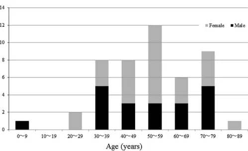
The subjects were 47 patients, including 20 men and 27 women, with a male-to-female ratio of 1:1.35. Ages ranged from six to 84 years old, with a mean of 52.9 years. We observed an age peak in the 50s (►Fig. 1). During the study
period, one to eight patients were diagnosed annually with EPTB in the head and neck, with a mean of 4.7 patients per year.
The most common site of EPTB in the head and neck was the cervical lymph nodes (30 patents; 13 men and 17 women), followed by the larynx (12 patients;five men and
seven women), the parotid gland (three patients; one man and two women), and the middle ear (two patients; one man and one woman) (►Fig. 2). The majority of EPTB cases (63.8%)
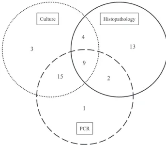
in the head and neck occurred in the cervical lymph nodes. We performed histopathological examination on 28 pa-tients. The examined sites included the cervical lymph nodes (20 patients), larynx (five patients), parotid gland (two patients), and middle ear (one patient). Of the 28 patients, 11 patients were PCR-positive (among them, two were culture-negative), while 13 patients were culture-positive (among them, four were PCR-negative). Nine patients were both culture- and PCR-positive, while 13 patients were both culture- and PCR-negative (►Fig. 3).
Of the 19 patients who did not undergo histopathological examination, 15 were both culture- and PCR-positive, two were only PCR-positive, and two were only culture-positive.
Fig. 1 Age and sex distributions of head and neck extrapulmonary tuberculosis in the present study. Larynx
12 cases (25.5%)
Cervical lymph nodes
30 cases (63.8%) Parotid gland
3 cases (6.4%)
Middle ear, 2 cases (4.3%)
Fig. 2 Site-specific distributions of head and neck extrapulmonary tuberculosis in the present study.
Before 2008, we performed TST for the supplemental diagno-sis of TB. After this time, we used IGRA instead of TST. We performed TST on nine patients, which resulted positive for all patients. We performed IGRA on 19 patients and it was positive for 18 patients.
The chief complaint of all patients with cervical lymph node TB was unilateral swelling of the cervical lymph nodes. The most commonly affected cervical lymph node was the supraclavicular node (18/30, 60.0%), followed by the internal jugular node (8/30, 26.7%), submandibular node (7/30, 23.3%), and posterior triangle node (5/30, 16.7%). Patients with multiple lymph node swelling accounted for 56.7% (17/ 30), while those with solitary lesion accounted for 43.3% (13/ 30) of patients. Of the patients with cervical lymph node TB, those with a painful lesion accounted for 26.7% (8/30). The chief complaints of the 12 patients with laryngeal TB included cough, laryngeal discomfort, laryngeal pain, and hoarseness. Almost all patients with laryngeal TB had lesions in the vocal cord, false cord, and epiglottis, which were characterized by the formation of granuloma-like or white-coated masses. One patient showed a whitish mass in the subglottis. The chief complaints of patients with parotid gland TB and middle ear TB were a unilateral painful parotid mass and refractory otorrhea, respectively.
We observed a history of TB in six patients. Complications considered to be associated with the decline in immune function were malignant tumors, type 2 diabetes mellitus, chronic renal failure requiring dialysis, multiple myositis treated with oral steroids, and asthma treated with inhaled steroid therapy in three, two, one, one, and one patients, respectively. No patients were human immunodeficiency virus (HIV)-positive. We observed concomitant PTB in 15 out of 47 patients (11 with laryngeal TB and four with cervical lymph node TB). No patients with parotid gland TB or middle ear TB had concomitant PTB.
The mean duration from thefirst visit to thefinal diagnosis of TB was 56 days (range, one day to three years). The shortest
duration was one day, for a patient with laryngeal TB with concomitant PTB. The longest was three years, for a patient with laryngeal TB with a chief complaint of hoarseness, in whom M. tuberculosis was detected by histopathological
examination of the biopsied specimen and sputum culture three years after the initial presentation.
Discussion
TB is still common in many parts of the world and is a serious, and often deadly, infection. Among the developed countries, Japan has a relatively high incidence of TB, although the incidence has been decreasing yearly. In addition, the inci-dence of TB is particularly high in the elderly and in the urban areas of Japan. Our hospital is located in Osaka city, which has the highest incidence of TB in Japan. According to 2013 data in Japan, 20,495 TB patients were newly notified. Among those, 4,274 had EPTB. Given that EPTB is frequently found in the head and neck region, otolaryngologists should be especially aware of the manifestations, diagnosis, and treatment of EPTB.
The male to female ratio was 20:27 (1:1.35) in the present study, which differed somewhat from the ratio reported in other previous studies, including 143:68 by Akkara et al.,1 115:189 by Ricciardiello et al.,2and 43:30 by Bruzgielewicz et al.3
The mean age of the subjects was 52.9 years and the peak incidence was observed at 50 years old. On the other hand, Akkara et al. reported that the peak incidence was noted in the 30s in their patient population,1while Ricciardiello et al. reported that the mean age at diagnosis was 16.5 years.2We consider that this may be related to the fact that many elderly people with low income are living in the endemic area around our hospital. However, there was also one six-year-old patient, suggesting that we must carefully monitor not only the older population, but also younger patients for the development of EPTB.
In the present study, the most common site of EPTB was the cervical lymph nodes, followed by the larynx, parotid gland, and middle ear. In the study by Akkara et al., the most common site in their 211 patients was the cervical lymph node (201 patients, 95.3%), followed by the middle ear (2.8%), larynx (1.4%), and nasal cavity (0.5%).1 In the study by Ricciardiello et al., the most common site was the cervical lymph nodes (94.12%), followed by the larynx (4.33%), pala-tine tonsil (0.62%), oral cavity (0.31%), middle ear (0.31%), and nasal cavity (0.31%).2Thus, while all studies reported that the cervical lymph nodes are the most commonly affected site, it should be noted that the present study also showed a relatively high proportion of laryngeal TB. In Japan, patients presenting with respiratory symptoms, such as cough, and laryngeal symptoms, such as hoarseness, usually undergo
fiberoptic laryngoscopy by otolaryngologists. This may be related to the frequent detection of laryngeal TB with concomitant PTB in the present study.
Among cervical lymph node TB cases, supraclavicular node swelling was the most common chief complaint in the present study, followed by internal jugular node, submandibular
Culture Histopathology
PCR
13 4
9
2
1 3
15
Fig. 3 The numbers of positive results of each examination. PCR, polymerase chain reaction.
International Archives of Otorhinolaryngology Vol. 20 No. 1/2016
node, and posterior triangle node swelling. On the other hand, Akkara et al. reported that the most commonly affected node was the posterior triangle node (87.6%);1 whereas, according to the report by Bruzgielewicz et al., the internal jugular node (15/26, 57.7%) and submandibular node (11/26, 42.3%) were the only affected nodes.3In our previous study,4 the most commonly affected sites were the posterior triangle node (26.5%), internal jugular node (24.5%), and supraclavic-ular node (18.4%). This indicates that these three regions are important. Furthermore, the ratios of solitary lesions were reported to be 4.0% by Akkara et al.,130.3% by Ricciardiello et al.,2and 14.3% by Iguchi et al.4; in the present study, the ratio was as high as 43.3%.
The chief complaints of the patients with laryngeal TB in the present study included cough, laryngeal discomfort/pain, and hoarseness. Those of patients with parotid gland TB included unilateral parotid mass and pain, while patients with middle ear TB complained of refractory otorrhea. These
findings were consistent with those of previous studies.3 Given that the incidence of concomitant PTB was high in patients with laryngeal TB, clinicians should be aware that patients with laryngeal TB are likely to have PTB.
In the present study, we diagnosed 28 patients (58.3%) based on histopathological examination. Similarly, Lee et al. reported that they often diagnosed EPTB based on histopath-ologicalfindings, including evidence of typical caseous gran-uloma and/or positive acid-fast stain.5 Accordingly, we consider that histopathological examination plays an impor-tant role in the diagnosis of EPTB; however, it should be noted that granulomatous hypoplasia can also occur in patients with immunodeficiency and that granuloma can be induced by other infectious pathogens, including fungus, brucella, and syphilis, among others.
We performed TST in nine patients, which resulted posi-tive for all patients. TST is a standard and economical screening tool for TB. However, the interpretation of the test results often requires careful consideration, because Bacille Calmette-Guérin vaccination may cause a false-posi-tive reaction to the test. In our institution, we started using IGRA in 2008, and the results were positive in 18 of the tested 19 patients. However, it should be noted that a negative IGRA test result does not always exclude the possibility of TB infection.5
It is well known that patients with immunocompromised conditions such as diabetes mellitus, chronic renal failure
(undergoing dialysis), HIV infection, immunosuppressive drug use, and malignancy are susceptible to TB infection. The subjects of the present study included three patients with a history of malignancy, two patients with diabetes mellitus, one patient with chronic renal failure, one patient taking oral steroids for multiple myositis, and one patient using inhala-tion steroids for asthma. No patients were HIV-positive. Notably, there is a recent report showing that, in Japan, 9% of HIV-positive patients are infected with TB, and that 3% of TB patients are HIV-positive.6Thus, clinicians should be aware of the close relationship between immunosuppressive condi-tions and TB infection.
Finally, studies have indicated that the duration from the
first visit to the final diagnosis of TB tends to be long in patients with EPTB who do not have concomitant PTB. In the present study, the patients with laryngeal TB with concomi-tant PTB were diagnosed earlier, while the patients with cervical lymph node TB tended to be diagnosed later, indicat-ing the importance of early differential diagnosis.
Conclusion
The symptoms of EPTB in the head and neck region are varied; hence, otolaryngologists should be aware of the manifesta-tions of EPTB to ensure early diagnosis to prevent TB out-breaks or nosocomial infections.
References
1 Akkara SA, Singhania A, Akkara AG, Shah A, Adalja M, Chauhan N. A
study of manifestations of extrapulmonary tuberculosis in the ENT
region. Indian J Otolaryngol Head Neck Surg 2014;66(1):46–50
2 Ricciardiello F, MartufiS, Cardone M, Cavaliere M, D’Errico P, Iengo
M. Otorhinolaryngology-related tuberculosis. Acta
Otorhinolar-yngol Ital 2006;26(1):38–42
3 Bruzgielewicz A, Rzepakowska A, Osuch-Wójcikewicz E, Niemczyk
K, Chmielewski R. Tuberculosis of the head and neck - epidemio-logical and clinical presentation. Arch Med Sci 2014;10(6):
1160–1166
4 Iguchi H, Wada T, Matsushita N, Teranishi Y, Yamane H. Clinical
analysis of 21 cases of cervical tuberculous lymphadenitis without
active pulmonary lesion. Acta Otolaryngol 2013;133(9):977–983
5 Lee JY. Diagnosis and treatment of extrapulmonary tuberculosis.
Tuberc Respir Dis (Seoul) 2015;78(2):47–55
6 Global tuberculosis report 2014 [Internet]. GenevaWorld Health
Organization2014. Available at: http://www.who.int/tb/publica-tions/global_report/en/. Accessed June 01, 2015