Universidade de Lisboa
Faculdade de Ciências
Departamento de Biologia Vegetal
The role of telomerase on cell proliferation in zebrafish
(Danio rerio): characterization of a tert mutant
Inês Moreira Tenente
Mestrado em Biologia Molecular e Genética
2010
Universidade de Lisboa
Faculdade de Ciências
Departamento de Biologia Vegetal
The role of telomerase on cell proliferation in zebrafish
(Danio rerio): characterization of a tert mutant
Inês Moreira Tenente
Mestrado em Biologia Molecular e Genética
2010
Dissertation advisors:
External advisor: Dr. Miguel Godinho Ferreira, Instituto Gulbenkian de Ciência , Oeiras (in colaboration with Dr. António Jacinto, Instituto de Medicina Molecular – FMUL, Lisboa) Internal advisor: Prof. Dr. Rui Gomes, FCUL-DBV, Lisboa
II
Agradecimentos/ Acknowledgements
“São o sonho e o acreditar que fazem o acontecer”
Quero agradecer a todos os que permitiram que atingisse mais esta etapa.
A special thanks to Dr. P. Chapouton, from the German Research Center for Evironmental Health, Germany, for kindly providing the terthu3430 mutant line.
Aos meus orientadores (Dr. Miguel Godinho Ferreira e Dr. António Jacinto), pela confiança que mostraram depositar em mim e nas minhas capacidades. Pela orientação atenta, pelas discussões importantes, pela motivação e entusiasmo. Obrigado por tudo, por me ensinarem a fazer Ciência.
Ao Prof. Rui Gomes, pela sua disponibilidade e orientação. Aos professores e funcionários de Biologia, da Faculdade de Ciências da Universidade de Lisboa, que me acompanharam nestes 5 anos. Guardo boas memórias e ensinamentos. Em especial à Prof. Rita Zilhão, à Prof. Graça Vieira e aos restantes professores da área de Biologia Molecular e Genética e da Biologia de Desenvolvimento, que me apresentaram a um mundo de perguntas fantásticas por responder. Aos restantes professores do meu percurso pré-universitário. À Isabel, à Hélia e a toda a unidade de Biologia Hematopoiética, por ocuparem o vosso tempo comigo no vosso laboratório. Aprendi muito e tenho por vós um grande apreço.
Aos amigos e colegas do IGC, pela ajuda técnica e discussões frutíferas. Em particular, ao pessoal do laboratório de Telómeros e Estabilidade Genómica, por me ensinarem os truques de laboratório e manterem a minha bancada limpa e desocupada! À Maria e à Sofia Azevedo por terem embarcado na aventura deste projecto no início e à Catarina, por acreditar nele, com entusiasmo, na fase final. To everyone at the UMO Unit (IMM), especially to fish people („little Lara‟ included) and fish facility technicians. Thanks for all the discussions and for introducing me to this remarkable model. Thanks for all the help during this year. I learned a lot! To everyone at IMM that helped me with technical issues during optimization, thank you!
Aos meus amigos de sempre, Catarina e Carlota, por partilharem comigo este ano tantas experiências e me apoiarem sempre. Ao Jeras, que estando longe continua perto de mim (via Skype). Aos meus colegas de BMG, em especial ao Tiago e à „pikinina‟ Ana Luísa, pela amizade em crescimento, apoio, discussões, enfim, tudo! À Guida, Rita, Ana Martins e à Tatiana, por serem assim, amigas, sinceras, e lindas! À Rita, Marco, Aninhas, Sara, Larita e Telmo, pelas aventuras tardias e pela amizade durante este ano. Vai deixar saudade!
À música, que preenche um vazio e de que nunca irei prescindir. Ao Paulo, Vasco e Catherine, por serem culpados de me encher desse bichinho que não desaparece, e pela amizade.
Às pessoas mais importantes, os meus pais e irmãos, e ao Samuel. Por aturarem os meus “stresses”, por estarem sempre do meu lado, e por me terem ouvido falar de telómeros, géis e peixes a toda a hora. Ao Samuel um obrigado especial pela ajuda com o MatLab. À minha família e aos que não sendo família é como se fossem. Por me apoiarem sempre. Obrigada!
III
Resumo
Os telómeros são as extremidades dos cromossomas lineares dos organismos eucarióticos. São regiões de cromatina com propriedades específicas, conferidas pela estrutura do seu ADN e pelas proteínas que a ele estão associadas. Em vertebrados, o ADN telomérico é constituído pela sequência TTAGGG, repetida lado-a-lado um número variável de vezes. Para além de uma região extensa em cadeia dupla, a parte terminal dos telómeros contém uma extermidade projectada a 3‟, rica em guanosinas, e as proteínas teloméricas estão associadas de forma específica a cada uma destas regiões.
Estas estruturas são essenciais para a manutenção da integridade do genoma, já que conferem protecção face à acção de exonucleases e à acção de enzimas de reparação de danos no ADN. Impedem também a perda de informação genética com a ocorrência da replicação, algo que está relacionado com o „problema da replicação terminal‟. Devido à incapacidade de as polimerases de ADN polimerisarem nucleótidos na ausência de um „primer‟, é impossível sintetizar a parte final da cadeia descontínua durante a replicação de moléculas lineares. Isto levaria a que a cadeia de ADN encurtasse, com cada ciclo replicativo, o que conduziria à perda progressiva de informação genética. Os telómeros, sendo estruturas não-génicas, impedem que tal aconteça. Sabe-se que os telómeros encurtam devido não só ao problema da replicação terminal mas também à ressecção adicional, regulada, que dá origem à extremidade 3‟ projectada. Para que tal não aconteça, as células apresentam mecanismos para alongar os telómeros que podem ser baseados em recombinação entre telómeros (chamados de Alternative Lengthening Mechanisms ou ALT) ou na acção da telomerase.
A telomerase é constituída por uma componente proteica com actividade de transcritase reversa (Tert) e uma componente de ARN (Terc). Esta nucleoproteína, bastante conservada entre os eucariotas, sintetiza repetições teloméricas a partir do molde que se encontra na componente de ARN, alongando telómeros curtos. A sua produção e actividade são controlados pelas proteínas e demais constituintes dos telómeros e a sua acção depende do estado celular. No Homem, a maioria das células somáticas não a expressa e existe uma correlação entre o tamanho dos telómeros e a idade, sendo os telómeros de pessoas de maior idade mais curtos do que os de pessoas mais jovens. A telomerase é expressa nas células da linha germinal, permitindo a manutenção do comprimento dos telómeros e a transmissão do genoma estável à próxima geração. Esta é também expressa nas células proliferativas de nichos de células estaminais em níveis que não são, porém, suficientes para impedir o encurtamento dos telómeros. As células somáticas não podem ser propagadas em cultura indefinidamente. Sabe-se que em cultura dividem um limitado número de vezes após o qual entram num estado de senescência. Este „limite de Hayflick‟
IV está relacionado com o facto de os telómeros encurtarem com cada divisão celular. Os telómeros criticamente curtos conduzem à activação de „checkpoints‟ dependentes de P53 e outros supressores de tumores, de modo a impedir a progressão do ciclo celular. Deste modo, as células sofrem uma série de alterações que caracterizam a senescência celular ou entram num processo apoptótico. Se tal não acontece, desencadeia-se uma resposta de reparação de danos no ADN, o que leva a instabilidade cromossómica, com a ocorrência de ciclos de fusões entre cromossomas e quebras no ADN e, em último caso, contribui para o surgimento de tumores.
É possível encontrar células senescentes em tecidos. Dado que a maioria das células somáticas não expressa telomerase é possível que o declínio na manutenção do funcionamento dos órgãos e funções biológicas, característico do processo do envelhecimento, esteja relacionado com a incapacidade de proliferação causada pelo encurtamento dos telómeros. Existem algumas doenças humanas que se manifestam em problemas na manutenção dos tecidos, envelhecimento precoce e tempo de vida diminuído. Uma destas doenças, a Disqueratose Congénita, é causada por mutações em componentes da telomerase, o que apoia o papel do encurtamento dos telómeros no processo do envelhecimento, juntamente com o facto de algumas variantes da proteína Tert estarem relacionadas com maior longevidade. Longevidade é o tempo em que é possível a um organismo manter as funções celulares, prevenindo alterações associadas com o envelhecimento. Apesar de o papel dos telómeros na senescência celular ser já reconhecido como importante, o seu papel no envelhecimento e longevidade permanece em investigação.
Estudos em ratinho, com vista a compreender o papel dos telómeros nestes processos, mostraram que a ausência da telomerase causa fenótipos de envelhecimento, como a degeneração de tecidos proliferativos, infertilidade e incapacidade de cicatrização de feridas, acompanhados de alterações citogenéticas de fusões e aneuploidias. No entanto, o ratinho apresenta várias características específicas que o afastam da realidade no Homem. São precisas gerações de cruzamentos entre ratinhos knock out, em que os seus telómeros são artificialmente encurtados a um ponto crítico, para que surjam fenótipos. Assim, o eixo telómeros/telomerase não parece ser fundamental para o controlo da proliferação celular e manutenção dos tecidos, ao contrário do que parece acontecer em humanos.
O peixe-zebra (Danio rerio) tem sido apresentado como um novo modelo de envelhecimento, e neste âmbito, poderá ser um bom modelo vertebrado alternativo para estudar o papel dos telómeros e da telomerase na proliferação celular e homeostasia de tecidos com o envelhecimento. Este modelo possui várias vantagens específicas: é fácil e barato de manter, o que permite seguir muitos indivíduos; envelhece gradualmente e manifesta características semelhantes às dos humanos; expressa vários marcadores
V celulares de senescência que permitem a procura de genes relacionados com o envelhecimento em larga escala; e é um modelo de excelência para manipulação genética e criação de transgénicos para microscopia em tempo real. O peixe tem a capacidade de regenerar quase todos os seus órgãos, ao contrário dos humanos, num processo que depende altamente de proliferação celular, e a expressão da telomerase tem sido apontada como uma das características necessárias para que tal aconteça. Tem também telómeros mais curtos que o ratinho, menor actividade da telomerase e várias observações apontam para um papel da manutenção dos telómeros na proliferação celular e sua implicação no processo de envelhecimento do peixe-zebra.
Neste contexto, pretendeu-se caracterizar um mutante em peixe-zebra, em que a actividade da telomerase foi abolida (terthu3430).
Determinou-se o tamanho médio dos telómeros de mutantes, em comparação com indivíduos selvagens, através da técnica de restrição dos fragmentos terminais seguida de hibridação Southern. O peixe-zebra parece ter, de facto, telómeros curtos, de cerca de 7 Kb, e o mutante apresenta logo aos 6 meses de idade uma diminuição significativa do tamanho dos telómeros no tecido da barbatana caudal, que não é especialmente proliferativo. Isto indica que a telomerase pode ser um mecanismo fundamental para a manutenção dos telómeros neste organismo. Os fenótipos apresentados são consistentes com um papel da telomerase e dos telómeros na proliferação celular e manutenção dos tecidos proliferativos do adulto (intestino, retina, músculo esquelético, brânquias, intestino e gónadas) com o tempo. Uma das primeiras consequências, associada à degeneração das gónadas, é a infertilidade dos machos e, mais tarde, das fêmeas. Embriões homozigóticos mutantes sem contribuição maternal são inviáveis, e apresentam fenótipos variados consistentes com problemas na proliferação celular e/ou apoptose. Assim, a telomerase parece ser fundamental em vários fenómenos que dependem da proliferação celular, em peixe-zebra.
Estes resultados aproximam-se da realidade em humanos, e apoiam a utilização do peixe-zebra como modelo vertebrado alternativo para compreender qual o papel dos telómeros no processo de envelhecimento, determinação de longevidade, e também para modelação do papel dos telómeros e da telomerase no cancro e no processo regenerativo.
Palavras-chave: telómero, telomerase, peixe-zebra, Danio rerio, senescência, envelhecimento, proliferação celular, células estaminais; células estaminais do adulto.
VI
Abstract
Telomeres, the tips of linear eukaryotic chromosomes, are responsible for protecting chromosome ends from exonucleases and DNA repair events. Together with the enzyme telomerase, they provide a way to elongate telomeres, preventing loss of genetic information with replication due to the end-replication problem and to additional resection of the 5‟ end. The progressive shortening of telomeres in the absence of telomerase, or other recombination-based mechanisms, leads to cellular senescence and it is hypothesized that this could be responsible for organismal ageing.
Zebrafish has been proposed as a promising vertebrate ageing model. It ages gradually and display many cellular features common to human senescence. It is currently used for the study of age-dependent changes in various organs, such as the muscle, skeleton and the eye. In contrast to inbred mice, they possess shorter telomeres of the same length of humans (5-10kb), which suggests similar mechanisms in the regulation of cell proliferation by telomeres.
An early nonsense mutation in telomerase component gene tert (terthu3430) results in fish that exhibit shorter telomeres observable by 6 months of age. This points telomerase as a fundamental mechanism for telomere maintenance in this organism, in contrast to inbred mice models. The phenotypes are consistent with a role of telomerase in cell proliferation and tissue maintenance in the adult, with impaired organ renewal and function leading to infertility and a decline in health status and precocious death by 1 year of age. A role for telomerase on cell proliferation is also evident on the developmental defects of maternal zygotic mutant embryos that make them unviable.
These results support the use of zebrafish as an alternative animal model for the role of telomeres and telomerase on cell proliferation events like development and regeneration, and tissue turn-over degeneration, which occurs during the ageing process.
Keywords: telomere, telomerase, zebrafish, Danio rerio, senescence, ageing, cellular
VII Index Agradecimentos/ Acknowledgements ... II Resumo ... III Abstract ... VI Introduction ... 1 Main goals ... 9 Experimental procedures ... 10
1. Zebrafish lines and husbandry ... 10
2. Genotyping of terthu3430 lines ... 10
3. Telomere Restriction Fragment (TRF) Analysis ... 11
4. Histological analysis ... 13
5. Adult fish imaging and histology slides’ image acquisition ... 14
6. Embryo imaging ... 14
Results ... 15
1. Zebrafish terthu3430 mutants have shorter telomeres by 6 months of age ... 15
2. Telomerase zygotic mutants are viable but display several proliferation-related phenotypes as adults and infertility ... 18
3. Maternal zygotic mutants are not viable due to gross abnormalities during embryonic development ... 21
Discussion ... 24
Final remarks ... 28
Bibliography ... 29
Annexes ... 33
Supplementary Materials and Methods ... 33
1
Introduction
1. Telomere structure and function
Telomeres (Gr. telos (end); meros (part)) are the chromosome components that stabilize its ends. Located at the tips of chromosomes, their importance was left unaddressed until Muller and McClintock[1,2] independently concluded that these terminal structures have unique properties that protect chromosomes from rearrangements and deleterious fusion events. Telomeric DNA is constituted by tandem arrays of G-rich motifs ((TTAGGG)n in
vertebrates) with a 3‟ overhang at the end. This structure is accompanied by a number of telomere-associated proteins (Figure 1) that bind either to specific sequences within double-stranded or single-double-stranded regions or to other telomeric proteins. These are responsible for telomere-specific properties and functions. Besides conferring protection, telomeres are essential for controlling terminal replication of linear chromosomes[3,4,5,6].
In mammals, this nucleoprotein complex on telomeres is called shelterin, with TRF1 and TRF2/RAP1 proteins lying on the double strand region, POT1/TPP1 on the single-stranded G-rich DNA and TIN2 bridging these two components (Figure 1)[7,8,9]
.
So far, most eukaryotes present an evident structural and functional conservation of telomere components[10]. Although telomere length varies widely both between and within species[10], this conservation supports the importance of telomeres and of the mechanisms responsible for maintaining their function on cellular and organism viability.Figure 1:The mammalian shelterin complex at telomeres[7].
It has recently become apparent that the chromatin at telomeres has also specific features that play an important role in telomere function. Non-coding RNAs transcribed from telomeres were also recently identified to play a role in telomere maintenance and regulation[11,12,13,14]. The discovery of this Telomere Repeat Associated Transcription (TERRA) changed the view of telomeres as transcriptionally silent, inert regions and, together with new insights on telomeric chromatin regulation and DNA methylation patterns, reinforced the view of telomeres as highly dynamic entities[9,15].
2
2. The end-replication problem and telomere elongation mechanisms
The end-replication problem was shown by Olovnikov[16] and Watson[17] in the 1970s, at the time of the discovery of the mechanism of DNA replication. In the assumed replication model, DNA polymerases require a 3‟OH primer to start incorporating nucleotides, usually provided by primases. In the case of linear DNA replication[17], this would mean that the very end of DNA molecules could never be replicated, therefore leading to an erosion of chromosomes with ultimate loss of genetic information in dividing cells (Figure 2). Without an elongating mechanism, mammalian telomeres shorten at a rate of 50–200 bp/ per population doubling (PD)[18] and not at 3 bp/PD as predicted by the end-replication problem[19]. This higher rate of telomere attrition comes from additional resection mechanisms (Figure 2) that involve enzymes from double strand break repair pathways and is regulated by telomeric proteins like POT1[20,21].
Organisms display various strategies of dealing with this problem. However, in most eukaryotes. telomeres and its associated mechanisms evolved to keep chromosome ends and avoid loss of genetic information[22]
.
Figure 2: Telomeres shorten with replication in the absence of telomere-elongating mechanisms (adapt.[23]).
- Telomerase: a solution to the problem
Blackburn started an important row of experiments that conduced to the discovery of telomerase[24]. Her group cloned Tetrahymena telomeres onto a linear plasmid construct from yeast provided by Szostak[25] and sequenced them after some culture time. For their surprise, they found that yeast-specific sequences were being added to the terminus of the initial telomeric sequences from Tetrahymena[26]. At the time some theoretical models for elongation of telomeres, which were based on recombination events, were already proposed.
3 These mechanisms, now referred to as Alternative Lengthening of Telomeres (ALT), are known to be present in various organisms and contexts[27]. Their results, however, implied that an active mechanism of de novo addition of sequences must exist, suggesting that there should be terminal transferase-like enzymes responsible for such event[26]. Later on, this terminal transferase-like activity was experimentally observed by newly developed assays in Tetrahymena protein extracts, by Greider and Blackburn[28,29]. The template behind specific-sequence polimerization at telomeres was later identified as an RNA component with a sequence complementary to the telomeric one[30]. This pointed to a mechanism of reverse-transcription for the de novo addition at telomeres[31,32]. The new enzyme, named telomerase[33], is composed by an RNA component (Terc) and a protein subunit (Tert) and was already described in most eukaryotes[10,34,35,36,37]. The mechanism of
de novo addition of telomere repeats in vivo relies on a telomerase activity cycle of binding of
the RNA component to telomere 3‟ overhang DNA; extension through its reverse transcriptase activity from the template; and either dissociation or translocation to repeat the cyclic polymerization. Filling of the other strand is then provided by common DNA polymerases using this template[38,39,40].
Apart from the canonical telomere-elongation function, telomerase may have non-canonical roles, controlling several cellular functions independently of telomere length changes. This can be by regulating the transcription of several genes involved in cell proliferation[41,42,43], survival (namely by controlling wnt and myc pathways)[44,45], and apoptosis[46], and hematopoietic differentiation[47]. However, this is still a controversial topic[48].
Much is now known about Tert structure and how it relates to its function and regulation in various organisms. Its structural organization can be divided into four functional domains: 1) N-terminal domain (with a conserved GQ block); Linker; 2) RNA-binding domain (with CP, QFP, and T blocks); 3) Reverse-transcriptase domain (with seven conserved RT motifs); and 3) a moderately conserved C-terminal extension. The N-terminal domain was shown to be involved in single-stranded DNA binding, and is important for the initial priming of the 3‟ overhang DNA at telomeres. It has an RNA-binding domain of unknown significance and is also important for enzyme processivity and activity in vivo. The C-terminus is highly variable and probably reflects species differences as it is dispensable for yeast enzyme activity in vivo but important for human and Tetrahymena telomerase activity. The most well studied domain, the Reverse-transcriptase one, contains seven RT motifs and a fingers-palm structure conserved with other reverse-transcriptases (as the HIV one)[38,40,49,50].
4
Figure 3:Schematic representation of Tert protein structure (domains and features)[40]
The role of telomerase on telomeres is highly regulated at many levels. tert gene transcription directly correlates with enzyme activity and several important signalling pathways impact on its expression levels. At the promoter, various components of oncogenic pathways (like the pair Myc/Mad) and growth factors act as activators and tumour suppressors (such as p53 and BRCA-1) as repressors[51]. In addition, tert expression levels are influenced by the chromatin status and epigenetic marks[51]. Besides this transcriptional-level regulation, telomerase holoenzyme production also involves regulated post-transcriptional events: production of splicing variants of Tert (of yet unknown significance)[50], ubiquitination and turnover of the protein[52], intracellular trafficking[53], phosphorilation and translocation to the nucleus and access to telomeres/enzymatic activity, by telomere-associated proteins[8,54].
3. The Hayflick limit, cell senescence and the cancer-ageing connexion
In the 1960s, cells were viewed as immortal as they were easily propagated in culture with appropriate media. This view changed after the work of Hayflick and others, who demonstrated that, instead, cells from a somatic tissue (human primary fibroblasts) could only divide a limited number of times in culture (the so-called Hayflick limit). After this, cells enter what was called Phase III or replicative cell senescence[55,56]. The immortal cells that were used for research purposes had oncogenic features, like heteroploidy or transformation by viruses, which accounted for their ability to divide indefinitely and permitted their establishment as cell lines[56]. Hayfick hypothesized that immortal cell lines can result from escaping the senescence state, through accumulation of some alterations that confer oncogenic potential[57]. This is in agreement with the fact that along with other modifications, over 90% of human cancers reactivate telomerase at some point and all of them have some kind of telomere elongating mechanism (like ALT) which accounts for their characteristic high proliferation rate[58].
The Hayflick‟s replicative limit is a programmed intrinsic property of cells, dependent on donor age (the limit is 50 cell divisions for embryonic-derived cultures and 20 for human adult somatic cells)[55,56]. This property was later localized in the nucleus[59,60,61] and in this
5 context, Olovnikov‟s theoretical predictions and the identification of the end-replication problem pointed to telomere shortening as the “replicometer” that counted the number of cell divisions[16,62].
Although cell senescence is well characterised, its implications in vivo are still an area of active investigation. Hayflick thought that replicative cell senescence was directly responsible for ageing[57]. Accordingly, one current model is that, as senescent cells accumulate in tissues, they limit the regenerative potential of stem cell pools thus conducing to failure in tissue maintenance, homeostasis and capacity to respond to physiological stress, which defines ageing[63,64]. Ageing can be seen as the consequence of failure in maintaining the active state of biomolecules due to malfunction of replacement and repair processes with time (one of them being telomere maintenance). The amount of time during which these processes can still maintain cellular functions and prevent age-associated changes account for longevity determination[65]. Therefore, in order to proceed with therapeutic approaches for slowing ageing and not just extending life with diminished health, one needs to increase both maximum longevity and median life-span (which is related to healthy live)[66].
There is a dramatic increase of cancer incidence with age, which reflects the close relationship between cancer and ageing in terms of mechanisms. A current model linking them is presented in Figure 4. Continuous division of somatic cells leads to dysfunctional telomeres and consequent arrest of proliferation, cellular senescence or cell death. Occasionally, cells escape this crisis by loss of sensors (like P53). This leads to telomere erosion and chromosomal instability. Re-activation of telomerase stabilizes telomere ends and allow for continuous tumour growth (Figure 1)[63,67,68,69].
Telomere and cell senescence implications on ageing are supported by a number of observations. Since most human somatic cells do not express telomerase[70], telomeres shorten with cell division in vitro[71] and in vivo[72]. Also, there is a strong correlation between age of cell donor/replicative capacity and initial telomere length[72]. Proliferative cell types, such as germ cells and proliferative cells in adult stem cell niches, have active telomerase and longer telomeres. However, this activity is not sufficient and telomere attrition still occurs in somatic stem cell niches with age[73,74,75,76]. Tert expression in human fibroblasts in culture allows continued cell division past the Hayflick limit and those cells look normal and non-oncogenic[77]. The study of telomere-associated inherited disorders, namely dyskeratosis
congenita (DC), further supports the implication of telomere shortening in tissue
degeneration. DC patients bear mutations either in the catalytic Tert component of telomerase and its regulatory subunits or in the gene for Terc, both required for the elongation of telomeres. These individuals have shortened telomeres, accelerated ageing and reduced longevity[75,78,79]. Additionally, some Ahskenazi centenarians have variations in
6 Tert that correlate with their longer telomeres[80]. However, a possible role of telomeres in human longevity determination still lacks experimental proof.
Figure 4: A model for the cancer-ageing connexion[68]
.
4. Mouse models for the sudy of whole organism ageing
To address these issues, mouse models were developed and extensively studied in the past years. A knock out model on the Mus musculus background was generated for the RNA component of telomerase (called mTR-/-). These mice lack telomerase activity in all tissues and only after three generations of incrossed strains (from G1 to G6) viability and fertility diminish together with the appearance of several telomere dysfunction-associated phenotypic features. The telomere length of primary mouse embryonic fibroblasts (MEFs) decreases with population doublings as in humans and this is also coincident with phenotypic changes associated with cellular senescence[81]. Accordingly, a decrease in telomere length eventually culminates in end-to-end chromosome fusions and aneuploidy by G4[82,83]. These molecular events correlate with progressive adverse phenotypes associated with highly proliferative organs or events. Reduced fertility is explained both by defective spermatogenesis (with increased apoptosis and decreased proliferation in the testis)[84] and by decreased viability of embryos, with prevalent neural tube defects[85].
In addition to reduced body size, many ageing-associated phenotypes are also present, like brain function decline[86], hair greying and alopecia[83,87], defective hematopoiesis[84], villi architecture disruption in the gut[83,87] and higher incidence of cutaneous lesions and reduced capacity of wound healing and liver regeneration[87,88]. These phenotypes were later shown to be linked to telomere shortening in stem cell niches[42,89]. These mice also exhibit higher frequency of early-onset spontaneous malignant tumours of epithelial proliferative tissues (teratocarcinomas and skin squamous cell carcinomas), and lymphomas, which are, interestingly, more frequent in elderly humans than in old mice[87]. On
7 the other hand, when in the presence of carcinogens or together with mutations in several tumour suppressors[90,91], the short telomeres of these late generation knockout mice are protective against tumour growth, reflecting the dual role of telomerase in cancer.
However, telomeres in all Mus musculus strains are uncommonly long and hypervariable, ranging from 10 to 60 Kb in length[81,82,92]. Unlike what happens in humans, it is possible to detect high levels of telomerase activity in several tissues, thereby maintaining long telomeres[81], whereas lack of telomerase activity is observed in other tissues, which correlates with telomere shortening with age[82]. The above-mentioned mouse models were built on these strains. The lack of observable phenotypes in the first generation knockout mouse conduced to further intercrossing of mTR-/- mice until telomeres were made more limiting and human-like (by G4) and telomere dysfunction ensured dramatic phenotypic effects. Although murine ageing appears to have many similarities to human ageing, it seems to occur largely independently of telomere length and/or dysfunction[93]. Moreover, replicative senescence is not important to escape crisis, since ALT mechanisms are present at a much higher frequency[94]. Wild-derived strains of Mus spretus have an average telomere length similar to humans (8-10 Kb)[81] and have been studied as a more relevant model to human ageing and to ageing-like syndromes (like DC)[95]. Telomere shortening in these mice, both in haploinsufficiency and intact for telomerase, leads to a degenerative decline in highly regenerative organs, including the intestine (villous atrophy) and the testes (hypocellularity), and in the haematopoietic system (pancytopenia)[79,96,97].
In order to fully understand ageing and the contribution of telomere dysfunction to it, it is increasingly important to characterize other multicellular models that fill some of the caveats still left with mouse models, taking advantage of the particular properties of other organisms to directly test hypothesis for whole organism ageing.
5. Zebrafish, ageing and telomeres
Zebrafish can be a complementary model for understanding these issues. It has recently emerged as a model for ageing research[98,99,100] , along with several other fishes[101]. It exhibits gradual ageing with a longer lifespan than mouse of approximately 5 years[98,102] and is currently used for the study of age-dependent changes in various organs, such as the muscle, skeleton and the eye[103,104]. Zebrafish was also shown to display senescence-associated-biomarkers that can be consistently used for characterization and for high-throughput screenings for novel senescence-associated players[105].
Although controversial in the literature, zebrafish seem to have telomeres with the same length as humans (~15-20kb)[47] and several reports point to a role of telomere length
8 in ageing in teleosts[106,107,108,109]. Supporting this idea, a mutant for TRF2 presents significantly higher senescence-associated markers in comparison to wild type controls, and this is accompanied by central nervous system necrosis and decreased life-span[105]. In vitro telomerase activity can be detected in several marine species. In zebrafish, telomerase activity is low, but present, in all tissues including germ cells (with higher activity), gills, muscle, skin, gut, brain and the eyes (with lower activity)[106,109,110,111]. The Tert subunit cDNA of zebrafish telomerase was cloned and described recently[111]. tert mRNA is present in all tissues and the expression levels correlate with its enzymatic activity, suggesting that transcriptional regulation of the tert gene is a major determinant of telomerase activity in zebrafish. Tert protein is detected in the nucleus of both proliferating and post-mitotic retina cells, supporting non-canonical functions for telomerase in non-proliferative cells[111]. The same was observed in the brain, where neurogenic zones display expression of both tert and
terc components although terc expression is more restricted with some differentiated cells
expressing tert alone[112].
It has been proposed that this prevalent telomerase activity in fish can be related to their continuous growth and regeneration capacity[113]. Many teleosts are able to regenerate a fully functional caudal fin and this process of epimorphic regeneration involves the formation of masses of undifferentiated cells near the amputated bony fin rays, called blastemas. This highly proliferative population of cells will later on differentiate to completely restore the fin to a functional architecture[114]. Further support for this hypothesis came from the observation that telomerase activity is enhanced at 7dpa and is accompanied by telomere elongation. This up-regulation may be important for maintaining telomere length and elongate shorter telomeres during the rapid cellular proliferation required during regeneration[110]. However, a conflicting recent report claims that both ageing and regeneration are unaffected by telomerase and telomere length[115]. Previous work has shown that knock-down of terc but not tert leads to a smaller blastema[116]. Interestingly, in the case of terc, these anti-sense molecules acted as direct antagonists of the enzymatic function, resembling what is being used in clinical trials for anti-telomerase chemotherapies.
A role for telomerase on embryonic development in zebrafish was also addressed using morpholino antisense oligonucleotides against both tert[116,117] and terc[47,116] components. While terc-directed morpholinos caused embryonic lethality and affected brain and porterior/notochord development associated with indicators of chromosomic instability[116], tert knock-down only conduced to embryonic hematopoiesis impairment and resulting pancytopenia without significant changes in telomere lengths[47].
9
Main goals
Recently, a telomerase mutant of the tert subunit was generated by a N-ethyl-N-nitrosourea-Tilling project at the Hubrecht Laboratory at the University of Utrecht, Netherlands, identified as terthu3430. The initial preliminary analysis of this mutant showed already shortened telomeres in the brain of 10 months-old zygotic mutants. The loss of half the dose of telomerase activity already resulted in a decrease of telomere length which indicates that the low telomerase activity is insufficient to compensate for telomere loss during ageing and/or cell doublings in the zebrafish brain[112].
The recent observations on the importance of telomeres and/or telomerase in zebrafish, combined with several technical advantages of using zebrafish make further characterization of this mutant and the establishment of zebrafish models in this scope highly relevant for our further understanding of whole organism ageing. Furthermore, zebrafish are relatively inexpensive to maintain, they are amenable for high throughput screens, and they allow the use of sophisticated genetic and imaging tools.
Therefore, my project aimed to achieve the following goals:
- To establish genotyping techniques for the terthu3430mutant and to maintain mutant lines; - To characterize telomere length distribution differences among genotypes;
- To assess the effects of telomerase absence and telomere status on cell proliferation-related contexts:
In the adult mutants, by addressing proliferative tissue maintenance with age;
10
Experimental procedures
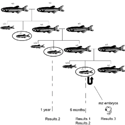
1. Zebrafish lines and husbandry
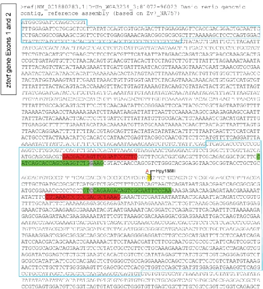
The terthu3430 mutation (see Figure S1,Figure S2Figure S3) was generated on a wild
type TL (tüpfel-long fin) background at the Ubrecht laboratory, Utrecht, Netherlands. Mutant
lines were maintained by intercrossing heterozygous individuals and background mutations were previously removed by outcrossing three times with wild type (AB strain) individuals at Dr. P. Chapouton laboratory at the German Research Center for Evironmental Health, Germany. Wild type and heterozygous siblings were used as controls for all experiments. Maternal zygotic mutant embryos were generated by crossing adult homozygous mutant couples. The crosses between adult homozygous zygotic mutants and respective controls were performed once a week. Each couple was put together overnight on appropriate crossing boxes and the embryos were collected and maintained in embryonic media 1x (diluted with system water from a stock 50x solution: 14,69g NaCl, 0,63g KCl, 2,43g CaCl2.2H2O, 4,07g MgSO4.7H2O – with 1mL of methylene blue/10L) which was changed
daily. Embryos were staged by time post fertilization (hours, hpf, or days, dpf) at 28ºC and by morphological criteria[118]. Wild type AB lines were used for optimization of techniques. All fish were kept on an aquarium system at 28ºC, except when recovering from caudal fin amputation or manipulation in which they were kept in crossing boxes on a 33ºC water bath.
2. Genotyping of terthu3430 lines
2.1. Caudal fin amputation and isolation of genomic DNA
The progenies of mutant heterozygote inter-crosses were separated after 3 months of age into breeding tanks and numbered for genotyping purposes. Each individual was separately anaesthetized with Tricaine 1x (160 mg/ml) (MS222, Sigma) and the caudal fin was amputated. The corresponding fin clip for each individual was collected into a 2mL eppendorf tube and incubated on a lysis buffer solution (Genomic DNA purification kit - #K0512, Fermentas) with 0,5mg/mL Proteinase K on a shaking thermomixer, overnight at 50°C. The extraction of genomic DNA was then performed as indicated on the Genomic DNA purification kit, but with the precipitation step performed overnight at -20ºC.
2.2. PCR, RFLP and sequencing
The genomic DNA was diluted in sterile nuclease-free water. After optimization, a fragment around the point mutation was amplified with proof-reading Phusion DNA-Polymerase (Finzymes, F-530S) and the following primers: Tert2A-1F
(5‟-CATCAGCACCAGCGAGGTCTGGAAG-3‟) and Tert2A-1R
(5‟-11
GACGACCAGTTCGGATCCCTTTC-3‟) and Tert2A-2R
(5‟-CTTTACCCTCCGCCGCTTTACC-3‟) for genotyping by both RFLP and sequencing methods (see Figure S2). The PCR conditions were the following: 98ºC for 1min; 98ºC for 10 sec., 65ºC for 30sec., 72ºC for 1min. (for 30 cycles); and a final elongation step of 72ºC for 10min. The amplified PCR products were separated from non-incorporated nucleotides and excess primers with a Wizard® SV Gel and PCR clean-up system (Promega, #A9281). Clean PCR reactions were sequenced (StabVida, Portugal) with both the forward and reverse primers (from the Tert2A-F/R-1 or Tert2A-F/R-2 sets), separately. Additionally, the genotype of some individuals was confirmed by RFLP: the samples were subjected to digest (Hpy188III for 2h at 37°C - New England Biolabs) and the fragments were visualized after electrophoresis on a 3% high resolution agarose gel (MetaPhor® - FMC BioProducts, #50180 or Sigma, #A4718).
3. Telomere Restriction Fragment (TRF) Analysis 3.1. Genomic DNA isolation and digestion
Genomic DNA from adult zebrafish fins was obtained as described above for genotyping purposes. The approximate DNA concentration was measured with Nanodrop equipment (Thermo scientific, ND 1000) and the DNA was visualized on a 1% agarose gel to control DNA integrity and RNA presence (in which case 1uL of RNase A (Sigma) was added to the samples, 5 min. at 37ºC) and for relative quantification of samples for further digestion. Approximately equal amounts of DNA (~1ug) were digested with HinfI (Invitrogen or Roche) and RsaI (Invitrogen) enzymes in a total volume of 30uL at 37ºC, overnight. Control undigested samples were produced by substituting enzymes by water.
3.2. Electrophoresis and Transference
The total volume of the reaction was loaded onto a 0,6% 25cm x 15cm agarose gel in 0,5x TBE, and separated for ~17h at 110 V and 4ºC. After electrophoresis, the gel was photographed and processed for transference: after an initial step in HCl 0,25N for 15 min., it was incubated on a denaturing Blot1 solution (NaOH 0,5M; NaCl 1,5M) for 30 min. and then neutralized in Blot2 (NH4Ac 1M; NaOH 0,02M) for 1 hour and washed with 6xSSC (NaCl
90mM; Trisodium Citrate 90mM). A 25cm x 15cm membrane (zeta-probe GT genomic tested blotting membrane, Biorad #162-0196) was soaked briefly on water and immersed 10min on Blot2 solution after which it was placed over 3 pieces of Blot2-embedded chromatography Whatman paper (Schleicher-Schuell, Sigma) on top of a stack of paper towels with the gel on top, wells down. The transference proceeded overnight and the DNA was crosslinked to the membrane by UV treatment (Stratalinker 1800, Stratagene).
12
3.3. Probe preparation, Hybridization and Detection
A telomere probe was obtained by random-primer labelling of a (CCCTAA)17
oligonucleotide (25 ng) with the Prime-it II random primer labelling kit (Stratagene) (10uL random primers were added to the oligonucleotide in a total volume of 24uL, put in 100ºC for 5min and then 5U of Klenow enzyme in buffer were added to dCTP-αP32 (5uL from a 10mCi/ml;3000Ci/mmol stock) for 30min. at 37ºC). The non-incorporated radioactive particles were removed by filtering the resulting product on a final volume of 100uL (with TE 1x) through a Sepharose G-50 spin column (GE Healthcare). The membranes were pre-incubated for at least 30min. on the hybridization Church-Gilbert buffer (1%BSA, 1mM EDTA, 7% SDS, 0,5M NaHPO4 pH 7,2) and then half the volume of the prepared probe was added
to one membrane, after boiling for 5 min., and allowed to hybridize overnight at 65ºC. The membranes were washed three times with a solution of 2xSSC; 1%SDS to remove non-hybridized probe and then exposed overnight on a phosphorimager screen (GE Healthcare) The signal was detected with a Storm 860 scanner, using the GE Healthcare‟s ImageQuantTM software selecting for a 100 micron pixel size and phosphor-image option to obtain quantifiable 16-bit digital images.
3.4. Data Analysis of Southern Blot
Images were analysed with ImageJ 1.43u. The pixel gray values (intensity) were measured along each lane and the mean per pixel x position was recorded on xy tables (where x is the position relative to well and y is the intensity value for each point). After calibrating the x axis with the ladder bands‟ position, the y values were normalized to a 0-1 scale:
Formula (1)
, with yn = normalized intensity; yi = intensity value for
table position i; ymin and ymax = minimum and maximum intensity value of the well (column), respectively.
Using Matlab 7.10.0.499 (R2010a) (under the guidance of Samuel Antão, MSc, Computer Engineer), normalized data was grouped in classes, producing histograms, in order to reduce the noise influence on calculations, as described[119]. The upper and lower ranges of molecular weight corresponding to telomere signal were analysed separately. Assuming that the TRF values can be described by a normal distribution, the most likely normal function was found and divided by the molecular weight distribution in order to determine the average molecular weight (MW) value for the mean TRF frequency. Independently, the relative frequency values were calculated from experimental data by dividing the normalized intensity values by the corresponding MW, as described[119], and presented graphically. The weighted and unweighted averages were also calculated directly
13 from the normalized histogram intensity values by applying the formulas below, as described[120] (as in TeloRun program, by Harley,C., Allsopp, R. and Vaziri, H.):
Formula (2) Weighted mean: , with ODi as the signal intensity
and Li as the DNA length at position i. It takes into account the dependency of the signal on
the length of molecules and corrects for this aspect.
Formula (3) Unweighted mean: with ODi as the signal
intensity and Li as the DNA length at position i.
(See supplementary materials and methods for implementation code).
4. Histological analysis
4.1. Fixation, processing and sectioning techniques
4.1.1. Fixation, processing and sectioning techniques for embryos Embryos older than 48hpf were anaesthetised in tricaine 1x (160 mg/ml) (MS222, Sigma) in embryonic media, before fixation. All embryos were fixed overnight in 4% paraformaldeyde (PFA) at 4ºC and then placed in 100% methanol at 4ºC before processing. After several washes in PBS (phosphate buffer saline) the embryos were cryoprotected on a sucrose 15%/PBS solution and then embedded in 7,5% pork skin gelatine (Sigma)/15% sucrose/PBS for one hour at 37ºC. The 1cm2 blocks were frozen in isopenthane/liquid nitrogen and stored at -80ºC until sectioning. The embedded samples were cut in 12um sections with a cryostat (Leica CM 3050S) either alternately for further paired Hematoxylin-Eosin (HE) stained- and non-stained slides or serially for staining only.
4.1.2. Fixation, processing and sectioning techniques for adults Fish were sacrificed in 25x Tricaine (MS222, Sigma) in system water and fixed overnight in 4% PFA at 4ºC. After decalcification overnight in 15% EDTA/H2O and washes in
dH20, they were dehydrated on an ethanol series (70%, 96%, 100%) and placed on xilol. The
samples were then embedded in paraffin on vacuum at 70ºC and oriented for sagittal sectioning on appropriate metallic scaffolds. The embedded samples were cut in 6um sections using a Minot microtome (Leica RM 2145). Sections were collected for further paired HE stained- and non-stained slides.
4.2. Hematoxylin-Eosin staining
4.2.1. Hematoxylin-Eosin staining for embryos
Slides were washed one hour in 1x PBS on a 37ºC water bath and then runned through a series of washing and staining steps: dH2O (5min.); Hematoxylin (5min.); dH2O
14 (briefly); 1% HCl (briefly); flowing H2O; NH4 (1-4 min.); 70% Ethanol (30sec.); Eosin (briefly); 70%/96%/100% Ethanol series (30sec.). The slides were mounted after application of xilol (10min.) in Entellan with 24 x 60 mm coverslips, and stored at room temperature.
4.2.2. Hematoxylin-Eosin staining for adults
Slides were deparaffinized in xilol for 15 minutes and rehydrated on a 100%/96%/70% Ethanol series (5min. each step). Then they were runned through a series of washing and staining steps: dH2O (5min.); Hematoxylin (10min.); dH2O (briefly); 1% HCl (briefly); flowing H2O; NH4 (1-4min.); 70% Ethanol (3sec.); Eosin (briefly); 70%/96%/100% Ethanol series (30sec. each). The slides were mounted with 24 x 60 mm coverslips in Entellan after application of xilol (10min.), and stored at room temperature.
5. Adult fish imaging and histology slides’ image acquisition
Images from adult fish were acquired using a regular photo cameraand the scale was set using a regular ruler on the image. Images from histology slides were acquired using a brightfield microscope (Leica DM2500). All images were processed with Adobe Photoshop® CS4 software and the image panels were created using Adobe Illustrator® CS4 software.
6. Embryo imaging
All images were collected with a Leica Z6 APO stereoscope with a Leica DFC 490 camera. The embryos were placed on top of a 1% agarose-coated dish in embryonic media or in Tricaine 1x (160 mg/ml) (MS222, Sigma) in embryonic media (for embryos older than ~48hpf). For one experiment of imaging of embryos below 24hpf, they were dechorionated and embedded in 3% Methyl cellulose (Sigma-Aldrich) in order to properly place them and obtain higher quality images. All images were processed with Adobe Photoshop® CS4 software and the image panels were created using Adobe Illustrator® CS4 software.
7. Statistics
The Graphpad Prism 5 program was used for statistical analysis. Comparisons between experimental and control results were made using paired a t-test with 0.95 confidence level. For p<0.05, differences were considered significant and the values were presented on the respective graph.
15
Results
1. Zebrafish terthu3430 mutants have shorter telomeres by 6 months of age
A PCR-based assay that measures telomerase activity in vitro from tissue protein extracts (Telomeric Repeats Amplification Protocol – TRAP) was performed by our lab, showing that these mutant animals have no telomerase activity in several organs, therefore confirming that the nonsense mutation terthu3430leads to a non-functional product.
The absolute length of zebrafish telomeres is controversial in literature (see Introduction-5.). In order to determine the absolute mean telomere length of this species, we optimized the method of Telomere Restriction Fragment (TRF) analysis by Southern Blot. We used 6 months-old animals, which are still young and have already reached sexual maturity in the case of wild types, and caudal fin tissue because it is easily available and analysed single fins in order to account for variability.
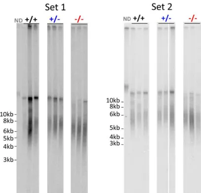
The result obtained by this technique is a smear signal that describes the distribution of telomere length in the chromosomes of all cells within the tissue. One can then calculate the average telomere length as well as observe the distribution of lengths within each sample. Duplicate samples of three individuals from each genotype were independently digested and separated, leading to two sets of results (Figure 5).
Figure 5: HinfI/RsaI TRF Southern Blot results for two duplicate sets of caudal fin samples from 6 months-old fish. Three samples per genotype were analysed in each set. Probe used: random primer-labelled (CCCTAA)12
32
16 Each lane was analysed in order to obtain densitometry profile data corresponding to the values of intensity on the graph varying according to the distance on the gel (Figure 6). This was then processed in order to give rise to distributions of relative frequency of TRF length for each sample (Figure 7).
As shown in Figure 5 and Figure 7, a smear from about 3 Kb to 10 Kb is present in wild
type samples. A higher molecular weight (MW) population of TRFs of about 13,5 Kb
(although the DNA ladder used does not allow for accurate measurements above 10 Kb) is also present. The overall distribution of TRFs is similar between wild type individuals and two populations within the lower molecular weight range are present, which can represent true distinct classes of telomere lengths in the tissue. Heterozygotes present roughly the same distribution and average telomere length as wild types except for the absence of the two clearly distinct populations on the lower MW range of telomeres. Homozygous mutants‟ samples present a TRF profile in which both higher and lower MW portions are shifted to a low MW value. Moreover, the higher MW band is compressed, suggesting that the longer molecules present in wild types are here shortened. The differences between genotypes are significant, as depicted in Figure 8. Mean TRF lengths are equivalent when calculated using weighted average formula (2) or by determining the best likely normal distribution (Figure 7, red line), dividing it by the MW distribution and finding its maximum (Figure 8). The shapes of the distribution of telomere lengths (Figure 6 and Figure 7) are also different between genotypes, with the homozygous mutant profiles shifted towards the left and with a sharper peak.
The results presented above indicate that, at least in this tissue, 6 months-old adult zebrafish individuals have short telomeres of about 6 to 7 Kb in average and that telomerase is important to maintain these values, since the telomerase mutants have clearly shorter telomeres with a difference of about 1 Kb in respect to wild type siblings. Having a clear distinction in mean telomere lengths between wild type and mutant siblings is particularly striking since the fin tissue is not an adult tissue with a particularly high proliferation rate (that in the absence of telomerase would be subjected to higher telomere attrition) neither these fish are particularly old (which would lead to increased telomere attrition in the absence of telomerase activity).
17
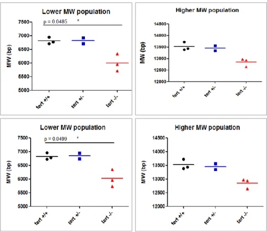
Figure 6:Densitometry graphs of intensity signals of the samples grouped by genotypes, for both sets
Figure 7: Relative frequency (Formula (1)) TRF length distribution of matched representative samples from each genotype from Set 1 (left) and Set 2 (right) blots.
18
Figure 8: Average TRF length as determined with (Formula (2)) (above) or x for y maximum of a Normal distribution/MW (below). Each point is average of duplicate values for each sample. See also
Figure S4 and Figure S 5.
2. Telomerase zygotic mutants are viable but display several proliferation-related phenotypes as adults and infertility
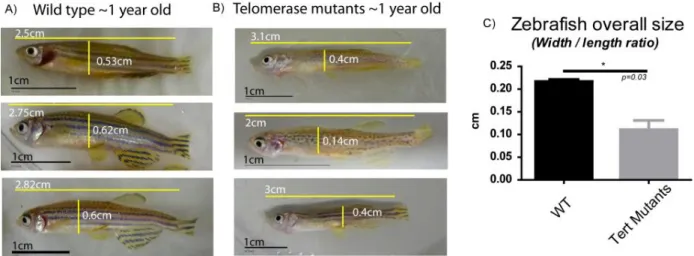
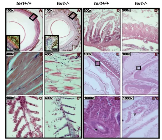
In order to determine the effects of lack of telomerase activity in zebrafish, we followed the health status of adult zygotic mutant fish through time. Some individuals displayed progressive deterioration including altered, less bright pigmentation, diminished activity and an extreme loss of body weight. This ultimately conduced to death by one year of age. In Figure 9, we can observe the extreme thinness of these fish, which is quantified on the graph in c) in form of a width/length ratio. We were able to sacrifice one individual for subsequent analysis, whereas the others died before we could collect them in a well-preserved condition. The results of the histology analysis of this individual‟s tissues are presented in Figure 10 (N=1). Here we can observe that all tissues have lost their structure and integrity. They are loose, with decreased cellularity in comparison with wild type. The gills‟ branches are thinner, with just one-row of cells on gill filaments (Figure 10C/C‟), the muscle fibres are disintegrated (Figure 10B/B‟) and the gut presents a profound atrophy of the villi with loss of intestinal crypts (Figure 10D/D‟). Interestingly, the retina has an abnormal shape and a disorganized agglomerate of cells is observed instead of the characteristic layering structure (Figure 10A/A‟). In the testis (Figure 10 E/E‟ and Eb/E‟b), it is possible to observe the lack of cellularity compared with the wild type sample, in which mature spermatozoids are present
19 inside seminiferous tubules (arrows in Figure 10 Eb/E‟b). These histological features are consistent with the poor health status of the individuals presented in Figure 9, although we could not conclude which was the first alteration to occur and what was their particular cause of death. Recent observations in the lab point to the testis as the first organs to show alterations, followed by the gut, since histological abnormalities in these organs are observed as soon as at 6 months of age. Although some slides were kept for subsequent immunohistochemistry, no subsequent studies were yet performed to address the telomere status of these samples in situ or for apoptosis. However, PCNA staining (which is a component of the DNA replication complex) performed in the lab showed that the mutant gut is devoid of proliferating cells whereas wild type crypts present a strong signal.
Figure 9: 1 year-old zygotic mutants displaying a „wasting phenotype‟ (A) and B)) and quantification of their different width/length ratios when compared with wild type siblings (C)).
Both male and female homozygous mutants have also lower fertility as soon as at 6 months-old. In order to count the number of eggs and the number of viable zygotes we crossed tert+/+; tert+/- and tert-/- couples. While tert+/+ and tert+/- females consistently produced large amounts of eggs, the female tert-/- was only able to produce them once every two weeks (~50% of the times relative to wild type couple) and in lower amounts (~50%), even when coupled with wild type males. This progressed to a state of complete infertility at 7 months of age, and this is accompanied by severe deterioration as it was observed in the 1-year old mutants of Figure 9. Most of the eggs produced by the female tert-/- also do not appear to be fertilized by any of the tert-/- males.
To investigate whether the low fertilization index of tert-/- eggs was caused by the mutant males or females, wild type and homozygous mutant individuals were crossed in different combinations. As we can observe in Figure 11, most of the eggs were fertilized when the tert-/- female was crossed with a wild type male, similar to wild type incrosses. On the other hand, most of the eggs were not fertilized when wild type females were crossed
20 with the tert-/- males, similarly to what happened in the mutant couple incross. These eggs that fail to develop are similar to non-fertilized eggs from wild type crosses (Figure 11)..
These results point to a role of telomerase in maintaining tissue renewal in the adult. The failure in keeping tissue homeostasis compromises organism viability in the mutant and translates into infertility.
Figure 10: Microscopy images from Hematoxylin-Eosin stained sections of several tissues from adult zebrafish. Matched wild type (tert+/+) and zygotic homozygous mutant (tert-/-) samples are presented side-by-side (X: wild type; X‟: mutant). A and A‟: Retina; B and B‟: Skeletal muscle from trunk region; C and C‟: Gill filaments; D and D‟: Midgut wall; E and E‟: Visceral region showing the placement of the testis (square); Eb and Eb‟: Amplified image of the region on the square in E and E‟, respectively, showing transversal sections of seminiferous tubules (arrow).
Figure 11: Phenotype of eggs resulting from a wild type cross (A) and from a cross between a wild type female and a homozygous zygotic mutant male (B).
21
Figure 12: Percentage of non-fertilized eggs per cross (mean nr. of non-fertilized eggs/total number of eggs produced by the female). Error bars represent standard deviation interval.
3. Maternal zygotic mutants are not viable due to gross abnormalities during embryonic development
As previously mentioned, tert-/- incrosses produced a low number of zygotes due to reduced fertility of mutants (Figure 12). However, the few maternal zygotic embryos generated were raised in order to characterize their development.
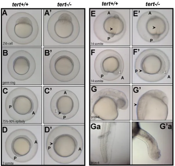
All mutant embryos were unviable and presented multiple phenotypes. They were delayed in development when compared to wild type control embryos fertilized at the same time. This delay was detected as soon as the first cell divisions start to occur (Figure 13 A/A‟). During gastrulation stages, mutant embryos showed a delay in epiboly movement, which corresponds to the movement of cell layers towards the vegetal pole of the embryo (Figure 13C‟ (arrows), when compared to Figure 13B‟). At the end of gastrulation, the tail region of mutant embryos appears abnormal and darker, suggesting the occurrence of apoptosis (Figure 13D‟ (arrow)). A few hours later, during segmentation stages, the posterior region of mutants also seems to be affected when compared to wild type and this translates into tail malformations, which are observable at 24hpf (Figure 13Ga/G‟a) and 48hpf (Figure 14C and Cb). The anterior brain region also shows defects in comparison to wild type (Figure 13G/G‟). Interestingly, we observed that later, at 3dpf, mutant embryos‟ neural tube fail to close (Figure 14I and Figure 14K/K‟), suggesting that tert has a role during brain development. The most common phenotype was a curvature of the body with associated bending of the notochord (Figure 13Ga/G‟a and Figure 14C, D) and improper somite/muscle development (Figure 13E/E‟ and Figure 14L/L‟). The posterior region anomalies were sometimes translated into severe truncations (Figure 14J). Also, even when no other striking phenotype was apparent, the eyes in the mutant were often smaller than in wild type (Figure 13F/F‟ and Figure 14 (white arrows)). In the case of one single mutant embryo that seemed
Infertility wt x wt wt fema le x tert -/- ma le wt ma le x tert -/- f ema le tert -/- x tert -/ -0 20 40 60 80 100 Crosses genotypes p= 0.0234 * N = 2 N = 3 N = 2 N = 3 0.95 No n fe rt il iz e d e g g s ( % )
22 normal, it was still delayed during development and failed to inflate the swim-bladder (Figure 14G‟). We also observed a reduced number of blood cells circulating near the trunk region (data not shown) and the heart (Figure 14B/B‟), which possibly explains the high incidence of heart edemas in mutant embryos (Figure 14, black arrows). Furthermore, the pattern of distribution and the shape of mutant pigment cells (melanophores) seem to be affected in mutants in comparison to wild type (Figure 14C and D). Interestingly, potential apoptotic cells were observed in some of the mutant embryos, which captured the methylene blue used as a pH indicator of the embryo medium (Figure 14C, Cb and E).
This broad manifestation of telomerase absence highlights its pleiotropic role in embryonic development, which relies greatly on cellular proliferation in order to proceed normally.
Figure 13: Time course of first hours of development of wild type (tert+/+) and maternal zygotic mutant (tert-/-) embryos. Wild type embryos were staged according to Kimmel et al., 1995 and corresponding images of mutant embryos for each time-point (A to G) is presented side-by-side. White arrow in C‟: EVL (enveloping layer); Black arrow in C‟: YSL (yolk sincitial layer); Black arrows in D‟ and F‟: embryo tail region looking abnormal; White arrow in F/F‟: developing eye; G and G‟: head region for G-corresponding time-point; Black arrow in G‟: particular aspect of cells near the head region, looking abnormal. Ga and G‟a: tail region for G-corresponding time-point. A: Anterior; P: Posterior.
23
Figure 14: 48hpf (left) and 5dpf (right) - images of maternal zygotic mutants (tert-/-) and wild type (tert+/+) embryos. A: whole body image of wild type 48hpf embryo; B and B‟: anterior region, showing the eye (white arrow) and the developing heart (black arrow); C: tert-/-embryo showing abnormal body curvature and methylene blue- likely apoptotic stained cells; Cb: close up of square in C. D: tert -/-embryo showing abnormal body curvature and abnormal distribution of melanocytes; E: tert-/- embryo showing heart edema (arrow) and methylene blue-stained brain region. F: whole body image of wild type 5dpf embryo. G and G‟: anterior region, showing the eye, brain, and developing internal organs. H, I and J: tert-/- embryos, showing small eyes (white arrows) and heart edemas (black arrow). I: tert-/- embryo showing opened neural tube. Transversal section of the head (line) is presented in K‟ (K: equivalent section from wild type). J: tert-/- embryo with severe truncation. L and L‟: transversal section of trunk region showing decreased cellularity in the muscle in the tert-/- embryo (L‟) relatively to wild type (L).
![Figure 1: The mammalian shelterin complex at telomeres [7] .](https://thumb-eu.123doks.com/thumbv2/123dok_br/15616971.1054426/9.892.307.589.690.847/figure-mammalian-shelterin-complex-telomeres.webp)
![Figure 4: A model for the cancer-ageing connexion [68] .](https://thumb-eu.123doks.com/thumbv2/123dok_br/15616971.1054426/14.892.239.701.165.463/figure-model-cancer-ageing-connexion.webp)