Daniela Patrícia Peneda Pacheco
Development of an Injectable PHBV microparticles-GG Hydrogel
Hybrid System for Tissue Engineering Applications
Dissertação do 2º Ciclo de Estudos Conducente ao Grau de Mestre
em Tecnologia Farmacêutica
Trabalho efetuado sob a orientação de:
Professora Doutora Maria Helena dos Anjos Rodrigues Amaral
Doutora Alexandra Margarida Pinto Marques
Doutor Vítor Manuel Correlo da Silva
É AUTORIZADA A REPRODUÇÃO INTEGRAL DESTA MONOGRAFIA
APENAS PARA EFEITOS DE INVESTIGAÇÃO, MEDIANTE DECLARAÇÃO
i
“Scientific work must not be considered from the point of view of the direct
usefulness of it. It must be done for itself, for the beauty of science”
Marie Curie (1867-1934)
iii
Acknowledgments
I would like to express my gratitude to PhD Professor Helena Amaral, my supervisor from Faculty of Pharmacy, for the support that she was giving me during this year both scientifically and academically.
I am completely grateful to my 3B’s Research Group supervisors. To Dr. Alexandra Marques, for all the brainstorms that she gave me with her knowledge, for always making me think forward, supporting me in all the ideas and aims. To the other member of the triad, Dr. Vitor Correlo, I want to say that it was a pleasure to work with him, and I want to express my indebted to him for sharing his “out of the box” ideas with me, that allowed the development of a unique work. I am especially thankful for both of them, for the good relationship, encouragement, availability, persistence, and fruitful discussions.
To Professor Rui L. Reis, for allowing me to conclude this stage of my academic life at his Research Group, allowing me the access to all the materials and equipment that were necessary to complete this important step of my academic years. Moreover, he provided me the contact with great scientists in this field, from whom I learned which is not yet written in books.
In this 3B’s journey, I had the opportunity to interact with unique people to who I want to express all my appreciation for the laugh, tears, culture and support. Dr. Praveen Sher I am deeply grateful for making me overcome the drug delivery systems boundaries with his tips which definitely made all the difference. To Marta Ondrésik (Márti), I want to thank her for all the car conversations. Definitely I have never met anyone that justifies the day to day things with biological pathways from tissues to molecules, I am grateful to you. To Dr. Manuel Alatorre for always spreading his happiness and smiles through the corridors, for the early morning rides and also for his knowledge on Physics, Biology and Chemistry that helped me a lot. To Dr. Marianti Mantha, the Greek that translated English to Portuguese for making me speak English. And finally, a special thanks to Sebastião van Uden, who definitely made this year easier and happier, who supported me in the bad and good moments.
To all the people of 3B’s Research group including technicians and friends, for their good work environment and friendship.
Um obrigada muito especial à minha família, nomeadamente aos meus pais, que definitivamente tanto se esforçaram para me dar a mim e aos meus irmãos os alicerces
iv
necessários para chegar até aqui, alcatroando as estradas para que o caminho a percorrer fosse mais fácil.
v Resumo
O design e síntese de sistemas eficazes de libertação de fármacos (SLD) é extremamente importante para a área da engenharia de tecidos e medicina regenerativa (TERM). Estruturas optimizadas com uma distribuição e uma cinética de liberação bem definidas são pré-requisitos para uma aplicação clínica segura e eficaz, evitando efeitos colaterais indesejáveis e toxicidade. Visando o desenvolvimento de SLD de aplicação minimamente invasiva, aliada a uma libertação sustentada, prolongada e localizada de sinais bioquímicos relevantes para a regeneração do tecido, foi desenvolvido neste trabalho um SLD injetável bifásico, o qual resulta da combinação de micropartículas (MPs) de poli(hidroxibutirato-co-hidroxivalerato) (PHBV) (MPs) com um hidrogel de goma gelana (GG).
A primeira etapa do trabalho apresentado nesta dissertação consistiu na produção de MPs de PHBV, com incorporação de moléculas modelo de proteínas e glucocorticóides. Para este efeito, foram incorporadas dentro de MPs de PHBV Albumina de Soro Bovino (BSA) e Dexametasona (Dex) usando um método de dupla emulsão A/O/A com evaporação do solvente, após algumas modificações. Visando o controlo adequado do perfil de liberação de moléculas bioactivas, como fase orgânica foram usadas diferentes proporções de clorofórmio e etanol. Algumas características do sistema de MPs foram afectadas pela modificação do método, nomeadamente o tamanho das partículas, bem como a topografia da superfície das mesmas. A influência da presença de etanol na fase orgânica também foi avaliada em termos de eficácia de incorporação das moléculas bioactivas, sendo que a sua presença conduziu a uma maior eficácia na incorporação de Dex, enquanto a incorporação de BSA não foi afectada. Por sua vez, os estudos de libertação in vitro revelaram que MPs produzidas na ausência de etanol na fase orgânica apresentam um perfil de libertação mais sustentado quando comparado com as MPs produzidas na presença de etanol. Estes resultados eram esperados uma vez que a adição de etanol à fase orgânica induziu a formação de poros maiores na superfície e no núcleo das MPs, proporcionando deste modo a penetração de água, e consequentemente a libertação mais rápida das moléculas bioactivas. Após a obtenção das MPs de PHBV com incorporação de moléculas de interesse para a área de TERM, o objectivo foi definir um sistema que actuaria como um sistema de transporte das MPs, contribuindo para aumentar o tempo de residência das MPs no local de injecção. Para este efeito, as diferentes MPs de PHBV foram incorporados nos hidrogéis injectáveis de GG, amplamente estudado no nosso grupo de investigação para diferentes aplicações de engenharia de tecidos. As MPs de PHBV incorporadas no hidrogel apresentam-se uniformemente distribuídos na matriz, demonstrando, também, uma forte integração com o polímero do hidrogel. Como esperado, a incorporação das MPs nos hidrogéis de GG influenciou fortemente o perfil de libertação, obtendo-se sistemas de libertação de ordem zero. Embora as propriedades do hidrogel possam obtendo-ser modificadas de um modo controlado, com o objectivo de ajustar o perfil de libertação de acordo com requisitos específicos dos tecidos e da patologia/lesão, o sistema proposto pode, certamente, ser explorado como uma SLD de longo prazo.
vi
Neste sentido, um SLD bifásico, que permite a libertação de sinais bioquímicos com diferentes características físico-químicas, bem como a sua libertação localizada, foi desenvolvido com sucesso e constitui uma ferramenta versátil de modulação de microambientes celulares instrutivos para aplicações em TERM.
A dissertação foi estruturada em diferentes capítulos: I) uma introdução que fornece uma visão geral dos conceitos básicos de sistemas de MPs, MPs poliméricas com base na sua formulação, mecanismos de entrega de moléculas bioactivas, e aplicações em TERM; II) a secção de Materiais e Métodos que descreve detalhadamente os materiais utilizados para o desenvolvimento das MPs e dos hidrogéis, bem como as respectivas metodologias de processamento e caracterização, III) o manuscrito completo que resultou do trabalho realizado, o qual se encontra estruturado num breve resumo, uma introdução mais concisa, o resumo dos materiais utilizados e das metodologias seguidas ao longo do trabalho, descrição dos resultados e subsequente discussão e uma breve conclusão; IV) as conclusões finais do trabalho e perspectivas futuras do sistema desenvolvido na área de TERM.
Palavras-chave: Sistemas de libertação de fármacos, Engenharia de Tecidos, poli(hidroxibutirato-co-hidroxivalerato), micropartículas, hidrogel injectável
vii
Abstract
Design and synthesis of efficient drug delivery systems (DDS) are of vital importance for tissue engineering and regenerative medicine (TERM). Optimized delivery structures and well-defined release kinetics appear to be logical prerequisites for safe and efficacious clinical application avoiding undesired side effects and toxicity. With a view toward developing DDS to be applied using minimally-invasive approaches, and that would provide sustained, prolonged and localized release of biochemical cues relevant for tissue regeneration, we designed a biphasic injectable DDS combining polyhydroxybutyrate-co-hydroxyvalerate (PHBV) microparticles (MPs) within a gellan gum (GG) hydrogel.
The production of PHBV MPs incorporating proteins and glucocorticoids model molecules was the first step of the work presented in this dissertation. For this purpose, Bovine Serum Albumin (BSA) and Dexamethasone (Dex) were incorporated inside PHBV MPs by using a double emulsification-solvent evaporation method with modifications. Aiming at tailoring the bioactive molecules release profile, different proportions of chloroform and ethanol, as organic phase, were proposed. This modification revealed to affect some features of the microparticulate system, namely particle size and surface topography. The influence of the presence of ethanol in the organic phase was also evaluated in terms of bioactive molecules incorporation efficiency. The presence of ethanol led to higher efficiency of Dex entrapment inside the MPs, while the BSA incorporation was not affected. In his turn, the in vitro release studies revealed that MPs produced in the absence of ethanol in the organic phase presented a more sustained profile when compared to the MPs produced in the presence of ethanol. This finding was expected since the addition of ethanol to the organic phase induced the formation of larger pores on the surface and in the core of the MPs thus facilitating water penetration, and consequently faster release of the bioactive molecule.
After obtaining the PHBV MPs loaded with the molecules of interest, the objective was to define a system that would act as carrier of the MPs and would enhance MPs residence time upon injection. For this purpose, the different PHBV MPs were embedded within injectable GG hydrogels, extensively studied in our Research Group as cell carriers for different tissue engineering applications. A uniform distribution of PHBV MPs across the GG matrix with a strong integration of the MPs with the polymer of the hydrogel was attained. As expected, the incorporation of the MPs into the GG hydrogels strongly influenced the profile, from one to nearly zero-order and the amount of released Dex and BSA. Although the properties of the hydrogel are prone to further improvement in order to tune the release profile according to tissue and pathology/injury specific requirements, the proposed system can certainly be explored as a long-term DDS.
In this sense, a biphasic DDS, which allows the release of biochemical cues with different physicochemical features, as well as its localized deliver, was successfully developed and represents a versatile tool to prepare instructive cell microenvironments for TERM.
viii
The dissertation was structured in different chapters: I) a General Introduction that provides an overview of the basics of microparticulate systems, polymeric microparticles based on their formulation, their mechanisms of drug delivery, and their applications in the TERM; II) a Materials and Methods section that describes with some detail the materials used to produce the MPs and the hydrogels as well as the respective processing and characterization methodologies; III) the complete manuscript that resulted from the work developed in which is provided a short abstract, a more concise introduction of the work, a resume of the materials used and methodologies followed along the work, the description of the results and subsequent discussion; IV) the final conclusions of the work and future perspectives for the area.
Keywords: Drug Delivery System, Tissue Engineering, polyhydroxybutyrate-co-hydroxyvalerate, microparticles, injectable hydrogel
ix
Table of Contents
I.
General Introduction ... 1
1. Drug Delivery Systems ... 1
2. Microparticles as Drug Delivery Systems ... 3
2.1. Microparticulate Systems in Tissue Engineering ... 4
2.1.1. Microparticulate Drug Delivery Systems ... 5
2.1.2. Microparticulate Drug Delivery Systems as part of Tissue Engineering Scaffolds 7 2.1.3. Microparticulate Drug Delivery Systems as Tissue Engineering Scaffolds ... 8
3. Final Remarks and Future Trends ... 9
4. Bibliography... 10
II.
Materials and Methods ... 17
1. Poly (hydroxybutyrate-co-hydroxyvalerate) Properties ... 17
2. Gellan Gum Properties... 17
3. Preparation of Poly (hydroxybutyrate-co-hydroxyvalerate) microparticles . 18 3.1. Loading Poly (hydroxybutyrate-co-hydroxyvalerate) microparticles with bioactive molecules ... 18
3.1.1. Bovine Serum Albumin-loaded Poly (hydroxybutyrate-co-hydroxyvalerate) microparticles ... 18
3.1.2. Dexamethasone-loaded Poly (hydroxybutyrate-co-hydroxyvalerate) microparticles………..19
4. Gellan Gum hydrogel preparation ... 19
4.1. Incorporation of Poly (hydroxybutyrate-co-hydroxyvalerate) microparticles in Gellan Gum hydrogel ... 19
5. Determination of Particles Production Yield ... 19
6. Efficiency of incorporation of bioactive molecules into the Poly (hydroxybutyrate-co-hydroxyvalerate) microparticles ... 20
6.1. Bovine Serum Albumin Quantification ... 20
6.2. Dexamethasone Quantification... 20
7. In vitro Release Studies ... 21
7.1. In vitro Release Studies from Poly (hydroxybutyrate-co-hydroxyvalerate) microparticles ... 21
7.2. In vitro Release Studies from Poly (hydroxybutyrate-co-hydroxyvalerate) microparticles embedded within Gellan Gum hydrogels ... 21
x
8. Laser Diffraction Spectrometry ... 21
9. Fourier Transform Infrared spectroscopy ... 22
10. Scanning Electron Microscopy ... 22
11. References ... 22
III. Development of an Injectable PHBV microparticles-GG Hydrogel Hybrid
System for Tissue Engineering Applications ... 25
1. Introduction ... 26
2. Materials and Methods ... 29
2.1. Preparation of Poly (hydroxybutyrate-co-hydroxyvalerate) microparticles ... 29
2.1.1. Bovine Serum Albumin-loaded Poly (hydroxybutyrate-co-hydroxyvalerate) microparticles ... 29
2.1.2. Dexamethasone-loaded Poly (hydroxybutyrate-co-hydroxyvalerate) microparticles…. ... 29
2.2. Gellan Gum hydrogel preparation ... 29
2.2.1. Incorporation of Poly (hydroxybutyrate-co-hydroxyvalerate) microparticles in Gellan Gum hydrogel... 30
2.3. Determination of Particles Production Yield ... 30
2.4. Efficiency of incorporation of bioactive molecules into the Poly (hydroxybutyrate-co-hydroxyvalerate) microparticles ... 30
2.4.1. Bovine Serum Albumin Quantification ... 30
2.4.2. Dexamethasone Quantification ... 31
2.5. In vitro Release Studies ... 31
2.5.1. In vitro Release Studies from Poly (hydroxybutyrate-co-hydroxyvalerate) microparticles ... 31
2.5.2. In vitro Release Studies from Poly (hydroxybutyrate-co-hydroxyvalerate) microparticles embedded within Gellan Gum hydrogels ... 31
2.6. Laser Diffraction Spectrometry ... 32
2.7. Fourier Transform Infrared spectroscopy ... 32
2.8. Scanning Electron Microscopy ... 32
3. Results ... 33
3.1. Poly (hydroxybutyrate–co–hydroxyvalerate) microparticles Process Production ... 33
3.2. Bovine Serum Albumin and Dexamethasone Entrapment Efficiency and Release Profile ... 36
3.2.1. Loaded Microparticles properties ... 36
3.2.2. Bovine Serum Albumin Release from Poly (hydroxybutyrate–co–hydroxyvalerate) microparticles ... 39
xi
3.2.3. Dexamethasone Release from Poly (hydroxybutyrate–co–hydroxyvalerate)
microparticles ... 41
3.3. Injectable Gellan Gum/ Poly (hydroxybutyrate–co–hydroxyvalerate) microparticles System ... 43
3.3.1. Injectable System Properties ... 43
3.3.2. Bovine Serum Albumin Release from the Injectable Gellan Gum/ Poly (hydroxybutyrate–co–hydroxyvalerate) microparticles System ... 44
3.3.3. Dexamethasone Release from the Injectable Gellan Gum/Poly (hydroxybutyrate– co–hydroxyvalerate) microparticles System ... 45
4. Discussion ... 46
5. Conclusion ... 50
6. References ... 51
IV. Final Remarks and Future Perspectives ... 59
V. Annex ... 61
1. Morphological Analysis ... 61
xii
List of Figures
I. General Introduction
Figure 1 – Requisites regarding the design of a new drug delivery system………..2 III. Development of an Injectable PHBV microspheres-GG Hydrogel Platform
Targeting Tissue Engineering Applications
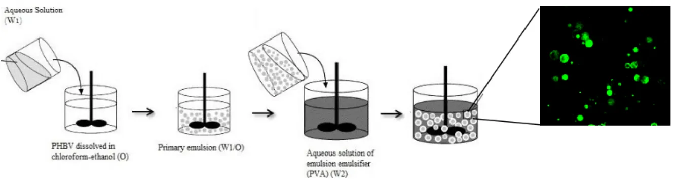
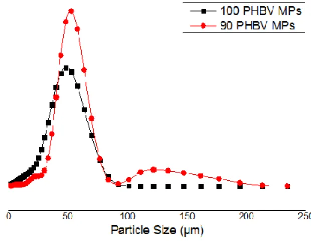
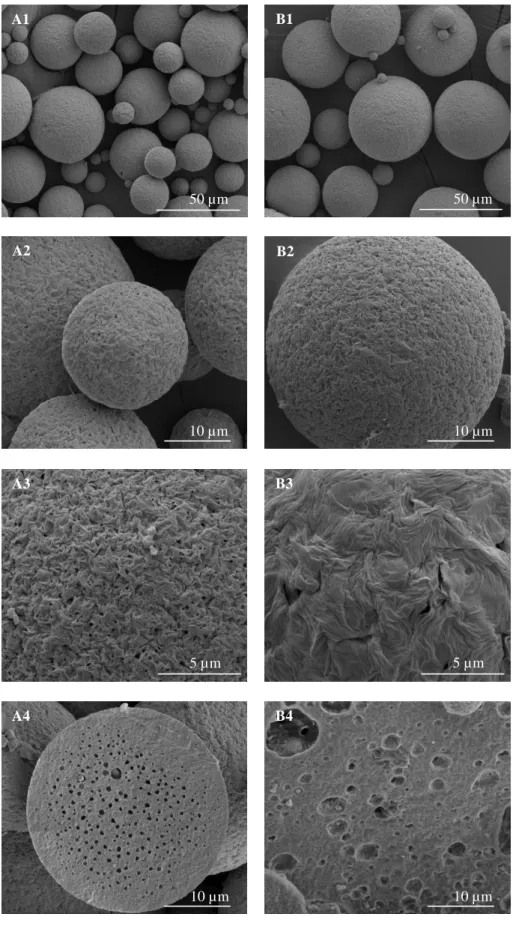
Figure 1 – Schematic depiction of double emulsification evaporation method………..33 Figure 2 – Particle size distribution of the PHBV MPs………34 Figure 3 - SEM micrographs of PHBV MPs obtained under different experimental conditions: (A)
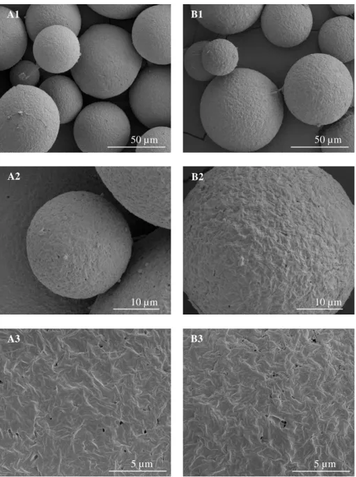
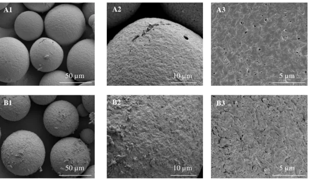
using 100% chloroform; and (B) a mixture of 90% chloroform: 10% ethanol. The sequential micrographs represent an overview of the obtained MPs showing the detail of their surface and respective close up of the internal morphology………35
Figure 4 - SEM micrographs of BSA loaded-PHBV MPs obtained under different experimental
conditions: (A) using 100% chloroform; and (B) 90% chloroform: 10% ethanol. The sequential micrographs represent an overview of the obtained MPs showing the detail of their surface……..37
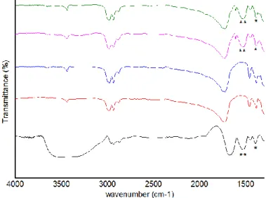
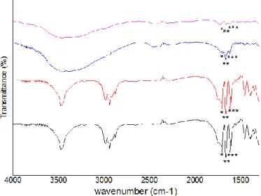
Figure 5 – FTIR spectra of BSA and PHBV MPs: BSA (black line); 100 unloaded-PHBV MPs (red
line); 90 unloaded-PHBV MPs (blue line); 100 BSA-loaded PHBV MPs (pink line); 90 BSA-loaded PHBV MPs (green line). The characteristics bands of BSA are marked (*)……….38
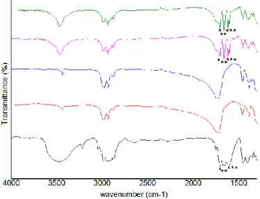
Figure 6 – FTIR spectra of Dex and PHBV MPs: Dex (black line); 100 unloaded-PHBV MPs (red
line); 90 unloaded-PHBV MPs (blue line); 100 Dex-loaded PHBV MPs (pink line); 90 Dex-loaded PHBV MPs (green line). The characteristics bands of Dex are marked (*)………..39
Figure 7 - In vitro release profile of BSA from BSA-loaded PHBV MPs obtained using a mixture of
chloroform and ethanol in different proportions: (100:0) % and (90:10) %...40
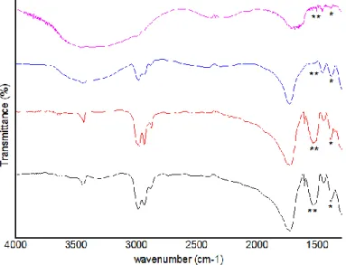
Figure 8 – FTIR spectra of BSA-loaded PHBV MPs: BSA-loaded 100 PHBV MPs (black line) and
BSA-loaded 90 PHBV MPs (red line) before release studies; and BSA-loaded 100 MPs (blue line) and BSA-loaded 90 MPs (purple line) after 21 days of in vitro release. The characteristics bands of BSA are marked (*)………...40
xiii
Figure 9 - SEM micrographs of BSA-loaded PHBV MPs obtained under different experimental
conditions: (A) using 100 % chloroform; and (B) with 90% chloroform: 10% ethanol, after 21 days of
in vitro release studies. The sequential micrographs represent an overview of the obtained MPs
showing the detail of their surface………...41
Figure 10 - In vitro release profile of Dex loaded-MPs obtained using a mixture of chloroform and
ethanol in different proportions: (100:0) %; and (90:10) %...42
Figure 11 – FTIR spectra of Dex-loaded PHBV MPs: Dex-loaded 100 PHBV MPs (black line) and
Dex-loaded 90 PHBV MPs (red line) before release studies; and 100 Dex-loaded MPs (line) and 90 Dex-loaded MPs (purple line) after 21 days of in vitro release. The characteristics bands of Dex are marked (*)………42
Figure 12 – Schematic representation of injectable fluorescein isothiocyanate labelled bovine serum albumin – loaded PHBV MPs embedded within GG hydrogel system………..43
Figure 13 - SEM micrographs of PHBV MPs embedded within injectable GG hydrogels. The
sequential images represent an overview of the distribution of the particles within the hydrogel, close up on the surface of the biphasic structure, and a cross-section………43
Figure 14 - In vitro release profile of BSA from GG hydrogels embedding PHBV MPs obtained
using a mixture of chloroform and ethanol in different proportions: (100:0) %; and (90:10) %...44
Figure 15 - SEM photographs of BSA loaded-MPs embedded within GG hydrogel, after 21 days of in vitro release studies. The sequential images represent an overview of the distribution of the
particles within the hydrogel, close up on the surface of the biphasic structure, and a cross-section……….45
Figure 16 - In vitro release of Dex from GG hydrogels embedding PHBV MPs obtained using a
mixture of chloroform and ethanol in different proportions: (100:0) %; and (90:10) %...45
V. Annex
Figure 1 - SEM micrographs of Dex loaded-PHBV MPs obtained under different experimental
conditions: (A) using 100% chloroform; and (B) 90% chloroform: 10% ethanol. The sequential micrographs represent an overview of the obtained MPs showing the detail of their surface……..61
xiv
List of Tables
III. Development of an Injectable PHBV microspheres-GG Hydrogel Platform Targeting Tissue Engineering Applications
Table I. Yield and Size of the PHBV MPs produced under different conditions………....33 Table II. Yield and Size of the PHBV loaded MPs produced under different conditions……..36 Table III. Incorporation Efficiency of Bovine Serum Albumin and Dexamethasone within PHBV
MPs………...38
List of Abbreviations
- weight of MPs– weight of the sum of polymer and bioactive substance
100 PHBV MPs – Microparticles obtained in the absence of ethanol 90 PHBV MPs - Microparticles obtained in the presence of ethanol
BMPs - Bone Morphogenetic Proteins BSA - Bovine Serum Albumin
DDS - Drug Delivery System
Dex - Dexamethasone EC - Endothelial Cells ECM - Extracellular Matrix FTIR - Fourier Transform Infrared GFs - Growth Factors
GG - Gellan Gum
IE - Incorporation Efficiency
IGF-1 - Insulin-like Growth Factor-1 MPs - Microparticles
Mw - Molecular Weight O – Organic phase
OPF - Oligo(poly(ethylene glycol) Fumarate
PGA - Poly(Glycolic acid) PHAs - Polyhydroxyalkanoates PHB – Poly (hydroxybutyrate) PHBV – Poly (hydroxybutyrate-co-hydroxyvalerate)
PLA – Poly (lactic acid)
PLGA – Poly (lactic-co-glycolic) acid PVA - Poly(vinyl alcohol)
RM - Regenerative Medicine
SEM - Scanning Electron Microscopy TE – Tissue Engineering
xv TGF - Transform Growth Factor
v/v % - Volume concentration
VEGF - Vascular Endothelial Growth Factor
w/v % - Mass concentration W1 – First aqueous phase W2 – Second aqueous phase W/O/W – Water/Oil /Water
1
I. General Introduction
1. Drug Delivery Systems
Drug Delivery is a field of vital importance for medicine and healthcare (Zhang, 2013). This field of pharmaceutical technology represents one of the most rapidly advancing areas of science in which chemists and chemical engineers are contributing to human health care (Jain, 2008).
Therapeutic efficacy and safety of drugs, administered by conventional methods, can be improved by more precise spatial and temporal placement within the organism, thereby reducing both the size and number of doses by using a controlled drug delivery system (DDS) (Bassyouni, 2013). Indeed, the DDS plays a vital role in controlling the pharmacological effect of the drug as it can influence its pharmacokinetic profile, release rate, the site and duration of its action and subsequently the side-effect profile (Perrie, 2010).
Controlled DDS improves bioavailability by preventing premature degradation and by enhancing uptake, maintains drug concentration within the therapeutic window by controlling the drug release rate, and reduces side effects by targeting diseased site and specific cells (Zhang, 2013). The release of the bioactive molecule is extended along with the time which is highly beneficial for molecules that are rapidly metabolized and eliminated from the body after administration (Jain, 2008). With this kind of release systems, the rate of drug release matches the rate of drug clearance and, therefore, the drug concentration is within the therapeutic window for the vast majority of the 24 hours period.
Tissue engineering (TE) was defined by Langer and Vacanti in 1993 as “an interdisciplinary field of research that applies the principles of engineering and life sciences towards the development of biological substitutes that restore, maintain, or improve tissue function”. In this research domain, the general strategy consists in developing biodegradable/biocompatible scaffold materials that play an important role as artificial extracellular matrix (ECM) in which cells are seeded/cultured, under pre-determined conditions, with defined biochemical and mechanical cues (Hutmacher, 2000). Nevertheless, they are often unable to create the exact/correct microenvironment during the engineered tissue development to promote the accurate in vitro tissue development. The emerging and promising next generation of engineered tissues is relying on producing scaffolds with functional cues (Malafaya, 2007) aiming at driving host healing
2
response at the site of injury. In order to produce biofunctional engineered tissues, researchers have been combining scaffolds with cells from assorted sources and/or growth factors (GFs) to stimulate cell migration, differentiation and tissue remodelling (Xiao, 2003; Kanczler, 2008; Anderson, 2009; Chen, 2010). With an improved understanding of the critical pathways involved in the development of integrated tissues, the role of GFs in tissue regeneration, and the expansion of their availability through recombinant technologies, the delivery of GFs by DDS is an increasingly important strategy to repair or regenerate damaged/diseased tissue and is a leading component of tissue engineering approaches.
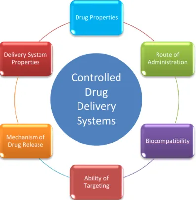
In the context of tissue engineering, an ideal DDS must have several general requirements, including being a biodegradable carrier material, possess high loading efficiency, a controlled release profile, the ability to target or be retained at the desired site of action, and ideally a certain degree of versatility that for example allows several bioactive molecules to be sequentially released in a manner that mimics the temporal profile of the healing process in vivo (Vasita, 2006; Chen, 2009a; Balasubramanian, 2010). Simultaneously, several factors, such as the properties of the drug/bioactive molecule, route of administration, nature of delivery vehicle, mechanism of drug release, ability of targeting, and biocompatibility must be taken in consideration (Figure 1) (Chiellini, 2008).
Figure 1 – Requisites regarding the design of a new drug delivery system (adapted from Chiellini,
2008). Drug Properties Route of Administration Biocompatibility Ability of Targeting Mechanism of Drug Release Delivery System Properties
Controlled
Drug
Delivery
Systems
3
The use of molecular or macromolecular entities and derived superstructures concerning the delivery of drugs has a long history. Antibodies, for instance, were suggested early in the last century as a mean to direct anticancer drugs to tumour cells in the body expressing the corresponding antigen (Ritz, 1982). Their use as monoclonal molecules is now in the vanguard of targeted therapy. Following advances in the discovery of cell receptors, receptor-binding macromolecules were added to the armamentarium of DDS for the targeting of drugs. In parallel, since the early 1970s, liposomes are at the forefront of drug and vaccine design owing to their well-documented abilities to act as delivery vehicles (Gregoriadis, 1993; Henriksen-Lacey, 2011). These superstructures, formed spontaneously from amphipathic lipid molecules, together with a diverse collection of other promising superstructures derived from a huge variety of natural and synthetic monomeric or polymeric units, have evolved to sophisticated versions of DDS. The incorporation onto their surface of macromolecules that contribute to optimal pharmacokinetics of actives and their delivery to where they are needed has been successively proposed (Vemuri, 1995; Fahy, 2009).
Recently, the advances in the peptide, proteins and drug designs and the emergence of gene therapy have been intensifying the need for the development of new controlled release strategies (Jain, 2008). Their clinical success is considered to be dependent on the controlled release devices design, which must ensure that the drug release will perfectly reach the targeted cells at a specific time. A number of DDS are currently under investigation to circumvent the limitations commonly found in conventional dosage forms and to improve the potential of the respective drugs. These DDS have been envisioned as liposomes (Drulis-Kawa, 2010; Monteiro, 2013), micelles (Wilson, 2013; Xu, 2013), polymeric micro and nanoparticles (Joshi, 2013; Liang, 2013; Kozielski, 2013; Wang, 2013), cyclodextrins (Chen, 2011; Dhule, 2012), among others (Oliveira, 2011a).
2. Microparticles as Drug Delivery Systems
There are various approaches to deliver a therapeutic substance to the target site in a sustained controlled release fashion. Of the different forms reported, microparticles attained much importance due to a tendency to accumulate in inflamed areas of the body (Puddu, 2010). The terminology used to describe microparticulate formulations can sometimes be inconsistent and confusing to readers unfamiliar with the field. Basically, the term “microparticle” refers to a particle with a diameter of 1-1000 μm, irrespective of the precise internal or external structure (Arshady, 1988; Freiberg, 2004). However, this criterion is very ubiquitous, and some researchers have been considering that, in terms of
4
diameter, microparticles cover a range between 0.1 and 2 μm (Shet, 2008; Puddu, 2010; Siljander, 2011, Press, 2012). Within the broad category of microparticles, microspheres specifically refers to core-shell microparticles, whilst the subcategory of microcapsules applies to microparticles which have a core surrounded by a material, distinctly different from that of the core, which can be solid, liquid, or even gas (Chemtob, 1986; Brazel, 2000; Berkland, 2004).
A number of different polymers, both synthetic and natural, have been exploited for the fabrication of biodegradable microparticles (Oliveira, 2011b). Synthetic polymers have the advantage of sustaining the release of the encapsulated therapeutic agent over a period of days to several weeks. In opposition, natural polymers have a relatively short duration of drug release and are in general limited by the use of organic solvents and relatively harsher processing conditions (Panyam, 2003).
The most frequently used polymers for this purpose include poly(lactic acid) (PLA), poly(glycolic acid) (PGA), their copolymers poly(lactic-co-glycolic) acid (PLGA), poly-ε-caprolactone, polyethylene, polymethyl methacrylate, oligo(poly(ethylene glycol) fumarate) (OPF), microparticulate systems; natural polymers such as poly(hydroxybutyrate) (PHB), its copolymers poly(hydroxybutyrate-co-hydroxyvalerate) (PHBV), alginate or gelatin (Vasita, 2006; Balasubramanian, 2010). In addition, inorganic materials such as low and high temperature calcium orthophosphates (including calcium phosphate cements and sintered ceramics), calcium sulphate cements and Bioglass have also been used as GF carriers (Chen, 2009b).
2.1. Microparticulate Systems in Tissue Engineering
Controlled release has many direct applications to TE. For example, local delivery of GFs can be accomplished by encapsulating the agent within a biocompatible polymer matrix or microparticle. The controlled-release polymer system is then implanted at the desired tissue site, where it releases the soluble factor directly into the interstitial space of the tissue (Saltzman, 2002).
GFs can be incorporated into the scaffolds by two approaches. The first approach involves adding a lyophilized factor like vascular endothelial growth factor (VEGF) to the polymeric microparticles prior to processing the polymer into a porous scaffold, which results in the factor being largely associated with the surface of the polymer. In this method, VEGF is subjected to rapid release (e.g., days to weeks in duration). The second approach involves pre-incorporation of a GF in microspheres. Therefore, fabricating scaffolds from these particles results in a more even distribution of factors throughout the
5
polymer, with release kinetics controlled by degradation rate of the polymer used to construct microparticles. The two approaches may be combined by mixing particulate polymers containing the first factor with microspheres containing a pre-encapsulated second factor to deliver two GFs with different release rates. The particulate and microparticles are then fused to form a homogeneous combined scaffold with an open-pore structure (Richardson, 2001).
2.1.1. Microparticulate Drug Delivery Systems
Therapeutic induction of tissue repair or function restoration in tissue engineering and regenerative medicine requires recapitulation of the spatial and temporal microenvironments presented by natural ECM, in many cases by providing exogenous instructive bioactive signals (Biondi, 2008). The identification and production of recombinant morphogens and GFs that play key roles in tissue regeneration have generated much enthusiasm. Pivotal among these signals are GFs, a group of proteins capable of acting on cell-surface receptors and directing cellular activities involved in wound healing, such as bone morphogenetic proteins (BMPs, particularly BMP2, 4 and -7), VEGF, epidermal growth factor, and fibroblast growth factor, which provide vital stimulation of cell recruitment, proliferation, morphogenesis, and differentiation (Tabata, 2000; Chen, 2010).
To be effective as a therapeutic agent, a GF has to reach the site of injury without degradation, and then, it has to remain in the target location sufficiently long to exert its action(s) (Chen, 2009b). GFs that are provided exogenously in solution into the site to be regenerated are generally not effective because GFs tend to diffuse away from injury sites and to be enzymatically digested or deactivated (Chen, 2009b; Chen, 2010). Interestingly, clinical trials of DDS that have shown benefit all contain a common denominator, the presence of a material carrier, strongly suggesting that spatiotemporal control over the location and bioactivity of factors after introduction into the body might be achieved by the use of those carriers (Lee, 2011).
In the search for vehicles that delivery without being damaged, microparticulate systems made of biodegradable polymers as reveal significant advantages when incorporating GFs. These systems have the potential to provide sustained release kinetics in remote parts of the body after implantation (Chen, 2010). The release profile of a GF can be altered either by tailoring the properties of the polymer or polymeric composition or even by adjusting the physical and chemical properties of the microparticles such as surface porosity, particle size, and its degradation rate (Chen, 2007; Chen, 2009b). Moreover, the
6
drug loading capacity of particulate systems can be greatly improved if a capsule is formed or if the core of each microsphere is designed to have an interconnected pore structure. Microparticles are not internalized by the cells and are retained in the tissue, providing prolonged bioactive substance release. Additionally, it is not easy for microparticles to diffuse out of the target tissue to other sites to cause deleterious side-effects in other tissues because of their size. Thus, they provide good control over the release rate and dose of GFs in a local microenvironment, yielding desirable concentrations over a period of days or months (Varde, 2004; Anitua, 2008; Mundargi, 2008).
Microparticles carrying GFs, small molecules, among others have been prepared by various techniques and their role has been investigated both in in vitro and in vivo. PLGA - based microcapsules containing GFs have been prepared by a double emulsion–solvent evaporation technique (Wenk, 2009). In 2008, Rocha and co-workers showed that the sustained delivery of VEGF encapsulated in PLGA microspheres interferes with wound healing cascade events and its release upregulates angiogenesis (Rocha, 2008). The degree of encapsulation and mechanism of growth factor entrapment are dependent on hydrophobic–hydrophobic or hydrophilic–hydrophilic interactions among the molecules and polymers. Blending of materials allow more complex structures and patterns of factor entrapment (Lee, 2011). In 2009, Zhu and collaborators have proposed a blend of PLGA and PHBV microparticles prepared by an emulsion technique to release of hepatocyte growth factor (Zhu, 2009). The final microparticulate structure showed a core-shell structure, where PHBV molecules, which are more hydrophobic and less degradable, were distributed within the shell. This prevents the loss of GFs during processing, resulting in a better encapsulation material than microspheres of PLGA- or PHBV-alone (Zhu, 2009). Recent studies with calcium phosphate cement /gelatin composite and diopside (CaMgSi2O6) ceramic microspheres showed that ceramic microparticle systems can be successfully used as injectable, biodegradable and osteoinductive delivery vehicles (Leeuwenburgh, 2010). Kirby and co-workers also demonstrated that PLGA/PLGA-PEG-PLGA microparticles entrapping and releasing BMP-2 in a sustained manner promote the differentiation of MC3T3-E1 cells to a greater extent than osteogenic supplements (Kirby, 2011).
7
2.1.2. Microparticulate Drug Delivery Systems as part of Tissue Engineering Scaffolds
When microparticles are injected in target tissues, they are continuously exposed to external mechanical deformation, leading to the uncontrollable displacements of particles (Fitzgerald, 1987; Griffith, 2000; Chan, 2005). This displacement greatly affects the concentration of bioactive macromolecules in target tissues, resulting in limited tissue regeneration (Lemperle, 2004). Moreover, when in free movement the particulate system can be expelled even before release of the therapeutic agent. Furthermore, these approaches for therapeutic tissue regeneration usually involve one-time delivery of single biochemical cues. Therefore, extensive efforts are being made to tackle these limitations while harnessing the advantages of particulate systems, including their minimally invasive delivery and controlled drug release.
The development of sophisticated drug delivery systems with multiple functionalities is one of the challenges of TE. Extraordinary progress has been made in the last years regarding the design of scaffolds with suitable multiscale hierarchical structure and toward the design of delivery systems able to release active molecules following a hypothetically complex delivery pattern (Biondi, 2008).
Current TE strategies are focusing on the development of porous scaffolds specifically designed to mimick tissues ECM at morphological but most importantly at biochemical level by incorporating bioactive components, selected to promote full regeneration (Guldberg, 2009). In this sense, the concept of particulate systems further associated with 3D scaffolds (Chen, 2007; Chung, 2007; Fei, 2008) serves two functions, as a scaffolding structure for cell attachment and tissue ingrowth, as well as a delivery platform for multiple bioactive molecules release. A variety of systems have been designed with this purpose. Chen and co-workers proposed a dual release system of BMP-2 and Insulin-like Growth Factor-1 (IGF-1) from gelatin microparticles embedded in a glycidyl methacrylated dextran hydrogel for periodontal TE purposes (Chen, 2009b). In this study, it was shown that the system allowed the release of both proteins in a sustained and independent fashion, which facilitated cell attachment, proliferation and osteoblastic differentiation of periodontal ligament fibroblasts in a synergistic manner. The effect of simultaneous, controlled release of VEGF and monocyte chemotactic protein-1 from dual particulate systems as a strategy for therapeutic vascularization was proposed by Jay (2010) and collaborators. Alginate microparticles loaded with both VEGF and MCP-1 with distinct release kinetics were integrated into a collagen/fibronectin gel used as endothelial cells (EC) carrier. The combined delivery of VEGF and MCP-1 increased functional vessel formation from transplanted EC and also led to a higher number of smooth muscle
cell-8
invested vessels than did EC therapy alone. Despite the well-known role of MCP-1 in inflammation, these beneficial effects were accomplished without a long-term increase in monocyte/macrophage recruitment or a shift to a pro-inflammatory (M1) macrophage phenotype. More recently, stem cells encapsulated in OPF hydrogels combined with transform growth factor (TGF)-β3 and/or IGF-1-loaded gelatin MPs were proposed for osteochondral tissue regeneration by Kim et al. (2013). In this study, a bilayered OPF hydrogel that intended to mimic the distinctive hierarchical structure of native osteochondral tissue was used to assess the effect of TGF-β3 with varied release kinetics on osteochondral tissue regeneration in a rabbit full-thickness osteochondral defect model. The in vivo study revealed that after 12 weeks post-implantation IGF-1 delivery alone contributed to enhanced cartilage repair in comparison to the dual GFs delivery. Additionally, no significant effects of the TGF-β3 release kinetics were observed on osteochondral tissue repair. The results suggested that the dual delivery of TGF-β3 and IGF-1 may not synergistically enhance the quality of engineered tissue, revealing that combined and complex approaches do not necessarily confer improved healing over the single delivery of GFs, and that most likely the approach to follow is highly dependent on the TE application. Thus, although current evidences suggest that well-chosen GFs cocktails can represent an advantage when associated in simultaneous delivery systems, uncertainties whether reciprocal or cooperative interactions exist as fare to be clear.
2.1.3. Microparticulate Drug Delivery Systems as Tissue Engineering Scaffolds
The use of microparticles as scaffolds is a relatively poorly explored TE approaches that has many advantages, mainly due to its versatility (Borden, 2002; Lu, 2003; Zhang, 2003; Ungaro, 2006). In fact, microspheres can be easily modified in a controlled manner (Hong, 2005) to introduce various ECM proteins aiming to tailor the cell-material interaction promoting cell adhesion. Simultaneously, GFs as well as other bioactive molecules can be easily incorporated and then further released in a controlled manner from the microspheres that acted as scaffolding structures themselves, thus regulating cell behavior (Tabata, 1999; Perets, 2003; Chen, 2003). The so-called microparticles aggregation method consists in their aggregation, by physical or chemical means, forming those 3D structures (Maquet, 1997) that can provide support for cell adhesion and also act as carriers of bioactive molecules (Gomes, 2005). For microsphere-based scaffolds, microsphere size is one of the major determinants of polymer degradation rate, governing the release kinetics of loaded molecules and providing the control over pore sizes and macro-porosity (Singh, 2008).
9
A different approach was developed by Nof and co-workers (Nof, 2002) which produced PLGA microspheres by double emulsification process incorporating a DNA plasmid and further designed using a gas foaming technique to create the 3D porous structures of different shapes. The obtained scaffolds exhibited a sustained plasmid release for at least 21 days with minimal burst effect during the initial phase, which differed from the first 24 hours release profile from individual microparticles. In a study by Malafaya et al., a novel approach was used envisioning osteochondral regeneration by effective differentiation of adipose tissue-derived mesenchymal stem cells in osteogenic and chondrogenic media. In this study, scaffolds were obtained by the agglomeration of chitosan microparticles using a heat-induced process. Osteochondral bilayered scaffolds were also developed consisting of a hydroxyapatite part and another made up of chitosan, linked by chemical crosslinking process, leading to an integrative bone and cartilage interface (Malafaya, 2005).
3. Final Remarks and Future Trends
Successful repair and regeneration strategies will require quantitative insight of tissue microenvironment and can be engineered via designing biomaterials, which provide quantitative adhesion, growth, or migration signals to direct cellular differentiation pathways. The use of microparticles in the health sciences has been adapted to TE strategies in several approaches. Advances in bioactive materials as well as DDS allow not only controlled release, but also protection of bioactive molecules from degradation. The combination of microparticles with TE scaffolds allow the control over local concentrations needed and/or local concentration gradients needed for successful tissue regeneration. Moreover these systems can be processed into a scaffold constituting a versatile platform for cell adhesion and bioactive molecules release.
From an upstream perspective, an effort is being made to develop scalable differentiation technologies and to study morphogen gradients in pluripotent stem cell differentiation by using localized delivery of GFs within multicellular aggregates from microparticle delivery vehicles. For this purpose, some advances are being made and researchers have reported gelatin-based microparticles to deliver GFs to specific areas of embryoid bodies as aggregates of differentiating stem cells. Although huge developments have being made, these release technologies in TE&RM is not yet delivering significant progress in terms of clinical outcomes and commercialization, this fascinating field of research is
10
bound to dramatically change clinical practice and the therapeutic choices made by clinicians, resulting in significant therapeutic commercial devices and in vitro models.
4. Bibliography
Anderson J. Biocompatility and bioresponse to biomaterials. In: Atala A, editors. Foundations of regenerative medicine: clinical and therapeutic applications. USA: Elsevier; 2009. p. 384. Anitua E, Sánchez M, Orive G, Andia I. Delivering growth factors for therapeutics. Trends
Pharmacol Sci 2008 Jan; 29 (1): 37-41.
Arshady R. Preparation of polymer nano- and microspheres by vinyl polymerization techniques. J Microencapsul 1988 Apr-Jun; 5 (2): 101-14.
Balasubramanian V, Onaca O, Enea R, Hughes D, Palivan C. Protein delivery: from conventional drug delivery carriers to polymeric nanoreactors. Expert Opin Drug Deliv 2010 Jan; 7 (1): 63-78.
Bassyouni F, ElHalwany N, Rehim M, Neyfeh M. Advances and new technologies applied in controlled drug delivery system. Res Chem Intermed 2013 Aug. doi: 10.1007/s11164-013-1338-2. [in press]
Berkland C, Kipper M, Narasimhan B, Kim K, Pack D. Microsphere size, precipitation kinetics and drug distribution control drug release from biodegradable polyanhydride microspheres. J Control Release 2004 Jan; 94 (1): 129-41.
Biondi M, Ungaro F, Quaglia F, Netti P. Controlled drug delivery in tissue engineering. Adv Drug Deliv Rev 2008 Jan; 60 (2): 229-42.
Borden M, Attawia M, Khan Y, Laurencin C. Tissue engineered microsphere-based matrices for bone repair: design and evaluation. Biomaterials 2002 Jan; 23 (2): 551-9.
Brazel C, Peppas N. Modeling of drug release from swellable polymers. Eur J Pharm Biopharm 2000 Jan; 49 (1): 47-58.
Chan B, So K. Photochemical crosslinking improves the physicochemical properties of collagen scaffolds. J Biomed Mater Res A 2005 Dec; 75 (3): 689-701.
Chemtob C, Chaumeil J, N'Dongo M. Tablets of metronidazole microcapsules: release characteristics. Int J Pharm Sci 1986 Mar; 29 (1): 83–92.
11
a
Chen F, Chen R, Wang X, Sun H, Wu Z. In vitro cellular responses to scaffolds containing two microencapulated growth factors. Biomaterials 2009 Oct; 30 (28): 5215-24.
Chen F, Shelton R, Jin Y, Chapple I. Localized delivery of growth factors for periodontal tissue regeneration: role, strategies, and perspectives. Med Res Rev 2009 May; 29 (3): 472-513. Chen F, Zhang M, Wu Z. Toward delivery of multiple growth factors in tissue engineering.
Biomaterials 2010 Aug; 31 (24): 6279-308.
Chen FM, Zhao YM, Sun HH, Jin T, Wang QT, Zhou W, et al. Novel glycidyl methacrylated dextran (Dex-GMA)/gelatin hydrogel scaffolds containing microspheres loaded with bone morphogenetic proteins: formulation and characteristics. J Control Release 2007 Mar; 118 (1): 65-77.
Chen RR, Mooney DJ. Polymeric growth factor delivery strategies for tissue engineering. Pharm Res 2003 Aug; 20 (8): 1103-12.
Chen Y, Zhou L, Pang Y, Huang W, Qiu F, Jiang X, et al. Photoluminescent hyperbranched poly(amido amine) containing β-cyclodextrin as a nonviral gene delivery vector. Bioconjug Chem 2011 Jun; 22 (6): 1162-70.
Chiellini F, Piras A, Errico C, Chiellini E. Micro/nanostructured polymeric systems for biomedical and pharmaceutical applications. Nanomedicine 2008 Jun; 3 (3): 367-93.
Chung YI, Ahn KM, Jeon SH, Lee SY, Lee JH, Tae G. Enhanced bone regeneration with BMP-2 loaded functional nanoparticle-hydrogel complex. J Control Release 2007 Aug; 121 (1-2): 91-9.
Dhule S, Penfornis P, Frazier T, Walker R, Feldman J, Tan G, et al. Curcumin-loaded γ-cyclodextrin liposomal nanoparticles as delivery vehicles for osteosarcoma. Nanomedicine 2012 May; 8 (4): 440-51.
Drulis-Kawa Z, Dorotkiewicz-Jach A. Liposomes as delivery systems for antibiotics. Int J Pharm 2010 Mar; 387 (1–2): 187–98.
Fahy E, Subramaniam S, Murphy R, Nishijima M, Raetz C, Shimizu T, et al. Update of the LIPID MAPS comprehensive classification system for lipids. J Lipid Res 2009 Apr; 50 (Suppl): S9–14.
Fei Z, Hu Y, Wu D, Wu H, Lu R, Bai J, et al. Preparation and property of a novel bone graft composite consisting of rhBMP-2 loaded PLGA microspheres and calcium phosphate cement. J Mater Sci Mater Med 2008 Mar; 19 (3): 1109-16.
12
Fitzgerald P, Hadgraft J, Wilson C. A gamma scintigraphic evaluation of the precorneal residence of liposomal formulations in the rabbit. J Pharm Pharmacol 1987 Jun; 39 (6): 487-90. Freiberg S, Zhu X. Polymer microspheres for controlled drug release. Int J Pharm 2004 Sep; 282
(1-2): 1-18.
Gomes M, Malafaya P, Reis R. Fiber bonding and particle aggregation as promising methodologies for the fabrication of biodegradable scaffolds for hard-tissue engineering. In: Reis RL, Román JS, editors. Biodegradable systems in Tissue Engineering and Regenerative Medicine. London: CRC Press; 2005. p. 53-65.
Gregoriadis G, Florence A. Liposomes in drug delivery. Clinical, diagnostic and ophthalmic potential. Drugs 1993 Jan; 45 (1): 15-28.
Griffith L. Polymeric biomaterials. Acta Materialia 2000 Jan; 48 (1): 263-77.
Guldberg RE. Spatiotemporal delivery strategies for promoting musculoskeletal tissue regeneration. J Bone Miner Res 2009 Sep; 24 (9):1507-11.
Henriksen-Lacey M, Korsholm K, Andersen P, Perrie Y, Christensen D. Liposomal vaccine delivery systems. Expert Opin Drug Deliv 2011 Apr; 8 (4): 505-19.
Hong Y, Gao C, Xie Y, Gong Y, Shen J. Collagen-coated polylactide microspheres as chondrocyte microcarriers. Biomaterials 2005 Nov; 26 (32): 6305-13.
Hutmacher D. Scaffolds in tissue engineering bone and cartilage. Biomaterials 2000 Dec; 21 (24): 2529-43.
Jain K. Drug delivery systems – an overview. In: Jain K, editors. Drug Delivery Systems. UK: Springer; 2008. p. 1-50.
Jay S, Shepherd B, Andrejecsk J, Kyriakides T, Pober J, Saltzman W . Dual delivery of VEGF and MCP-1 to support endothelial cell transplantation for therapeutic vascularization. Biomaterials 2010 Apr; 31 (11): 3054-62.
Joshi R, Nelson C, Poole K, Skala M, Duvall C. Dual pH- and temperature-responsive microparticles for protein delivery to ischemic tissues. Acta Biomater 2013 May; 9 (5): 6526-34.
Kanczler J, Oreffo R. Osteogenesis and angiogenesis: the potential for engineering bone. Eur Cell Mater 2008 May; 15: 100-14.
13
Kim K, Lam J, Lu S, Spicer PP, Lueckgen A, Tabata Y, et al. Osteochondral tissue regeneration using a bilayered composite hydrogel with modulating dual growth factor release kinetics in a rabbit model. J Control Release 2013 Jun; 168 (2): 166-78.
Kirby G, White L, Rahman C, Cox H, Qutachi O. PLGA-Based Microparticles for the Sustained Release of BMP-2. Polymers 2011 Mar, 3 (1): 571-86.
Kozielski K, Tzeng S, Green J. Bioengineered nanoparticles for siRNA delivery. Wiley Interdiscip Rev Nanomed Nanobiotechnol 2013 Sep; 5 (5): 449-68.
Langer R, Vacanti J. Tissue engineering. Science 1993 May; 260 (5110): 920-6.
Lee K, Silva E, Mooney D. Growth factor delivery-based tissue engineering: general approaches and a review of recent developments. J R Soc Interface 2011 Feb; 8 (55): 153-70.
Leeuwenburgh S, Jo J, Wang H, Yamamoto M, Jansen J, Tabata Y. Mineralization, biodegradation, and drug release behavior of gelatin/apatite composite microspheres for bone regeneration. Biomacromolecules 2010 Oct; 11 (10): 2653-9.
Lemperle G, Morhenn V, Pestonjamasp V, Gallo R. Migration studies and histology of injectable microspheres of different sizes in mice. Plast Reconstr Surg 2004 Apr; 113 (5): 1380-90. Liang C, Li H, Tao Y, Peng L, Gao J, Wu J, et al. Dual release of dexamethasone and transforming
growth factor β3 from polymeric microspheres for the stem cell matrix accumulation in a rat disc degeneration model. Acta Biomater 2013 Aug. doi: 10.1016/j.actbio.2013.08.019. [in press]
Lu HH, El-Amin SF, Scott KD, Laurencin CT. Three-dimensional, bioactive, biodegradable, polymer–bioactive glass composite scaffolds with improved mechanical properties support collagen synthesis and mineralization of human osteoblast-like cells in vitro. J Biomed Mater Res A 2003 Apr; 64: 465-74.
Malafaya P, Silva G, Reis R. Natural-origin polymers as carriers and scaffolds for biomolecules and cell delivery in tissue engineering applications. Adv Drug Deliv Rev 2007 May; 59 (4-5): 207-33.
Malafaya PP, Pedro AJ, Peterbauer A, Gabriel C, Redl H, Reis RL. Chitosan particles agglomerated scaffolds for cartilage and osteochondral tissue engineering approaches with adipose tissue derived stem cells. J Mater Sci Mater Med 2005 Dec; 16 (12): 1077-85. Maquet V, Jerome R. Design of macroporous biodegradable polymer scaffolds for cell
14
Monteiro N, Martins A, Ribeiro D, Faria S, Fonseca N, Moreira J, et al. On the use of dexamethasone-loaded liposomes to induce the osteogenic differentiation of human mesenchymal stem cells. J Tissue Eng Regen Med 2013 Oct. doi: 10.1002/term.1817. [in press]
Mundargi R, Babu V, Rangaswamy V, Patel P, Aminabhavi T. Nano/micro technologies for delivering macromolecular therapeutics using poly(D,L-lactide-co-glycolide) and its derivatives. J Control Release 2008 Feb; 125 (3): 193-209.
Nof M, Shea LD. Drug-releasing scaffolds fabricated from drug-loaded microspheres. J Biomed Mater Res 2002 Feb; 59 (2): 349-56.
a
Oliveira J, Sousa R, Malafaya P, Silva S, Kotobuki N, Hirose M, et al. In vivo study of dendronlike nanoparticles for stem cells "tune-up": from nano to tissues. Nanomedicine 2011 Dec; 7 (6): 914-24.
b
Oliveira M, Mano J. Polymer-based microparticles in tissue engineering and regenerative medicine. Biotechnol Prog 2011 Jul; 27 (4): 897-912.
Panyam J, Labhasetwar V. Biodegradable nanoparticles for drug and gene delivery to cells and tissue. Adv Drug Deliv Rev 2003 Feb; 55 (3): 329-47.
Perets A, Baruch Y, Weisbuch F, Shoshany G, Neufeld G, Cohen S. Enhancing the vascularization of three-dimensional porous alginate scaffolds by incorporating controlled release basic fibroblast growth factor microspheres. J Biomed Mater Res A 2003 Jun; 65 (4): 489-97. Perrie Y, Rades T. Controlling drug delivery. In: Perrie Y, Rades T, editors. Pharmaceutis: drug
delivery and targeting. London: Pharmaceutical Press; 2012. p. 1-24.
Press JZ, Reyes M, Pitteri SJ, Pennil C, Garcia R, Goff BA, et al. Microparticles from ovarian carcinomas are shed into ascites and promote cell migration. Int J Gynecol Cancer 2012 May; 22 (4): 546-52.
Puddu P, Puddu G, Cravero E, Muscari S, Muscari A. The involvement of circulating microparticles in inflammation, coagulation and cardiovascular diseases. Can J Cardiol 2010 Apr; 26 (4): 140-5.
Richardson T, Peters M, Ennett A, Mooney D. Polymeric system for dual growth factor delivery. Nat Biotechnol 2001 Nov, 19: 1029 – 34.
Ritz J, Schlossman S. Utilization of monoclonal antibodies in the treatment of leukemia and lymphoma. Blood 1982 Jan; 59 (1): 1-11.
15
Rocha F, Sundback C, Krebs N, Leach J, Mooney D, Ashley S, et al. The effect of sustained delivery of vascular endothelial growth factor on angiogenesis in tissue-engineered intestine. Biomaterials 2008 Jul; 29 (19): 2884-90.
Saltzman W, Olbricht W. Building drug delivery into tissue engineering. Nat Rev Drug Discov 2002 Mar; 1 (3): 177-86.
Shet A. Characterizing blood microparticles: Technical aspects and challenges. Vasc Health Risk Manag 2008 Aug; 4 (4): 769–74.
Siljander P. Platelet-derived microparticles - an updated perspective. Thromb Res 2011 Jan; 127 Suppl 2: S30-3.
Singh M, Morris CP, Ellis RJ, Detamore MS, Berkland C. Microsphere-based seamless scaffolds containing macroscopic gradients of encapsulated factors for tissue engineering. Tissue Eng Part C Methods 2008 Dec; 14 (4): 299-309.
Tabata Y, Hijikata S, Muniruzzaman M, Ikada Y. Neovascularization effect of biodegradable gelatin microspheres incorporating basic fibroblast growth factor. J Biomater Sci Polym Ed 1999; 10 (1): 79-94.
Tabata Y. The importance of drug delivery systems in tissue engineering. Pharm Sci Technolo Today 2000 Mar; 3 (3): 80-89.
Ungaro F, Biondi M, d'Angelo I, Indolfi L, Quaglia F, Netti PA, et al. Microsphere-integrated collagen scaffolds for tissue engineering: effect of microsphere formulation and scaffold properties on protein release kinetics. J Control Release 2006 Jun; 113 (2): 128-36.
Varde N, Pack D. Microspheres for controlled release drug delivery. Expert Opin Biol Ther 2004 Jan; 4 (1): 35-51.
Vasita R, Katti D. Growth factor-delivery systems for tissue engineering: a materials perspective. Expert Rev Med Devices 2006 Jan; 3 (1): 29-47.
Vemuri S, Rhodes C. Preparation and characterization of liposomes as therapeutic delivery systems: a review. Pharm Acta Helv 1995 Jul; 70 (2): 95-111.
Wang Y, Cooke M, Sachewsky N, Morshead C, Shoichet M. Bioengineered sequential growth factor delivery stimulates brain tissue regeneration after stroke. J Control Release 2013 Aug; 172 (1): 1-11.
Wenk E, Meinel A, Wildy S, Merkle H, Meinel L. Microporous silk fibroin scaffolds embedding PLGA microparticles for controlled growth factor delivery in tissue engineering. Biomaterials 2009 May; 30 (13): 2571-81.
16
Wilson D, Zhang N, Silvers A, Forstner M, Bader R. Synthesis and evaluation of cyclosporine A-loaded polysialic acid-polycaprolactone micelles for rheumatoid arthritis. Eur J Pharm Sci 2013 Sep; 51 C: 146-56.
Xiao Y, Qian H, Young W, Bartold P. Tissue engineering for bone regeneration using differentiated alveolar bone cells in collagen scaffolds. Tissue Eng 2003 Dec; 9 (6): 1167-77.
Xu L, Xu X, Chen H, Li X. Ocular biocompatibility and tolerance study of biodegradable polymeric micelles in the rabbit eye. Colloids Surf B 2013 Jul; 112 C: 30-4.
Zhang S. Building from the bottom up. Mater Today 2003 May; 6 (5): 20–27.
Zhang Y, Chan HF, Leong K. Advanced materials and processing for drug delivery: the past and the future. Adv Drug Deliv Rev 2013 Jan; 65 (1): 104-20.
Zhu X, Wang C, Tong Y. In vitro characterization of hepatocyte growth factor release from PHBV/PLGA microsphere scaffold. J Biomed Mater Res A 2009 May; 89 (2): 411-23.
17
II. Materials and Methods
1. Poly (hydroxybutyrate-co-hydroxyvalerate) Properties
Polyhydroxyalkanoates (PHAs) are produced by a wide variety of microorganisms as an internal carbon and energy storage, as part of their survival mechanism (Doi, 2002; Jung, 2005). Poly (hydroxybutyrate-co-hydroxyvalerate) (PHBV) is a member of PHAs, which together with is homopolymer poly(hydroxybutyrate) (PHB) are the most widely used in Tissue Engineering and Drug Delivery Systems. In terms of physicochemical features, PHBV is thermoplastic optically active polyester which is insoluble in water and exhibit a high degree of polymerization (Scandola, 1997; Kang, 2001). These polymers also present some interesting characteristics, namely antioxidant (i.e. inhibits the oxidation of other molecules) and piezoelectric properties, i.e. the capacity of a material to suffer electric polarization due to mechanical stress (Chen, 2000). PHB and PHBV, which are members of PHAs family, degrade into d-3-hydroxybutyrate acid, which is a normal constituent of human blood, which may justify their low toxicity (Tokiwa, 2004; Choi, 2005; Cheng, 2006). PHB is very crystalline and brittle, whereas the copolymers of PHB with hydroxyvaleric acid (PHBV) are less crystalline (Srithep, 2013), more flexible, and more processable (Barham, 1984; Yu, 2006).
2. Gellan Gum Properties
Gellan gum (GG) is one of the widely used fermentation materials, which offers a solution to many problems encountered in gelling agents, which is resistant to heat and acid (Prajapati, 2013). GG is an extracellular microbial anionic heteropolysaccharide consisting of glucose–glucuronic acid–glucose–rhamnose as a repeating unit and that forms a gel in the presence of metallic ions (Kang, 1982; Jansson, 1983). It is commercially available in two forms, acetylated and deacetylated both forming thermo-reversible gels with different mechanical properties in the presence of metallic ions and upon temperature decrease. The key advantages of GG are its high gelling efficiency and its ability to produce a wide spectrum of mechanical properties (Sworn, 2009). GG also requires concentrations two to three times less than those of gelatin and agar to achieve the similar levels of mechanical strengths (Morris, 2012). Moreover, GG based hydrogels have been shown to efficiently sustain the deliver and growth of human cells for different tissue engineering applications (Silva-Correia, 2012).
18
3. Preparation of Poly (hydroxybutyrate-co-hydroxyvalerate) microparticles
The PHBV (molecular weight (Mw) = 425,692 g mol-1) (PHB Industrial S.A., Brazil) microparticles were prepared by using a modification of the double emulsion solvent evaporation method previously described by Ogawa (Ogawa, 1988). The W/O/W double emulsion technique was followed since it offers the possibility of incorporating both hydrophobic and hydrophilic molecules, allowing the incorporation of several agents (Freiberg, 2004). The first step of this method is the formation of water in oil (W1/O) emulsion where the aqueous solution (W1) can contain or not the hydrophilic active component and the organic phase (O) contains a hydrophobic polymer. In detail, 3 ml of a 0.5% w/v Poly(vinyl alcohol) (PVA) (Sigma-Aldrich, Germany) aqueous solution (W1) was mixed with 15 mL of a 3% w/v PHBV solution in chloroform-ethanol (O). Two different ratios of chloroform-ethanol were used in order to verify its effect over the release profile of the bioactive substances, namely 100:0 chloroform:ethanol (100 PHBV MPs) and 90:10 chloroform:ethanol (90 PHBV MPs). The mixture was emulsified for 3 minutes using an ultra-turrax T18 (IKA-Werke, Germany) in order to obtain the first emulsion (W1/O). The primary emulsion was then dripped into 150 mL of 1% w/v PVA solution (W2) and homogenized (RW 16 basic IKA-Werke, Germany) during 5 minutes forming the secondary emulsion (W1/O/W2). The final W1/O/W2 emulsion was left to evaporate under magnetic stirring for 4–5 h. The obtained microspheres were collected by centrifugation, washed at least three times with deionized water and freeze-dried.3.1. Loading Poly (hydroxybutyrate-co-hydroxyvalerate) microparticles with bioactive molecules
After optimizing the procedure of preparation of PHBV MPs it was proceeded to the incorporation of Bovine Serum Albumin (BSA) and Dexamethasone (Dex), respectively as protein and hydrophilic bioactive substance model, and glucocorticoid as well as hydrophobic molecule model.
3.1.1. Bovine Serum Albumin-loaded Poly (hydroxybutyrate-co-hydroxyvalerate) microparticles
BSA-loaded PHBV MPs were also prepared following the same protocol previously described. In this sense, BSA (Sigma-Aldrich, Germany) was used at a concentration 0.1% (w/v), being dissolved within the first aqueous phase (W1).
19
3.1.2. Dexamethasone-loaded Poly (hydroxybutyrate-co-hydroxyvalerate) microparticles
A similar procedure described above was followed aiming at incorporate Dex (Sigma-Aldrich, Germany) within the PHBV microparticles (MPs). Regarding this, Dex was dissolved in the organic phase at a final concentration of 3.3x10⁻³ % (w/v).
4. Gellan Gum hydrogel preparation
For the preparation of the injectable GG hydrogels the procedure previously described by Silva (Silva, 2013) was followed. Gelzan CM (Sigma-Aldrich, Germany) powder was mixed at room temperature with deionized water at a concentration of 1.25% (w/v) under constant stirring. The solution was heated at 90ºC and kept at this temperature for 30 min. Subsequently, CaCl2 (VWR, USA) was added to the GG solution at a concentration of 0.18% (w/v) to act as a crosslinker. Finally, the solution was left at room temperature during approximately one hour, allowing the formation of the gel, which was maintained in a phosphate-buffered saline (PBS) solution.
4.1. Incorporation of Poly (hydroxybutyrate-co-hydroxyvalerate) microparticles in Gellan Gum hydrogel
To incorporate PHBV MPs into the GG hydrogel, a similar procedure previously described was followed. In this regard, 30 mg of PHBV MPs were dispersed into 20 mL of a 1.25% (w/v) GG solution. This solution was posteriorly cross-linked with 0.18% (w/v) of CaCl2 and left at room temperature during approximately one hour, being maintained in a PBS solution.
5. Determination of Particles Production Yield
The yield of the particles prepared under the different conditions defined in section 3 was calculated using Equation 1.