Molars and Orthodontic Treatment After
En Bloc Resection of Conventional
Ameloblastoma
Rafael Lima Verde Osterne, DDS, MSc,*
Jos
e Jeov
a Siebra Moreira Neto, DDS, MSc, PhD,y
Augusto Darwin Moreira de Ara
ujo Lima, DDS, MSc,z
and Renato Luiz Maia Nogueira, DDS, MSc, PhDx
Ameloblastoma treatment can lead to significant bone defects; consequently, oral rehabilitation can be challenging. We present the case of a 14-year-old girl diagnosed with a conventional ameloblastoma in the mandible who was treated using en bloc resection and rehabilitated with autotransplantation of the immature third molars and orthodontic treatment. The lesion was in the region of the lower left canine and premolars, and en bloc resection resulted in a significant alveolar bone defect. Autotransplantation of the lower third molars to the site of the lower left premolars was performed. After 2 years, the upper left third molar was transplanted to the site of the lower left canine. During the orthodontic treatment period, considerable alveolar bone formation was observed in the region of the transplanted teeth, and roots developed. To the best of our knowledge, this is the first reported case of alveolar bone formation induction caused by tooth transplantation after ameloblastoma treatment.
Ó2015 American Association of Oral and Maxillofacial Surgeons
J Oral Maxillofac Surg 73:1686-1694, 2015
Ameloblastoma is a benign odontogenic tumor of epithelial origin that represents 19.3 to 41.5% of all odontogenic tumors.1-6 Although benign, this lesion is locally aggressive and involves the mandible more frequently than the maxilla (at a ratio of 10:1).1This tumor can arise from the enamel organs, the remains of dental lamina, basal cells of the oral mucosa, or epithelial cells in odontogenic cysts. Ameloblastoma is one of the most clinically significant odonto-genic tumors.
Ameloblastoma occurs most frequently in the second to fourth decades of life. Although some investi-gators have stated that it has no gender predilection,3 ameloblastoma was more frequently diagnosed in
females in some studies.1,7 The current classification of ameloblastoma includes 4 types, 3 of which are intraosseous and the last of which is a rare peripheral variant. Intraosseous ameloblastomas are classified as solid/multicystic or conventional ameloblastoma, unicystic ameloblastoma, or desmoplastic ameloblastoma. Solid/multicystic ameloblastoma is the most common variant, comprising 54 to 85% of all types.1,8
Intraosseous ameloblastomas are aggressive, slow-growing lesions that can become very large and cause bone destruction. On a panoramic radiograph, con-ventional ameloblastoma can show unilocular or (more frequently) multilocular radiolucencies that
*Assistant Professor, Department of Pathology, Fortaleza University School of Medicine, Fortaleza, Brazil; PhD Student, Federal University of Ceara School of Dentistry, Fortaleza, Brazil.
yAssociated Professor, Department of Dental Clinic, Discipline of Pediatric Dentistry, Federal University of Ceara School of Dentistry, Fortaleza, Brazil.
zPhD Student, Department of Dental Clinic, Federal University of Ceara School of Dentistry, Fortaleza, Brazil.
xAssociated Professor, Department of Dental Clinic, Discipline of Oral and Maxillofacial Surgery and Stomatology, Federal University of Ceara School of Dentistry, Fortaleza, Brazil; Oral and Maxillofacial
Surgeon, Department of Oral and Maxillofacial Surgery, Memorial Batista Hospital, Fortaleza, Brazil.
Address correspondence and reprint requests to Dr Osterne: Department of Pathology, Fortaleza University School of Medicine, Av Washington Soares, 1321, P.O. Box 1258, Edson Queiroz, 60811-905 Fortaleza, Ceara, Brasil; e-mail:rlimaverde@unifor.br
Received November 5 2014 Accepted May 12 2015
Ó2015 American Association of Oral and Maxillofacial Surgeons 0278-2391/15/00598-4
http://dx.doi.org/10.1016/j.joms.2015.05.014
are usually described as ‘‘soap bubbles.’’ Radicular resorption is not an unusual finding, and nonerupted teeth can be involved in the lesion.9,10 Pain, swelling, malocclusion, and paresthesia are more frequently associated with larger lesions.9
Evidence from microscopy studies showed that the tumor cells of conventional ameloblastomas usually infiltrate the trabecular spaces beyond the radio-graphic extent of the tumor11; therefore, radical sur-gery is the treatment of choice for this variant. Large mandibular defects with or without significant facial disfiguration can result and occasionally require multi-ple surgeries and long-term treatment. Conventional ameloblastoma, therefore, represents a challenge in the field of oral rehabilitation.
We present the case of a 14-year-old girl who was diagnosed with a conventional ameloblastoma in the mandible. The patient was treated using en bloc resec-tion and was rehabilitated with autotransplantaresec-tion of 3 teeth with incomplete root formation and orthodon-tic treatment.
Case Report
A 14-year-old girl was referred to the oral and maxil-lofacial surgery department of the Hospital Batista Memorial because of an asymptomatic lesion in the mandible that was incidentally discovered on a pano-ramic radiograph taken for orthodontic treatment. Her medical history was not significant. The physical
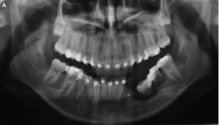
extraoral examination findings were unremarkable, and facial deformity was not observed. A physical in-traoral examination revealed an asymptomatic and un-obtrusive bone expansion near the lower left canine and premolars. A radiographic examination showed a well-defined multilocular radiolucent area with a ‘‘soap bubble’’ appearance (Fig 1). With the patient un-der local anesthesia, an incisional biopsy of the lesion was performed without complications. The histopath-ologic examination revealed a benign odontogenic lesion characterized by the presence of epithelial nests presenting with peripheral columnar cells with reversed polarity and hyperchromatic nuclei. The cen-tral cells were loosely arranged to resemble the stellate reticulum and occasionally presented with cysts. The stroma was composed of mature connective tissue. The final diagnosis was conventional ameloblastoma.
In September 2008, under general anesthesia, the patient underwent marginal en bloc resection of the lesion extending from the lower left canine to the lower left first molar. The postoperative period was uneventful; however, a substantial mandibular bone defect was created (Fig 2). In April 2009, oral rehabilitation was raised as a possibility, with auto-transplantation of the lower third molar to the bone defect site.
Because of the lack of root development, the auto-transplantation was not performed until March 2010. The lower third molars were transplanted to the first molar and second premolar regions, respectively.
FIGURE 1. A radiograph showing a well-defined multilocular radiolucent area with a ‘‘soap bubble’’ appearance.
Osterne et al. Teeth Autotransplantation After Treatment of Ameloblastoma. J Oral Maxillofac Surg 2015.
With the patient under local anesthesia, mucoperios-teal flaps were raised in the mandibular defect and third molar areas. The donor inferior third molars were carefully extracted using elevators (keeping the radicular part intact) and transplanted to the first molar and second premolar areas without extraoral storage. Because of the bone defects at the recipient site, no additional bone was removed to create a socket; instead, only a horizontal crestal incision was made, and the teeth were placed into the soft tissue. The transplanted teeth were splinted to the lower second molar with acid-etch composite resin. Inferior third molars showed approximately one third of the to-tal root development; these teeth were left in
infraoc-clusion. A liquid diet was recommended for 30 days. The postoperative period was uneventful; gingiva formed and attached, and the splint was removed approximately 60 days after transplantation. In September 2010, 6 months after transplantation and after visualization of initial bone formation, the trans-planted teeth were released for orthodontic move-ment. After very slow orthodontic movement, both transplanted teeth were in occlusion (January 2011;
Fig 3).
In April 2012, the upper left third molar was trans-planted to the site of the lower left canine. The surgical technique and method of postoperative care were the same as those used with the first autotransplantation. FIGURE 2. Mandibular bone defect after a marginal en bloc resection of the lesion extending from the lower left canine to the lower left first molar.A, Clinical intraoral view.B, Radiographic view.
The upper left third molar was selected because of the more appropriate mesiodistal length, which adapted better to the receptor site. In December 2012, the transplanted tooth was released for orthodontic treat-ment with very slow movetreat-ment (Fig 4).
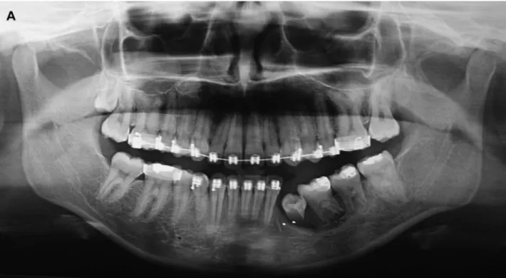
During orthodontic treatment, considerable alveolar bone formation was observed in the region of the trans-planted teeth (Fig 5). Root development occurred in all the transplanted teeth; however, the root growth of the tooth transplanted into the canine region was decreased compared with that of the other trans-planted teeth. All the transtrans-planted teeth were in masti-catory function with no discomfort, mobility, or pathologic conditions. The gingival contour, color, and depth of the pocket were all normal. Also, all the transplanted teeth responded to thermal pulp testing, although partial pulp obliteration was present. In July 2014, the orthodontic treatment was completed.
Discussion
Conventional ameloblastoma has a high potential for recurrence after curettage, and the radiographic margin of the lesion will not correspond to the histo-logic margin. The tumor is usually found 2 to 8 mm (mean 4.5) beyond the radiographic margin; thus, radical surgery with a bone margin of 1 to 1.5 cm is
rec-ommended.11When doubt exists regarding the bone margins, a radiograph of the resected surgical spec-imen or the use of a frozen section biopsy can assist the surgeon.11The jaw is important, not only from a functional viewpoint regarding dentition and occlu-sion, but also for facial aesthetic reasons. The face has a central importance in daily interactions. Thus, whenever possible, en bloc resection with preserva-tion of the base of the mandible (which could assist in future oral rehabilitation) should be planned for mandibular odontogenic tumors.
Several methods can be used in oral rehabilitation, including prosthodontics or dental implants, which can be preceded by bone grafts or osteogenesis distraction.12However, patients are not always candi-dates for these procedures because of age-related or financial reasons. In such cases, autotransplantation could be an option.13
Most investigators have stated that a tooth trans-plantation candidate should have one-half to three-fourths root formation to facilitate tooth stabilization for periodontal healing and open the apex for pulp revascularization.14-19 However, success can be achieved in cases of one-third or complete root formation.20,21
To avoid extending cell damage to the root surface, the tooth should be removed gently from the donor FIGURE 3. Radiographs after transplantation of the lower third molars to the region of the first molar and second premolar.A, Radiograph taken after 7 days.(Fig 3 continued on next page.)
Osterne et al. Teeth Autotransplantation After Treatment of Ameloblastoma. J Oral Maxillofac Surg 2015.
site without extensive osteotomies or severe disloca-tions.22Minimizing the duration that a donor tooth is out of the mouth and the duration of root manipula-tion during autotransplantamanipula-tion are keys for success; however, in some cases, dentoalveolar ankylosis, root resorption, or even compromised root growth can occur. In the present patient, all the transplanted teeth had approximately one-third root formation, and the autotransplantations were considered success-ful because undesirable complications were not observed, the roots developed, and the teeth were in occlusion and functional. Damage to the periodontal ligament, root resorption, and pulp necrosis are more likely to occur in teeth with complete root for-mation than in teeth transplanted during root develop-ment.16-19
The minimal apical foramen diameter needed to obtain vital pulp in an autotransplanted tooth is 1 mm. In cases with a smaller diameter, pulp revascu-larization will be unpredictable, although an animal
study demonstrated that in 10 teeth with apical diam-eters varying from 0.24 to 0.53 mm, 50% presented with vital tissue in more than two-thirds of the pulp chamber.23In the present patient, all the transplanted teeth were in the root development period; thus, the roots had an open apex with a good chance of apical revascularization and pulp vitality. Endodontic treat-ment is not usually necessary in cases of an open apex unless clinical or radiologic symptoms of periapi-cal inflammation or root resorption occur.
Tooth transplantation can be performed at a fresh extraction site; however, when complete postextrac-tion regenerapostextrac-tion of the alveolar socket has occurred, a recipient site can be created with burs. Some inves-tigators have demonstrated similar results for trans-plantations into both prepared and fresh sockets.24 In atrophied jaw sections, free bone grafts and a split-ting osteotomy of the alveolar process can be per-formed during tooth transplantation; however, these procedures have had a lower success rate.24 In the FIGURE 3 (cont’d). B, Radiograph taken after 60 days.C, Radiograph taken after 6 months.D, Radiograph taken 1 year after transplan-tation showing considerable alveolar bone formation.
present patient, significant loss of the alveolar process had occurred, and burs were not used to create a socket. Instead, the teeth were placed into the soft tis-sue and splinted to the distal tooth with acid-etch com-posite resin.
To our knowledge, this is the first case of alveolar bone regeneration in mandibular bone defects after ameloblastoma resection and tooth autotransplanta-tion without bone grafting. Considerable alveolar bone formation was observed after transplantation. A previous study showed that tooth transplantation can induce bone formation.25 Additionally, a recent in vivo study demonstrated that Hertwig’s epithelial root sheath cells can stimulate osteogenic differentia-tion of dental follicle cells by way of cell to cell communication26 and could play a role in alveolar bone formation. Tooth autotransplantation after ame-loblastoma treatment was described by Lim and Chun27 in an 18-year-old woman who presented with a unicystic ameloblastoma. The patient was treated using enucleation and immediate bone graft-ing, followed by autotransplantation and orthodon-tic treatment.
In addition to its predictability, tooth autotransplan-tation can offer a good aesthetic result, especially in the germ phase. Tooth autotransplantation can also induce alveolar bone growth and re-establish the alveolar process.25,28-30 The risk of ankylosis after
transplantation of an unerupted tooth germ is reduced, because the dental follicle protects the root surface from injury. Additionally, transplantation of unerupted teeth has been associated with a high likelihood of pulp revascularization, generation of normal periodontal tissue, and root growth.31It has previously been shown that apical pulp-derived cells have the potential to induce hard tissue formation, sug-gesting that a tooth with an immature apex is an effec-tive source of cells for hard tissue/periodontal complex regeneration,32,33 and some clinical results have supported this finding.25,31 Bone defects secondary to trauma or resulting from surgical resection could benefit from the autotransplantation of immature teeth, even during maxillomandibular development.
Several techniques can be used for the rehabilitation of mandibular defects. The use of dental implants or fixed prosthodontics during maxillomandibular devel-opment could interfere with bone growth, because os-teointegrated implants will not grow with the patient’s changing jaw, resulting in an infraocclusion. Osteo-genic distraction could be used, although it has usually been associated with greater morbidity than that with tooth transplantation. Additionally, it requires longer follow-up periods and greater patient cooperation, and a risk exists of deviation of the distraction vector.34 Recombinant human bone morphogenetic protein 2 could be another option; however, this method is FIGURE 4. Radiographs after transplantation of the upper third molar to the canine region.A, Radiographs taken after 7 days.(Fig 4 continued on next page.)
Osterne et al. Teeth Autotransplantation After Treatment of Ameloblastoma. J Oral Maxillofac Surg 2015.
FIGURE 4 (cont’d). B, Radiographs taken after 7 days.C, Radiograpsh taken at 15 months after transplantation showing alveolar bone for-mation in the canine region.
Osterne et al. Teeth Autotransplantation After Treatment of Ameloblastoma. J Oral Maxillofac Surg 2015.
FIGURE 5. A,B, Clinical intraoral views showing all transplanted teeth in occlusion.(Fig 5 continued on next page.)
more expensive, and after bone formation, the use of dental implants or fixed prosthodontics would be necessary.
Accordingly to Kumar et al,13tooth transplantation can be considered successful if the tooth is fixed in its socket without residual inflammation, masticatory function is satisfactory with no discomfort, the tooth is not mobile, no pathologic conditions are apparent from radiographic examinations, the lamina dura is radiographically normal, the tooth shows radiographic evidence of root growth and pulpal regeneration, and the depth of the pocket, gingival contour, and gingival color are all normal. In the present patient, these criteria for successful autotransplantation were ful-filled, with the exception of the partial root growth in first premolar, and all transplanted teeth responded to thermal pulp testing, although partial pulp oblitera-tion was present. Even if this tooth were to be lost in
the future, the bone gain that occurred in the region would facilitate rehabilitation at the site.
In conclusion, tooth autotransplantation is well suited to growing individuals. In the case of failure, dental implants can be inserted in patients before the completion of pubertal growth. Long-term follow-up should be emphasized in the present patient owing to the possibility of ameloblastoma recurrence and root resorption of the transplanted teeth.
References
1. Osterne RL, Brito RG, Alves AP, et al: Odontogenic tumors: A 5-year retrospective study in a Brazilian population and anal-ysis of 3406 cases reported in the literature. Oral Surg Oral Med Oral Pathol Oral Radiol Endod 111:474, 2011
2. Gaitan-Cepeda LA, Quezada-Rivera D, Tenorio-Rocha F, Leyva-Huerta ER: Reclassification of odontogenic keratocyst as tumour: Impact on the odontogenic tumours prevalence. Oral Dis 16:185, 2010
FIGURE 5 (cont’d). C, Radiograph showing alveolar bone formation and continuous root formation of all teeth with the exception of partial formation of the tooth transplanted to the canine region.D, Three-dimensional reconstruction of the mandible showing alveolar bone formation.
Osterne et al. Teeth Autotransplantation After Treatment of Ameloblastoma. J Oral Maxillofac Surg 2015.
3. Avelar RL, Antunes AA, Santos T, et al: Odontogenic tumors: Clin-ical pathology study of 238 cases. Braz J Otorhinolaryngol 74: 668, 2008
4. Jing W, Xuan M, Lin Y, et al: Odontogenic tumours: A retrospec-tive study of 1642 cases in a Chinese population. Int J Oral Max-illofac Surg 36:20, 2007
5. Luo HY, Li TJ: Odontogenic tumors: A study of 1309 cases in a Chinese population. Oral Oncol 45:706, 2009
6. Tawfik MA, Zyada MM: Odontogenic tumors in Dakahlia, Egypt: Analysis of 82 cases. Oral Surg Oral Med Oral Pathol Oral Radiol Endod 109:e67, 2010
7. Fernandes AM, Duarte EC, Pimenta FJ, et al: Odontogenic tumors: A study of 340 cases in a Brazilian population. J Oral Pathol Med 34:583, 2005
8. Buchner A, Merrell PW, Carpenter WM: Relative frequency of central odontogenic tumors: A study of 1,088 cases from North-ern California and comparisons to studies from other parts of the world. J Oral Maxillofac Surg 64:1343, 2006
9. Barnes L, Eveson JW, Reichart P, Sidransky D (eds): World Health Organization Classification of Tumours. Pathology and Genetics of Head and Neck Tumours. Lyon, IARC Press, 2005, pp 296–300
10. Fregnani ER, da Cruz Perez DE, de Almeida OP, et al: Clinicopath-ological study and treatment outcomes of 121 cases of amelo-blastomas. Int J Oral Maxillofac Surg 39:145, 2010
11. Carlson ER, Marx RE: The ameloblastoma: Primary, curative sur-gical management. J Oral Maxillofac Surg 64:484, 2006
12. O’Fearraigh P: Review of methods used in the reconstruction and rehabilitation of the maxillofacial region. J Ir Dent Assoc 56:32, 2010
13. R1 Kumar, Khambete N, Priya E: Successful immediate autotrans-plantation of tooth with incomplete root formation: Case report. Oral Surg Oral Med Oral Pathol Oral Radiol 115:e16, 2013
14. Henrichvark C, Neukam FW: Indikation und Ergebnisse der au-togenen Zahntransplantation. Dtsch Zahnarztl Z 42:194, 1987
15. Kallu R, Vinckier F, Politis C, et al: Tooth transplantations: Descrip-tive retrospecDescrip-tive study. Int J Oral Maxillofac Surg 34:745, 2005
16. Andreasen JO, PaulsenH U, Yu Z: A long-term study of 370 auto-transplanted premolars: Part I—Surgical procedures and standard-ized techniques for monitoring healing. Eur J Orthod 12:3, 1990
17. Andreasen JO, Paulsen HU, Yu Z, Schwartz O: A long-term study of 370 autotransplanted premolars: Part II—Tooth survival and pulp healing subsequent to transplantation. Eur J Orthod 12:14, 1990
18. Andreasen JO, Paulsen HU, Yu Z, Schwartz O: A long-term study of 370 autotransplanted premolars: Part III—Periodontal heal-ing subsequent to transplantation. Eur J Orthod 12:25, 1990
19. Andreasen JO, Paulsen HU, Yu Z, Bayer T: A long-term study of 370 autotransplanted premolars: Part IV—Root development subsequent to transplantation. Eur J Orthod 12:38, 1990
20.Yoshino K, Kariya N, Namura D, et al: Survival rate in autotrans-planted premolars with complete root formation: A retrospec-tive clinical survey. Bull Tokyo Dent Coll 54:27, 2013
21.Lai FS: Autotransplantation of an unerupted wisdom tooth germ without its follicle immediately after removal of an impacted mandibular second molar: A case report. J Can Dent Assoc 75: 205, 2009
22.Sch€utz S, Beck I, K€uhl S, Filippi A: Results after wisdom tooth transplantation: A retrospective study. Schweiz Monatsschr Zahnmed 123:303, 2013
23.Laureys WG, Cuvelier CA, Dermaut LR, De Pauw GA: The critical apical diameter to obtain regeneration of the pulp tissue after tooth transplantation, replantation, or regenerative endodontic treatment. J Endod 39:759, 2013
24.Bauss O, Engelke W, Fenske C, et al: Autotransplantation of immature third molars into edentulous and atrophied jaw sec-tions. Int J Oral Maxillofac Surg 33:558, 2004
25.Hjortdal O, Bragelien J: Induction of jaw bone formation by tooth autotransplantation. Nor Tannlaegeforen Tid 88:319, 1978
26.Yang Y, Ge Y, Chen G, et al: Hertwig’s epithelial root sheath cells regulate osteogenic differentiation of dental follicle cells through the Wnt pathway. Bone 63:158, 2014
27.Lim WH, Chun YS: Orthodontic treatment combined with auto-transplantation after removal of ameloblastoma. Am J Orthod Dentofacial Orthop 135:375, 2009
28.Lima JP, Neto JJ, Beltr~ao HC, et al: Esthetic considerations for re-shaping of autotransplanted premolars replacing maxillary cen-tral incisors: A case report. Dent Traumatol 25:631, 2009
29.Maia RL, Vieira AP: Auto-transplantation of central incisor with root dilaceration. Technical note. Int J Oral Maxillofac Surg 34: 89, 2005
30.Czochrowska EM, Stenvik A, Zachrisson BU: The esthetic outcome of autotransplanted premolars replacing maxillary incisors. Dent Traumatol 18:237, 2002
31.Plakwicz P, Wojtaszek J, Zadurska M: New bone formation at the site of autotransplanted developing mandibular canines: A case report. Int J Periodontics Restorative Dent 33:13, 2013
32.Abe S, Yamaguchi S, Watanabe A, et al: Hard tissue regeneration capacity of apical pulp derived cells (APDCs) from human tooth with immature apex. Biochem Biophys Res Commun 371:90, 2008
33.Xu L, Tang L, Jin F, et al: The apical region of developing tooth root constitutes a complex and maintains the ability to generate root and periodontium-like tissues. J Periodontal Res 44:275, 2009


