Longitudinal evaluation of dental arches
individualized by the WALA ridge method
Márcia de Fátima Conti*, Mário Vedovello Filho**, Silvia Amélia Scudeler Vedovello***, Heloísa Cristina Valdrighi***, Mayury Kuramae***
Introduction: The mandibular arch form is considered one of the main references among the diagnostic tools because the maintenance of this arch form and dimension is an important factor for stability of orthodontic treatment. Objectives: to evaluate the changes in man-dibular intercanine and intermolar widths during orthodontic treatment and 3 years of post treatment, in which the WALA ridge was used for individualization of the mandibular arch form. Methods: The sample comprised 20 patients (12 women and 8 men), with a mean age of 20.88 years. The dental casts of the initial, final and post-treatment evaluations were used for measurement of the intercanine and intermolar distances in the center of the facial surface of the clinical crown and in the width of the WALA ridge. Data were analyzed by means of ANOVA test followed by Tukey test (p<0.05). Results: There was a statistically significant difference in intercanine and intermolar distances among the three stages evaluated. These distances increased significantly with treatment, and presented a reduction in the post-treat-ment period, however not reaching the initial values. Conclusions: the WALA ridge method used in this study for construction of the individualized diagrams and for measurement of the intercanine and intermolar distances was shown to be valuable, allowing the individualization of the dental arches and favoring the post-treatment stability.
Abstract
Keywords: Malocclusion. Angle Class I. Orthodontics. Relapse.
* Specialist in Orthodontics, “Centro Universitário Hermínio Ometto - UNIARARAS / SP”.
** PhD in Dentistry, Coordinator of the Graduate Course in Orthodontics of the “Centro Universitário Hermínio Ometto-UNIARARAS”. *** PhD in Orthodontics, Professor of the Graduate Program in Dentistry, Area of Concentration Orthodontics, “Centro Universitário Hermínio
Ometto - UNIARARAS / SP”.
INTRODUCTION AND LITERATURE REVIEW
The purpose of orthodontics is to correct mal-occlusions, and place the teeth in their ideal po-sitions and in equilibrium with their bony bases. Esthetics and function is enhanced provided that the periodontal tissues and support structures
will avoid periodontal problems, such as gingival retractions, instability and deficiencies in the es-thetic results.11 The form of the mandibular dental
arch is considered one of the main references dur-ing treatment, as its maintenance is an important factor for the stability of orthodontic treatment.20
In an ideal occlusion, the teeth are positioned in the greatest possible degree of harmony with their bony bases and with the surrounding tissues. Thus, preservation of the form and dimensions of the dental arches must be one of the first objects of orthodontic treatment. Various factors may in-fluence the morphology of the dental arches, such as the facial type, genetics, type of occlusion, mus-culature and ethnicity. The mandibular canines and molars are considered determinant factors in the arch width and movements of incisors in the buccal direction should be avoided.18
Dental arch form studies began in 1889 with Bonwill,6 who developed the first diagram which
was used in orthodontics by other research-ers. On the basis of his postulations, Hawley13
constructed a diagram denominated Bonwill-Hawley, for orthodontic purposes. From then onwards, various diagrams were drawn with the aim of helping in the construction of metal arch-es used during treatment. In addition to form, the dimension of the dental arch was also a rea-son for concern.8,11,16 It is known that when
al-terations are made in the distance between the canines and molars during treatment, there is a great tendency towards relapse.19,25
Andrews and Andrews2 suggested the use of
an anatomic reference such as a parameter with the object of centralizing the roots of teeth in the basal bone, which they denominated the WALA ridge, of which the initials mean Will Andrews + Lawrence Andrews. The WALA ridge is the strip of soft tissue immediately above the mucogingival junction of the mandible, at the level of the line that passes through the centers of rotation of the teeth or close to it, and is exclusive to the mandi-ble. The mandibular dental arch will present this
requirement and will be with the ideal form when the midpoint of the vertical axes of the facial sur-faces (“FA” points) of the central and lateral in-cisors, canines, 1st premolars, 2nd premolars, 1st and 2nd molars are 0.1 mm; 0.3 mm; 0.6 mm; 0.8 mm; 1.3 mm; 2.0 mm and 2.2 mm, respec-tively, from the WALA ridge. In the authors’ per-ception, after eruption the crowns of the perma-nent teeth are subject to alterations as a result of “environmental” forces. These forces may tip the teeth around their centers of rotation. Hypotheti-cally, when this occurs, the centers of rotation of the mandibular teeth, which remain in the cen-ter of the basal bone, do not alcen-ter, however, the crowns and root apexes may be altered. Therefore, the center line of rotation (hypothetical line that passes through the horizontal center of rotation of each tooth) would be the line that best conserves the original and supposedly ideal form of the den-tal arch. Thus, the ideal form of the maxillary and mandibular dental arches would be dictated by the form of the basal bone of the mandible. When the form of the mandibular dental arch is correct, the wire that unites the bracket slots of “straight-wire” brackets should have the same shape as that of the WALA ridge.17
By means of the “Six Elements of Facial Har-mony”, defined as a classification of the objectives and goals of orthodontic treatment, Andrews and Andrews3 determined that Element 1 referred to
The WALA ridge concept would keep a close relationship with the “Six Keys of Perfect Occlu-sion”1 and was consolidated as a real and true
ref-erence for determining the individual morphol-ogy of the dental arches.
PROPOSITION
To perform a longitudinal evaluation of dental casts of patients submitted to orthodontic treat-ment, who had their dental arches individualized by the WALA ridge method with regard to the following aspects:
• alterationsinthemandibularintercaninedis -tance and between the mandibular molars;
• transversal alterations in theWALA ridge, in
the region contiguous to the mandibular inter-canine and intermolar distances;
• reliabilityofthemethodfortheindividualiza -tion of dental arches.
MATERIAL AND METHODS Material
The materials were used in accordance with the regulations of the National Council of Health, No. 196/1996, Ministry of Health, and this study was approved by the Research Ethics Committee of UNIARARAS Protocol No. 219/2007.
The sample was obtained from the files of pa-tients who received orthodontic treatment at a private clinic in Curitiba/ PR, Brazil. It was com-posed of 20 plaster models, obtained from the same clinic, taken at the stages of pre-treatment (T1), post-treatment (T2) and 3 years after the end of treatment (T3) of patients ranging be-tween the ages of 13 years and 11 months and 39 years and 1 month, among whom 8 were men and 12 women.
For selecting the T1 sample, the following fac-tors were evaluated:
• Presence of complete permanent dentition,
except third molars.
• ClassImalocclusion,determinedbytherela -tionship of the first molars and premolars.
• Absenceofdiastemas.
• Teethandalveolarridgevisibleinplastermod -els, the latter being compatible and checked against the morphology of the WALA ridge clinically presented by the patient.
• Slightmandibularcrowding(-1mmto-4mm).
For selecting the T2 sample, the following fac-tors were evaluated:
• ClassI,determinedbytherelationshipofthe
canines, premolars and first molars; correct in-tercuspation provided by the first molar cusp-sulcus relationship and premolar cusp imbra-sure relationship, evaluated from the lingual perspective.
• Overjetof0to2mmandoverbiteof1to2mm. • Angulationandinclinationofthecrownsac -cording to Andrews method of Keys II and III, respectively.1
• Absenceofdiastemas.
• CurveofSpeedepthof0to2.5mm.
• Teethandalveolarridgevisibleinplastermod -els, the latter being compatible and checked against the morphology of the WALA ridge clinically presented by the patient.
The corrective orthodontic treatment was performed according to the following proto-col: without extractions; finishing objectives in accordance with Andrews’ six keys method;1
straight-wire technique, Andrews standard pre-scription (“A” Company, California, USA) with slot 0.022 x 0.028-in; wire contour individual-ization for leveling and alignment defined by the WALA ridge form, observed from the occlusal perspective of the mandibular plaster model and adapted to a diagram recommended by Andrews and Andrews.2
incisors, as 3 cases presented Bolton’s discrepancy due to excess maxillary tooth size.
Plaster models of patients that presented the following anomalies were excluded from the sample: Dental agenesis; supernumerary and ex-tranumerary teeth; teeth with alterations in shape, dental mutilations and bony bases compromised in the sagittal direction.
Methods
To mark the axes, points and reference ridges and to obtain dimensions on the plaster models, the following equipment was used: Black pencil (model t5.5v Regent 1250 6B, Faber Castell, SP) and a digital caliper with a resolution of 0.01 mm and exactness of approximately 0.02 mm (Mitu-toyo Sul Americana Ltda., Brazil). The measure-ments were made exclusively by the researcher.
Axis, WALA ridge, points and their demarcations
A single examiner, using a pencil, marked the axes, points and WALA ridge on the maxillary plaster models for T1, T2 and T3, by the visual method, according to the following description: a) Facial-Axis of the Clinical Crown (FACC):
The most prominent portion of the central lobe of the facial surface of all teeth crowns, except for the molars, which corresponds to the sulcus that separates the two large facial cusps. Demarcation was done with a graph-ite on the crowns of mandibular canine and first molar teeth.
b) WALA Ridge: Soft tissue ridge located be-low the gingival margins of mandibular tooth crowns and immediately above the mucogingi-val junction.
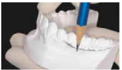
c) Facial-Axis Point (FA point): Point on FACC that separates the gingival half of the clinical crown from the occlusal half. De-marcation was done with a graphite tip on the crowns of mandibular canine and first molar teeth (Fig 1).
d) WALA Ridge Point (point WR): Demarcation of the WALA ridge was made with the graph-ite surface (Fig 2); the most prominent point on the curve of the WALA ridge adjacent to each tooth was denominated Point WR (Fig 3). Demarcation was done with a graphite tip contiguous to the mandibular canine and first molar teeth.
Measurement of the linear variables (mm)
These were made with the use of a digital cali-per directly on the mandibular plaster models and noted on a specific chart.
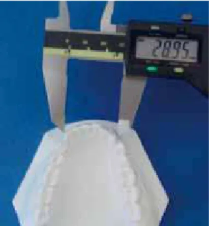
a) Mandibular intercanine distance (IC): distance between the mandibular right and left canines,
ontherespectiveFApoints(Fig4).
b) Mandibular intermolar distance (IM): distance between the mandibular right and left canines,
ontherespectiveFApoints(Fig4).
c) Intercanine distance at the width of the WALA ridge (IC WR): transversal dimension between the points of the WALA ridge of the mandibu-lar canines (Fig 6).
FIGURE 1 - Delimitation of Points FA with a graphite tip.
FIGURE 2 - Demarcation of the WALA ridge made with a graphite surface.
d) Intermolar distance at the width of the WALA ridge (IM WR): transversal dimension between the points of the WALA ridge of the mandibu-lar momandibu-lars (Fig 7).
After every measurement taken, the caliper was reset in the initial position (zero), in order to avoid an erroneous readout. The caliper was placed on the reference points, using the tips of the measurement probes, taking care to keep it parallel to the occlusal plane during each mea-surement to ensure the recordings were made only in the horizontal direction.
Statistical Analysis
Method Error
To calculate the intra-examiner error, 7 pairs of models for each evaluated stage (T1, T2 and T3) were randomly selected, for a second demarcation of the points and linear variable measurements, totaling 21 pairs of plaster models. The approxi-mate interval between the first and second mea-surement was 15 days.
The formula proposed by Dahlberg9 (Se2
=∑ d2/2n) was applied to estimate the order
of variables of the casual errors, while the
FIGURE 4 - IC distance at the respective FA points.
FIGURE 6 - IC WR distance at the respective WR points.
FIGURE 5 - IM distance at the respective FA points.
systematic errors were analyzed by the appli-cation of the paired “t” test, according to Hous-ton14. The level of significance was established
at 5% (p<0.05).
Statistical Method
Descriptive statistics were performed of all the data obtained from the sample: age at the begin-ning of treatment (T1); treatment time (T2-T1); post-treatment evaluation time (T3-T2), as well as for the studied variables (IC, IM, IC WR and IM WR), in all the stages and periods studied: T1, T2, T3, T2-T1, T3-T2 and T3-T1.
The dependent ANOVA test was used, and when there was a significant result, the Tukey test was performed to observe whether there
was significant alteration in the studied variables between the initial, final and post-treatment stages. The level of significance was established at 5% (p<0.05).
RESULTS
Table 1 presents the descriptive statistics (mean, standard deviation, minimum and maxi-mum) initial age, treatment time and post-treat-ment evaluation time.
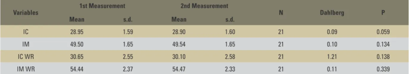
Table 2 presents the results of the systematic and casual and error evaluations by means of the paired t test and the Dahlberg formula,9 applied
to the studied variables. There were no system-atic errors and the casual errors were considered acceptable, and it could be affirmed that the WALA ridge method was an easily reproducible method, since there was no difference between the two measurements of the variables IC WR and IM WR performed by the same examiner at two different times.
The results of the descriptive statistical analy-sis for the variables IC, IM, IC WR and IM WR are shown in Tables 3, 4, 5 and 6, respectively, in all the studied times: T1, T2, T3, T2-T1, T3-T2 and total Variables Mean s.d. Minimum Maximum
Initial age 20.88 7.86 13.91 39.08
Time of treatment 2.47 0.57 1.36 3.17
Post-treatment
evaluation time 3.20 0.32 3.05 4.17 TABLE 1 - Descriptive statistics of initial age, time of treatment and post-treatment evaluation time (mm).
TABLE 3 - Descriptive statistics of the variable IC (mm). TABLE 4 - Descriptive statistics of the variable IM (mm). TABLE 2 - Results of the estimate of systematic and casual errors applied to the variables IC, IM, IC WR and IM WR.
Variables 1st Measurement 2nd Measurement N Dahlberg P
Mean s.d. Mean s.d.
IC 28.95 1.59 28.90 1.60 21 0.09 0.059
IM 49.50 1.65 49.54 1.65 21 0.10 0.134
IC WR 30.65 2.55 30.10 2.58 21 1.21 0.138
IM WR 54.44 2.37 54.47 2.33 21 0.11 0.339
Variables Mean s.d. Minimum Maximum
IM T1 48.07 2.14 44.00 52.20
IM T2 50.30 1.77 47.20 53.70
IM T3 49.30 2.08 44.00 52.90
IM T2-T1 2.22 1.73 -0.90 5.30
IM T3-T2 -0.99 1.15 -4.60 0.60
IM T3-T1 1.23 1.13 -0.50 3.50
Variables Mean s.d. Minimum Maximum
IC T1 29.29 1.62 25.70 31.80
IC T2 30.42 1.57 27.60 33.30
IC T3 29.79 1.68 26.10 32.50
IC T2-T1 1.12 1.06 -0.70 3.10
IC T3-T2 -0.62 0.69 -2.30 0.20
Variables Mean s.d. Minimum Maximum
IC WR T1 30.06 2.23 25.30 33.20
IC WR T2 30.82 1.60 26.90 33.40
IC WR T3 30.39 1.88 26.00 33.30
IC WR T2-1 0.76 0.90 -1.60 1.90
IC WR T3-2 -0.43 0.52 -1.60 0.50
IC WR T3-1 0.33 0.56 -1.10 1.70
TABLE 5 - Descriptive statistics of the variable IC WR (mm).
TABLE 7 - Results of the ANOVA test and Tukey test for the variables IC, IM, IC WR and IM WR, among the 3 evaluation times T1, T2 and T3.
Different letters indicate statistically significant differences (p<0.05).
TABLE 6 - Descriptive statistics of the variable IM WR (mm).
Variables Mean s.d. Minimum Maximum
IM WR T1 54.18 1.94 50.50 57.30
IM WR T2 54.79 1.97 51.10 58.40
IM WR T3 54.51 1.90 50.70 57.50
IM WR T2-1 0.61 1.08 -2.40 2.50
IM WR T3-2 -0.28 0.75 -1.50 2.50
IM WR T3-1 0.32 0.72 -1.50 2.10
Variables Initial (T1) Final (T2) Post-treatment (T3) P
Mean (s.d.) Mean (s.d.) Mean (s.d.)
IC 29.29 (1.62)A 30.42 (1.57)B 29.79 (1.68)C
0.000*
IM 48.07 (2.14)A 50.30 (1.77)B 49.30 (2.08)C
0.000*
IC WR 30.06 (2.23)A 30.82 (1.60)B 30.39 (1.88)A
0.004*
IM WR 54.18 (1.94)A 54.79 (1.97)A 54.51 (1.90)A 0.074
alteration between the initial stage and the post-treatment evaluation stage (T3-T1).
Table 7 demonstrates the results of the depen-dent ANOVA test and Tukey test for the variables IC, IM, IC WR and IM WR, among the 3 evalua-tion times.
The results of the dependent ANOVA test for the variables IC and IM indicated that there was statistically significant difference among the three studied stages. This demonstrates that these vari-ables increased significantly with the treatment (T2-T1), and presented a reduction in the post-treatment period (T3-T2); that is, a return to the pre-treatment values, however, not attaining the initial values.
The variable IC WR presented an increase during treatment, and also presented a significant relapse post-treatment, returning to the initial values. The variable IM WR did not change sig-nificantly with the treatment or during the post-treatment period.
DISCUSSION
According to Houston,14 in order for the
precision of a methodology to be adequately
analyzed, a minimum of 20-30% of the sample must be re-evaluated. Therefore, for the intra-examiner error evaluation, new measurements of the four studied variables were taken in 7 ran-domly selected study models, totaling 21 pairs of models, measured about 15 days after the first measurements were taken. The results of the two measurements were then submitted to the for-mula proposed by Dahlberg,9 to obtain the
ca-sual errors. To obtain the systematic errors the paired t test was applied. Some degree of judg-ment and subjectivity on the part of the examin-er may occur during measurement of the plastexamin-er models,24 which emphasizes the importance of
the methodological error analysis in the case of measurements taken from plaster models.
and objectivity of the measurements of the study models, making the WALA ridge diagram method extremely reliable and easily reproducible.
Of the evaluated sample, 17 patients presented an increase in IC during the treatment and 3 pre-sented a slight reduction. On an average, the increase was 1.12±1.06 mm (Table 3). IC showed a slight but statistically significant increase during treatment, perhaps due to the fact that 8 patients were treated by having rapid maxillary expansion performed. The change in the post-treatment period—hat is, be-tween the final and the post-treatment evaluation stages—also referred to as relapse, was slight, but sta-tistically significant (Table 7). This change in IC in the post-treatment period occurred in the direction of the initial position occupied by the canines; that is, there was a reduction of –0.62±0.69 mm in this distance after the conclusion of treatment (Table 3). This reduction was shown to be significant, however, it did not attain the values obtained at the beginning of treatment (Table 7).
These results may support the concept of maintenance of the original intercanine distance in orthodontic treatment, as it tends to return to the initial values, as has been described in the liter-ature9,19. Some authors17,21,26 have also concluded
that the increase in IC could lead to a deficiency in the results. However, it is difficult to distinguish between what is relapse or what is a natural re-duction in this distance as the years pass.4,5,22,23
As regards IC WR, this presented a significant alteration during treatment, as well as a signifi-cant relapse in the post-retention period (Tables 5 and 7). Nevertheless, these alterations were well reduced, representing an increase of 0.76±0.90 mm during treatment and a reduction of only
-0.43±0.52 mm in the post-treatment period
(Table 5). Clinically, these alterations may be con-sidered insignificant. Moreover, these alterations in IC WR were shown to be smaller than the al-terations of IC measured at the FA points; that is, in the center of the facial surface of the clinical crown of the mandibular canines.
There was a slight increase in IM during the treatment stage, and a relapse tending to a re-duction in this distance in the post-treatment pe-riod. Only 2 patients presented a reduction in IM during the treatment. The mean increase during treatment was 2.22±1.73 mm. The relapse was small, a mean of -0.99±1.15 mm, however, it was significant since the alteration in the treatment was also shown to be significant, and it occurred in the direction of the initial position occupied by the mandibular molars but did not attain the
pre-treatmentvalues(Tables4and7).Thevari -able IM presented greater differences between the beginning and end of treatment than be-tween the final and post-treatment evaluation stages, which demonstrated that this dimension presented a relative longitudinal stability.22,23 The
relapse found in the post-retention stage for the intermolar distance was small, similar to that found in some studies.12,21
The variable IM WR presented no significant alteration during treatment and in the post-treat-ment period (Tables 6 and 7). Due to the non-significant results, one could consider that IM WR was not altered during the treatment and remained stable during the post-retention period. The alterations in IM WR were shown to be more stable than the alterations in the intermolar dis-tance measured in the FA points.
Before examining the models of dental arches with the objective of evaluating their form, spe-cifically for determining the diagram, it is neces-sary to have a dagnosis and the general treatment goal defined. On this point, the dental arches had probably been examined and influenced the di-agnosis and treatment plan. Only after this is it possible to have parameters for judging the man-dibular arch, and evaluating its form.7,11
other question would be the standardization of the method of evaluating the position of the teeth in the dental arch.
During orthodontic treatment, the intercanine distance can be increased,15 but many authors have
observed that any alteration in the mandibular in-tercanine width was unstable.19,25 Therefore, the
original width needs to be maintained to increase the long term stability. According to the results obtained, it can be affirmed that the WALA ridge method2,10 was shown to be valid and allowed the
individualization of dental arches in order to favor post-treatment stability.
Consequently, evaluating the form of dental arches with the object of defining the form of the arches to be used in dental treatment in an indi-vidualized manner is a mandatory procedure. This study, therefore, supports the affirmation that there is true individualization only when it allows treat-ment intentions, interacting with the anatomic characteristics, to define the form of the arches.7
CONCLUSIONS
It was concluded that:
» IC and IM increased with treatment and underwent a statistically significant re-duction in the post-treatment period, al-though they did not return to the initial values. The alterations were small and clinically insignificant.
» IC WR increased with treatment and un-derwent a reduction in the post-treatment period, although the alterations were clinically insignificant. IM WR were not altered during treatment and remained stable during the post-retention period. » Clinically, the WALA ridge method used
Contact address
Mário Vedovello Filho
Av. Maximiliano Baruto, 500 Jd. Universitário - CEP: 13.607-339 - Araras / SP, Brazil
E-mail: [email protected]
1. Andrews LF. The six keys to normal occlusion. Am J Orthod. 1972 Sep;62(3):296-309.
2. Andrews LF, Andrews WA. Syllabus of Andrews philosophy and techniques. 8th ed. San Diego: Lawrence F. Andrews Foundation; 1999.
3. Andrews LF, Andrews WA. The six elements of orofacial harmony. Andrews J. 2000 Winter;1(1):13-22. 4. Barrow DB, White JR. Developmental changes of the
maxillary and mandibular dental arches. Angle Orthod. 1952 Jan;22(1):41-6.
5. Bishara SE, Jakobsen JR, Treder JE, Stasi MJ. Changes in the maxillary and mandibular tooth size-arch length relationship from early adolescence to early adulthood. A longitudinal study. Am J Orthod Dentofacial Orthop. 1989 Jan;95(1):46-59.
6. Bonwill WGA. Scientiic articulation of human teeth as founded in geometric mathematical laws. Dent Items. 1889; 21:617-43, 873-80.
7. Capelozza Filho L, Capelozza JAZ. DIAO: diagrama individual anatômico objetivo. Uma proposta para escolha da forma dos arcos na técnica de straight-wire, baseada na individualidade anatômica e nos objetivos de tratamento. Rev Clín Ortod Dental Press. 2004 out-nov;3(5):84-92. 8. Carrea JU. Ensayos odontométricos [tese]. Buenos Aires
(ARG): Escuela de Odontologia de la Facultad de Ciências Médicas; 1920.
9. Dahlberg G. Statistical methods for medical and biological students. New York: Interscience; 1940.
10. Fengler A. Estudo das alterações transversais do arco dentário inferior e da distância transversal da Borda WALA no pré e pós-tratamento ortodôntico [dissertação]. São Bernardo do Campo (SP): Universidade Metodista de São Paulo; 2007.
11. Garbui IU, Boeck EM, Nouer DF, Pereira Neto J. Diagrama ortodôntico individualizado (Klontz- Merriield). Rev Assoc Paul Cir Dent. 2003 jan-mar;1(1):43-9.
12. Glenn G, Sinclair PM, Alexander RG. Nonextraction orthodontic therapy: posttreatment dental and skeletal stability. Am J Orthod Dentofacial Orthop. 1987 Oct;92(4):321-8.
REfERENCES
13. Hawley CA. Determination of the normal arch and its implication to Orthodontia. Dent Cosmos. 1905 May;47(2):541-52.
14. Houston WJB. The analysis of errors in orthodontic measurements. Am J Orthod. 1983 May;83(5):382-90. 15. Howes A. Expansion as a treatment procedure - where does
it stand today. Am J Orthod. 1960;46:515-34.
16. Interlandi S. Diagrama de contorneamento ortodôntico para a técnica do arco contínuo (Straight Wire). Ortodontia. 2002 jan;35(1):91-105.
17. Johnson KC. Cases six years postretention. Angle Orthod. 1997 Jul;47(3): 210-21.
18. Kanashiro LK, Vigorito JW. Distância entre as faces vestibulares dos arcos dentários e o rebordo alveolar em diferentes tipos de oclusão. Ortodontia. 2007 abr-jun; 40(2):115-24.
19. Little RM, Wallen TR, Riedel RA. Stability and relapse of mandibular anterior alignment: irst premolar extraction cases treated by traditional Edgewise orthodontics. Am J Orthod. 1981 Oct;80(4):349-65.
20. Raberin M, Laumon B, Martin JL, Brunner F. Dimensions and form of dental arches in subjects with normal occlusions. Am J Orthod Dentofacial Orthop. 1993 Jul;104(1):67-72. 21. Rossouw PE, Preston CB, Lombard CJ, Truter JW. A
Longitudinal evaluation of the anterior border of the dentition. Am J Orthod Dentofacial Orthop. 1993 Aug;104(2):146-52.
22. Sinclair PM, Little RM. Dentofacial maturation of untreated normals. Am J Orthod. 1985 Aug;88(2):146-56.
23. Sinclair PM, Little RM. Maturation of untreated normal occlusions. Am J Orthod. 1983 Feb;83(2):114-23. 24. Tang EL, Wei SH. Recording and measuring malocclusion:
a review of the literature. Am J Orthod Dentofacial Orthop. 1993 Apr;103(4):344-51.
25. Uhde MD, Sadowsky C, BeGole EA. Long-term stability of dental relationships after orthodontic treatment. Angle Orthod. 1983 Jul;53(3):240-52.
26. Williams S, Andersen CE. Incisor stability in patients with anterior rotational mandibular growth. Angle Orthod. 1995;65(6):431-42.
Submitted: October 2008