Average Arch Widths and Associated Changes
between Initial, Post-treatment and Post-retention
Measurements
Tatjana DOSTALOVA1 Jaroslav RACEK1 Eva TAUFEROVA1 Vladimír SMUTNY2
1Charles University, 1st Medical Faculty, General Faculty Hospital, Department of Stomatology,
Prague, Czech Republic
2Center for Machine Perception, Faculty of Electric Engineering, Czech Technical University,
Prague, Czech Republic
Computer image monitoring was used for evaluation of dental arch changes. A new special device captured geometrically calibrated images permitting comparison of several different dental casts. In the first part of this study 792 sets of study casts were screened. Measurements of dental arch width between reference points of canines, first premolars and first molars were made: upper jaw: men: canines: 35.1 ± 0.13 mm; first premolars: 37.5 ± 0.13 mm; first molars: 48.1 ± 0.19 mm; women: canines: 33.4 ± 0.13 mm; first premolars: 35.6 ± 0.15 mm; first molars: 46.7 ± 0.19 mm. In the second part of the study, changes between initial, post-treatment and post-retention alignment (5-years after orthodontic therapy) of upper and lower dental arch of 36 subjects were analyzed. Upper and lower arch compression in first premolars and molars area was visible before treatment. We conclude that computer image monitoring can be used for evaluation of dental arch changes during the different steps of treatment.
Key words: dentistry, orthodontics, computer image monitoring.
Correspondence: Tatjana Dostalova, Professor, MD, PhD, Charles University, 1st Medical Faculty, GFH, Department of Stomatology, Katerinská 32, 128 01 Prague 2, Czech Republic. Fax: +420-2-2491-6573. e-mail: [email protected]
INTRODUCTION
Proper diagnosis is important in both prosthetics and orthodontics. The diagnostic procedure commences with the initial examination, during which a large num-ber of individual findings and analyses of the etiology and specifics of the occlusion or malocclusion are clarified. The objective of this study was to describe the morphological and functional characteristics of each patient using specific guidelines, and then to provide a prognosis of therapy.
Due to possible unexpected developments and reactions during the course of treatment, this initial diagnosis should be reviewed regularly. The data ob-tained during the initial diagnosis is again evaluated to ensure that it is still relevant. The objective of periodic examinations is to reassess the success of the therapeu-tic method and to establish whether further procedures
are advisable. Sometimes the treatment plan is changed. This is called continuing diagnosis. The main objective of the final check-up is to assess the stability of the treatment results.
arch form could be accurately represented by an ellipti-cal curve.
With present-day knowledge, the study cast analysis as a whole is often considered to have a limited diagnostic value (4). A new method of computer imag-ing and measurements on a dental stone cast is a ubiq-uitous tool in dentistry and helps to record precise information. Currier(5) compared the arch form of 25 dental casts with parabolic and the elliptic curves. By computerized analysis, he discovered that the curve of the incisal edge of the incisors and canines, together with the buccal cusps of premolars and molars, could be expressed as an ellipse in both arches. Brader (6), on the other hand, maintained that the teeth were arranged in formation as in the constricted end of a trifocal ellipse. De La Cruz et al. (7)identified the lower arch form as an ellipse in 96% of subjects. BeGole and Lyew (8) devel-oped a method, using cubic spline function, to analyze change in dental arch form pre- and post-treatment and post-retention. The spline curves were within normal limits for dental arch form.
Currently computers are widely used for numer-ous tasks in the modern dental practice, including word processing, practice management, diagnosis and treat-ment planning, and recording pertinent information for restoration machining (9).
Computer image monitoring in dentistry is one of the methods which is helpful for both diagnosis and therapy evaluation. Typical systems use a digitizing tablets for landmark coordinate input; a computer with graphic capabilities for display, analysis, and manipu-lation and a special printer for image generation. Video
or CCD cameras are the second tool for capturing images in the human mouth. This type of image is converted to a digital representation using a frame grabber, which provides a computer interface. The third method of acquiring images is scanning. It is important for all these techniques to acquire images, so the dentist can utilize them – e.g. measure the changes, check the surface morphology, or to simulate the therapeutic effect (4).
The aim of the present study was to use computer imaging of dental stone casts for evaluation of dental arch changes.
MATERIAL AND METHODS
Technical Description of Device
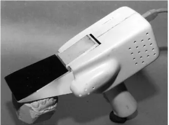
A special device (research prototype of the Czech Technical University) was prepared (Figure 1). It con-sists of an optical head, a power supply, and a PC computer. The optical head contains a digital color camera, a source of illumination, and a removable prism. The optical head is a handheld device. Its front part consists of a removable prism, which is put into the mouth. The device is manufactured in various sizes and shapes to fit all varieties of arch sizes. The optical prism can be sterilized by heat as well as chemically. The central part of the device forms a handle, which con-tains the light source and control buttons. The back part is occupied by the camera. The connection to the com-puter and power supply is by a soft cable. The digital camera is connected to PC via FireWire to allow live image display.
Software Description
The software is designed to work on a PC run-ning MS Windows 2000. It can display live images from the camera as well as archived images. The func-tionality supports all common image enhancing tech-niques like contrast/brightness control, palette chang-ing or zoomchang-ing. The camera is calibrated by reference samples placed in front of the camera, after which the images captured have a known scale and geometric measurements can be performed on them for specific distances, distance ratios, angles, and areas.
Two overlapping images can be displayed in the same scale for comparison, and the user can also con-Figure 1. Computer device - research prototype of the Czech Technical
trol their relative position by translation and rotation of one image with respect to the other. Special palettes allow easy discrimination of differences between the images being compared.
Database Interface
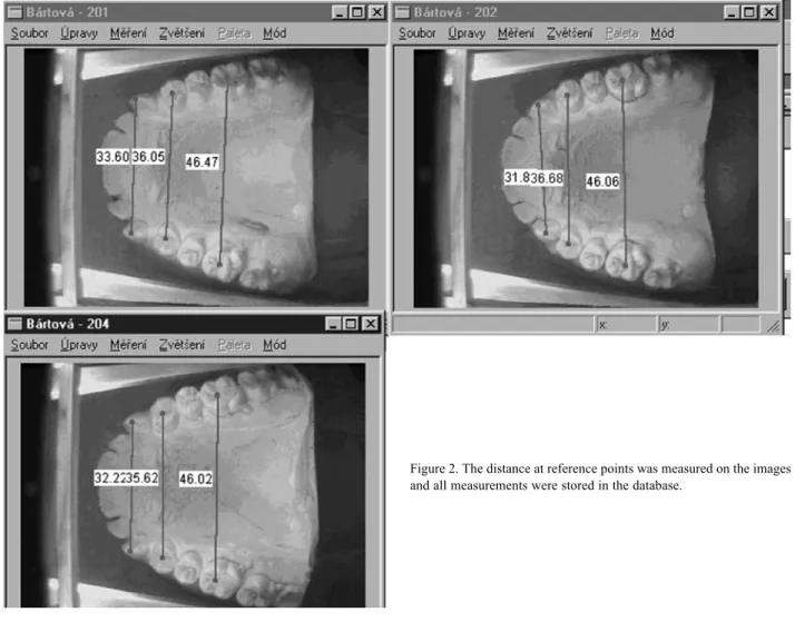
All measurements performed on images are stored in the database (Figure 2). The copied images are prompted to “archive” them and new captured images overwrite the archived images according to FIFO (first in, first out) strategy. The images are stored in JPEG format. The image processing part also allows export and import in other formats like BMP, etc.
Sample
In the first part of the study, measurements of dental arch width between the canines, first premolars
and first molars were made on 792 sets of study casts (Charles University students).
In the second part of the study, the association between initial, post-treatment and post-retention align-ment (5-years after orthodontic therapy) of upper and lower dental arch was analyzed for 36 patients with 108 sets of study models.
The reference points were found, and the dis-tance was measured (Figure 2). Definition of reference points: maxillae: distance between a) cusps of canines; b) lower-most points of the transverse fissure of the first premolars; c) points of intersection of the transverse fissure with the buccal fissure of the first permanent molars; mandible: distance between a) cusps of ca-nines; b) lower-most points of the transverse fissure of the first premolars (second part of study – 108 study models); c) points of intersection of the fissures of the first permanent molars (second part of study – 108 study models).
The metric analysis of dental arch form was made. The models were evaluated side-by-side (juxta-position), the reference points were found and the dental arch width was measured.
RESULTS
For the control group of 792 sets of study casts (Charles University students – control group), the fol-lowing dental arch width measurements were recorded between reference points of canines, between the first premolars, and between the first molars respectively: upper jaw: men: canines: 35.1 ± 0.13 mm; first premolars: 37.5 ± 0.13 mm; first molars: 48.1 ± 0.19 mm; women: canines: 33.4 ± 0.13 mm; first premolars: 35.6 ± 0.15 mm; first molars: 46.7 ± 0.19 mm (Table 1). The ideal values from healthy students were compared with orthodontic patient values before and after treatment. The correlation analysis of the 5-year recall was prepared and the sets of upper and lower dental arch study cast were analyzed (Table 1). Nonstructural data were also collected. Upper and lower arch compression in first premolar and molar area was visible before treatment: upper jaw: men: canine: 34.59 ± 1.28 mm and 37.65 ± 0.78; first molar: 43.7 ± 0.81 mm and 44.28 ± 1.01; women: canine: 34.91 ± 0.46 mm and 36.06 ± 0.30 mm; first molar: 44.12 ± 0.45 mm and
43.86 ± 0.55.
The size and shape of each dental arch was checked 5 years post-therapy (overlapping of images and measurements between reference points) (Table 1). Correlation analysis revealed that the pattern was the same. The shape of dental arch was wider mainly in the anterior region. A significant increase was found be-tween canines (men: from 35.29 ± 0.83 to 36.39 ± 0.96; women: from 33.62 ± 0.39 to 34.53 ± 0.39) and first premolars (men: from 34.59 ± 1.28 to 37.65 ± 0.78; women: from 34.91 ± 0.46 to 36.06 ± 0.30).
Application of fixed (24 patients) or removable (12 patients) appliance had no influence on treatment stability.
Metric analysis of arch form shows a significant difference between patients before and after treatment – see values in Table 1. The arch form width in men was larger than in women. A significant difference was found in the distance between the first molars.
DISCUSSION
Study cast analysis is a three-dimensional as-sessment of the maxillary and mandibular dental arches and the occlusal relationships (10). The actual results of the individual cases were compared with the standard measurements of the “normal arch”. Human arch form
varies considerably. The study of Cassidy et al. (11) analyzed the size and shape of the maxillary and man-dibular dental arches of 320 adolescents. Arch dimen-sions were significantly larger in boys than in girls, both mediolaterally and anteroposteriorly, a sex difference largely established prior to the onset of the adolescent growth spurt. We observed a similar result and found that a certain correlation between arch length, width, and mesiodistal dimensions existed. Contradictory find-ings on shape and size of dental arch as a risk factor for relapse of optimal alignment have been discussed in the literature (12,13).
Measurements of the dental arch width between reference points of canines, first premolars, and first molars were made. In students, we found similar dis-tances as observed in standard metric analyses of Faber (14). Photocopy of the dental arch and computer analy-sis during treatment was able to help to check the dental arch form. The occlusograms superimposed at refer-ence marks allowed the transfer of referrefer-ence crosshairs to following tracing during treatment. The measure-ments were not done. Our evaluation confirmed that the length of the dental arch can be modified by inclining the incisors without varying the inter-canine width. Our results indicate an expansion of the maxillary anterior arch segment during active treatment. Fixed or remov-able orthodontic appliances did not influence treatment stability. Orthodontic treatment was stable over a long-term period. We found that several irregularities of anterior teeth were observed in 7% of our 36 patients. We conclude that computer image monitoring can be used for evaluation of dental arch changes. Geometrically calibrated images help to compare sev-eral different steps of the treatment. The analysis of arch shows a significant difference between patients before and after treatment.
RESUMO
Monitoramento de imagem computadorizada foi usado para avaliar as mudanças no arco dental. Um novo e especial dispositivo capturou imagens calibradas geometricamente permitindo a comparação de vários modelos de gesso. Na primeira parte desta pesquisa, 792 modelos de estudo foram medidos. Medidas de largura do arco dental entre pontos referentes aos caninos, primeiros pré-molares e primeiros molares foram feitas: arco superior: homens - caninos: 35,1 ± 0,13 mm; primeiros pré-molares: 37,5 ± 0,13 mm; primeiros pré-molares: 48,1 ± 0,19 mm; mulheres - caninos: 33,4 ± 0,13 mm; primeiros pré-molares: 35,6 ± 0,15 mm; primeiros molares: 46,7 ± 0,19 mm. Na segunda parte
do estudo, mudanças entre o alinhamento inicial, pós-tratamento e pós-retenção (5 anos depois do tratamento ortodôntico) dos arcos superior e inferior de 36 pacientes foram analisadas. A compressão no arco superior e inferior na área dos primeiros pré-molares e pré-molares foi visível antes do tratamento. Concluiu-se que o monitoramento de imagens computadorizadas pode ser usado para avaliação de mudanças na arcada dentária durante as diferentes fases do tratamento.
ACKNOWLEDGEMENTS
This research was supported by the Grant Agency of the Ministry of Health of the Czech Republic (No. 8112-3).
REFERENCES
1. Tweed CH. The Frankfort mandibular incisor angle in orthodon-tic diagnosis, treatment planning and prognosis. Angle Orthodon-tics 1954;24:1212-1269.
2. MacConaill MA, Scher E. The ideal form of the human dental arcade, with some prosthetic application. Dent Record 1949;69:285-302.
3. Izard G. New method for the determination of the normal arch by the function of the face. Int J Orthodontia 1927;13:582-595. 4. Graber TM, Vanarsdall RL. Orthodontics. 2nd ed., St. Louis:
Mosby, 1994;268-274.
5. Currier JH. A computerized geometric analysis of human dental arch form. Am J Orthodon 1969;56:164-179.
6. Brader AC. Dental arch form related with intraoral forces: PR = C. Am J Orthodon 1972;61:541-561.
7. De La Cruz AR, Sampson P, Little R, Firtun J, Shapiro PA. Long-term changes in arch form after orthodontic treatment and reten-tion. Am J Orthodon Dentofac Orthoped 1995;107:518-530. 8. BeGole EA, Lyew RC. A new method for analyzing change in
dental arch form. Am J Orthodon Dentofac Orthoped 1998;113:394-401.
9. Rakosi T, Jonas T, Graber TM. Orthodontic diagnosis. Georg Tieme Stuttgart: Verlag; 1993; p 207-217.
10. Surbeck BT, Artun J, Hawkins R, Leroux B. Associations be-tween initial, posttreatment, and postretention alignment of max-illary anterior teeth. Am J Orthodon Dentofac Orthoped 1998;113:185-196.
11. Cassidy KM, Harris EF, Tolley EA, Keim RG. Genetic influence on dental arch form in orthodontic patients. Angle Orthodontist 1998;5:445-454.
12. Filser F, Kocher P, Weibel F, Luthy H, Scharer P, Gauclker LJ. Reliability and strength of all ceramic dental restorations fabri-cated by direct ceramic machining. Int J Computerized Dent 2001;4:89-104.
13. Corrucini RS. Mouth breathing, occlusion, and modernization in a north Indian population. An epidemiologic study. Angle Orthod 1985;55:190-196.
14. Faber DS. Occlusograms in orthodontic treatment planning. Am J Orthodon Dentofac Orthoped 1992;6:1-13.
15. Herberger RJ. Stability of mandibular intercuspid width after long periods of retention. Angle Orthodontist 1981;51:78-83. 16. Asanami S, Kasazaki Y. Expert third molar extractions.
Quintes-sence 1990;21:21-22.