O R I G I N A L P A P E R
Central giant cell lesion of the jaws: study of
CCND1
gene
amplification and p16
INK4aprotein levels
Renato Luiz Maia Nogueira•Ma´rio Henrique Gira˜o Faria•
Rafael Lima Verde Osterne• Roberta Barroso Cavalcante•
Ronaldo Albuquerque Ribeiro•Cassiano Francisco Weege Nonaka•
Silvia Helena Barem Rabenhorst
Received: 15 February 2013 / Accepted: 7 March 2013 / Published online: 16 March 2013 ÓSpringer Science+Business Media Dordrecht 2013
Abstract Central giant cell lesions (CGCLs) are uncommon benign jaw lesions with uncertain etiology and a variable clinical behavior. In neoplasms, alterations in molecules involved in the G1/S checkpoint are frequently found. Loss of p16INK4a expression or overexpression of cyclin D1 may stimulate cell proliferation. The purpose of this study was to analyzeCCND1 gene amplification and the expression of p16INK4ain CGCLs. Structural analysis of the CCND1 was performed using chromogenic in situ hybridization. Immmunohistochemistry was used to iden-tify p16INK4a protein levels. Statistical analysis correlated the two biomarkers with clinical behavior and between each other. Twenty-four lesions were included, being 11 aggressive and 13 non-aggressive. Moderate/high-level
CCND1 amplification was found in 12 lesions. Also, immunoreactivity for p16INK4a was present in 12 cases, mainly in mononuclear cells. There was a significantly higher level of p16INK4aexpression in mononuclear cells of
non-aggressive lesions and lesions with moderate/high-level CCND1 amplification in mononuclear cells. It could be speculated that some CGCLs may develop as a true benign neoplasm. The higher expression of p16INK4a in non-aggressive lesions and in cases with moderate/high-levelCCND1 amplification may show that these molecules have a role in CGCLs.
Keywords Giant cell lesionCCND1 amplification
p16INK4a
Introduction
Central giant cell lesions (CGCLs) are rare intraosseous lesions that occur almost exclusively in the jaws and are more common in adolescents and young adults (Jundt
2005). Microscopically, CGCL is characterized by the
R. L. M. Nogueira
Department of Dental Clinic, Discipline of Oral and Maxillofacial Surgery and Stomatology, Federal University of Ceara School of Dentistry, Fortaleza, Brazil
R. L. M. Nogueira
Department of Oral and Maxillofacial Surgery, Memorial Batista Hospital, Fortaleza, Brazil
M. H. G. Faria
Department of Pathology and Forensic Medicine, Molecular Genetics Laboratory (LABGEM), School of Medicine, Federal University of Ceara´, Fortaleza, Brazil
R. L. V. Osterne (&)
Department of Pathology, Fortaleza University School of Medicine, Av. Washington Soares, 1321, Edson Queiroz, P.O. Box 1258, Fortaleza, Ceara´ 60811-905, Brazil e-mail: [email protected]
R. B. Cavalcante
Department of Oral and Maxillofacial Pathology, Fortaleza University School of Dentistry, Fortaleza, Brazil
R. A. Ribeiro
Department of Physiology and Pharmacology, Federal University of Ceara School of Medicine, Fortaleza, Brazil
C. F. W. Nonaka
Department of Dentistry, Center for Biological and Health Sciences, State University of Paraiba, Paraı´ba, Brazil
S. H. B. Rabenhorst
presence of multinucleated giant cells (MGCs) in a cellular background composed of mononucleated stromal cells (MSCs) with ovoid or spindle-shaped nuclei, which are fibroblastic in origin (Jundt2005; de Lange and Van den Akker 2005; Kruse-Losler et al. 2006). The giant cells themselves may vary in size, shape, and number, probably representing osteoclasts or macrophages in origin (Regezi
2002; Jundt 2005; de Lange and Van den Akker 2005; Kruse-Losler et al. 2006). Although little is know about pathogenesis of CGCLs, some studies have shown that in the MSCs component are the proliferative cells in CGCLs (O’Malley et al.1997; Liu et al.2003).
The CGCL was classified in 2005 by the World Health Organization as an idiopathic benign lesion with a variable clinical behavior ranging from a slowly growing, asymp-tomatic radiolucent lesion to an aggressive process asso-ciated with pain, root resorption, cortical bone destruction, and a tendency to recur after treatment (Chuong et al.1986; Jundt 2005; de Lange and Van den Akker 2005; Kruse-Losler et al. 2006). The pathogenesis of CGCL is not completely understood. There is not even agreement as to whether CGCL is a neoplasm or a reactive process (Souza et al.1999,2000; Kauzman et al.2004; Nogueira et al. 2010; Nogueira et al. 2012). Some authors regard CGCLs as lesions related to giant cell tumors (GCTs) of the bones, representing different ends of a clinical-pathological spectrum of the same diseases process (Auclair et al.1988; Kauzman et al.2004).
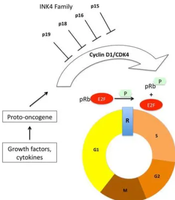
An imbalance in cell proliferation control is an important characteristic of aggressive lesions, in which molecules involved in the G1/S checkpoint are frequently altered (Malumbres and Barbacid 2001). The late G1 cell cycle checkpoint is controlled by a complex of proteins that include p16INK4a, cyclin D1, cyclin-dependent kinases (CDKs) 4/6, and retinoblastoma protein (pRb) (Fang et al. 1998; Diehl
2002). These proteins are components of the pRb cell cycle control pathway; cyclin D1 stimulates the phosphorylation of pRb by association with CDKs, and p16INK4a binds to CDK4/6, blocking their association with D-type cyclins (Fig.1). Thus, the loss of p16INK4a expression or overex-pression of cyclin D1 cause pRb pathway dysfunction and stimulate cell proliferation (Serrano et al. 1993; Mittnacht
1998). While the three D-type cyclins are almost indistin-guishable biochemically, only cyclin D1 is frequently over-expressed in cancers. TheCCND1gene is a proto-oncogene located on chromosome 11q13 and encodes cyclin D1, and the amplification of this gene is a common mechanism that leads to aberrant overexpression of cyclin D1 (Diehl2002). Only one study (Kauzman et al.2004) has analyzed the amplifica-tion of theCCND1gene in CGCLs, and the expression of p16INK4ahas never been studied in CGCLs.
Gathering more information regarding the genetic events involved in CGCL is an important task in
understanding this lesion. Due to the role of cyclin D1 and p16INK4a in pRb pathway, and considering that these molecules are frequently involved in tumorigenesis, the purpose of this study was to analyzeCCND1gene ampli-fication and the expression of p16INK4a in CGCLs of the jaws.
Materials and methods
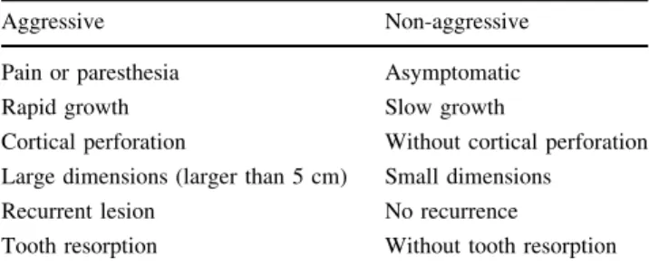
For this study, formalin-fixed paraffin-embedded blocks of 27 consecutive cases of primary CGCLs from patients treated at Memorial Batista Hospital were retrieved from the archives of the Department of Oral and Maxillofacial Pathology, Fortaleza University, Fortaleza, Brazil. Samples were excluded based on associated diagnoses (such as cherubism or hyperparathyroidism, or Brown’s tumor), insufficient clinical data to classify the behavior of the lesions as aggressive or non-aggressive, or inadequate specimen for molecular or biochemical analysis. Based on clinic and radiographic data, all cases were classified according to the criteria established by Chuong et al. (1986) (Table1).
Fig. 1 The pRb pathway. Growth factors lead to the accumulation of
Chromogenic in situ hybridization (CISH) for CCND1 gene amplification
CCND1 gene amplification was performed as previously described (Reis-Filho et al. 2006) using the ready-to-use digoxigenin-labeled SpoT-Light Cyclin D1 amplification probe (Zymed, South San Francisco, CA, USA). Heat pretreatment of deparaffinized sections consisted of incu-bation for 15 min at 98°C in CISH pretreatment buffer (SPOT-light tissue pretreatment kit, Zymed) and digestion with pepsin for 7 min at room temperature according to the manufacturer’s instructions. An appropriateCCND1 gene-amplified breast tumor control was included.
CISH analysis
The CISH results were evaluated by optical microscopy at high magnification (4009). Only unequivocal signals were counted. Morphologically unequivocal cells were assessed for the presence of the gene probe signals. Amplification was defined as[5 signals per nucleus in more than 50 % of
lesion cells or as the presence of large gene copy clusters. High-level gene amplification was defined as more than 10 discrete copies per nucleus or as large gene copy clusters (observed as confluent masses containing more than 10 signals) in more than 50 % of the nuclei evaluated. Low-level amplification was defined as 5–6 copies per nucleus in more than 50 % of the cells. Moderate-level amplifica-tion was defined as 7–10 copies per nucleus in more than 50 % of the cells. Unaltered gene copy number was defined as 1–5 copies per nucleus.
Immunohistochemistry
Immunostaining was performed according to a previously described protocol (Faria et al.2007). For antigen retrieval, deparaffinized sections were pretreated by heating them in a microwave oven in 10 mM citrate buffer, pH 6.0, for 15 min. After cooling, the sections were immersed in PBS containing 3 % hydrogen peroxide for 10 min to block
endogenous peroxidase activity. Sections were then incu-bated in a humid chamber (4 h, 4O) with a primary antibody against anti-p16INK4a (CINtec HistologyÒ, clone E6H4Ò dilution, MTM Laboratories, Heidelberg, Germany). After rinsing with PBS, slides were incubated with the secondary antibody followed by the streptavidin-peroxidase complex, both for 30 min at room temperature with a PBS wash between each step (LSAB? system; DakoCytomationÒ, Glostrup, Denmark). The slides were developed with diaminobenzidine-H2O2(DAB?system; DakoCytomation
Ò , Glostrup, Denmark), counterstained with Harry’s hematox-ylin and mounted. Positive controls consisted of sections of colon cancer; the negative control consisted of replacing the primary antibody with non-immune mouse serum.
Immunostaining analysis
The immunohistochemical staining in the nuclei was assessed using a direct light microscope. A differential count was performed for MSCs and multinucleated giant cells (MGC). Staining was quantified through manual counting of at least 1.000 MSC in 10 different fields at a magnification of 4009. In the same 10 high-power fields, all MGC were counted to a maximum of 1.000 cells. The labeling index (LI) was expressed as the percentage of positive cells with nuclear staining for p16INK4a in each section (Landber and Roos1993).
Statistical analysis
Data were analyzed using SPSS 13.0 (SPSS Inc., Chicago, IL, USA). The results were expressed as the mean. To compare the marker LI with respect to clinical forms and the treatment result groups, the non-parametric Mann– Whitney test was used. Significance was established at a
p valueB0.05.
The present study was approved by the Ethics Com-mittee of the Hospital Complex of the Federal University of Ceara´ under protocol 13/08, respecting Resolution 196/96 of the National Council of Health—Ministry of Health/Brazil.
Results
Of the 27 selected cases of CGCL, 24 have enough bio-logical material to perform all reactions. Eleven (45.8 %) were in male patients and 13 (54.2 %) in females, with a median age of 19 years (range from 5 to 50 years). Thir-teen (54.2 %) lesions were classified as non-aggressive and 11 (45.8 %) as aggressive CGCL. The age, sex, location, and aggressiveness status of the samples are presented in Table2.
Table 1 Aggressiveness status of CGCL as defined by Chuong et al.
(1986)
Aggressive Non-aggressive
Pain or paresthesia Asymptomatic
Rapid growth Slow growth
Cortical perforation Without cortical perforation
Large dimensions (larger than 5 cm) Small dimensions
Recurrent lesion No recurrence
Tooth resorption Without tooth resorption
CISH analysis
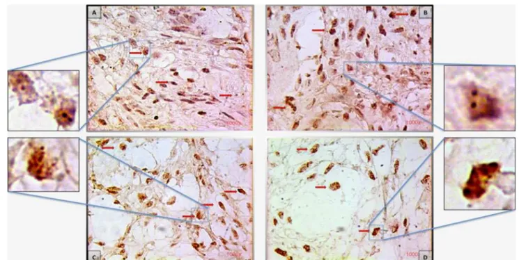
Normal or low-levelCCND1gene amplification in mono-nuclear cells (Fig.2) was found in 14 (58.4 %) lesions and moderate or high-level amplification in 10 (41.6 %) CGCLs (Table3). No statistical significance was found (p=0.697) with respect to association ofCCND1 ampli-fication in mononuclear cells and aggressiveness of the CGCL (Table4). Also, moderate or high-level CCND1 amplification in multinucleated giant cells was found in 10 (41.6 %) of CGCL, again no association was found with aggressiveness (p =0.697) (Table5). Eight of the cases withCCND1 amplification in mononuclear cells also pre-sented amplification in multinucleated giant cells.
Immunostaining analysis
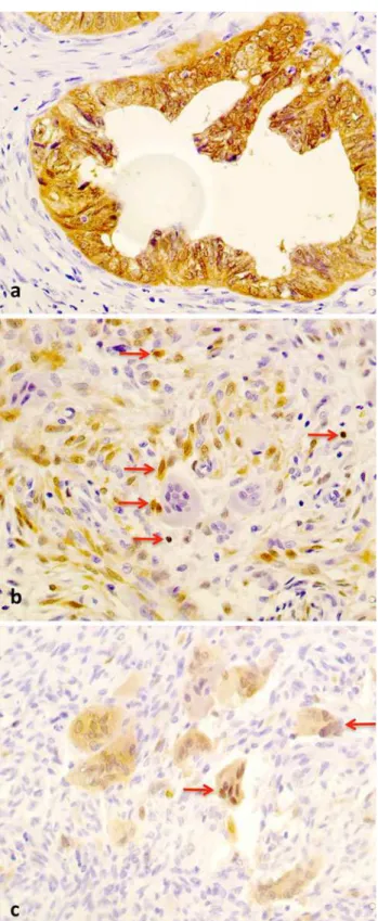
Immunoreactivity for p16INK4a was detected in 12 cases (50.0 %) in mononuclear cells and only in 5 cases (20.8 %) in multinucleated giant cells (Fig.3; Table3). The non-parametric Mann–Whitney test revealed a significantly (p=0.044) higher level of expression of p16INK4a in mononuclear cells in non-aggressive CGCLs (Table6). Considering only immunostainig in multinucleated giant
cells, 4 positive cases occurred in non-aggressive CGCL, and 1 case in an aggressive lesion.
Correlation between the biomarkers
Analyzing the correlation between CCND1 gene amplifi-cation and p16INK4a immunoreactivity revealed that there was significantly (p =0.012) higher p16INK4a expression in mononuclear cells of CGCLs with moderate/high-level
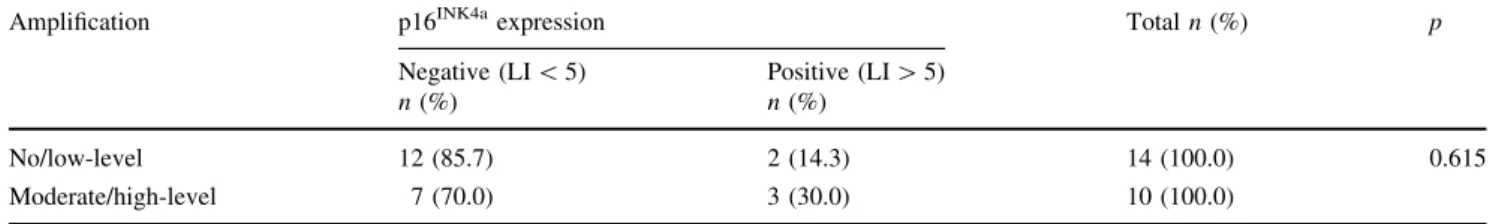
CCND1 gene amplification in mononuclear cells (Table7). Although only 5 cases showed positive immunoreactivity for p16INK4ain multinucleated giant cells, 3 of these cases occurred in CGCL with CCND1 gene amplification in multinucleated giant cells (p =0.615) (Table8).
Discussion
Some authors have stated that CGCL is an uncommon reactive process that affects the jaw bone and that may be related to trauma, such as dental extraction (Unal et al.
2006). Aggressive CGCL, with rapid growth and high recurrence rates, are believed to be neoplastic in nature, but the etiopathognesis is still uncertain (Nogueira et al.2012). Analyses of cell cycle regulatory proteins have been used to distinguish neoplastic from reactive conditions and to predict the biological behavior of tumors (Souza et al.
1999). Few studies regarding cell cycle protein alterations in CGCL have been published (O’Malley et al. 1997; Souza et al. 1999,2000; Kauzman et al.2004). The iden-tification of molecular markers characteristic of a lesion and understanding the nature and behavior of a lesion may allow clinicians to better classify and eventually treat such lesions.
In this study, moderate to elevated CCND1 gene amplification in MSC and/or MGC was found in 12/24 cases. Although the authors knows that gene amplification doesn’t necessarily, but usually, leads to high protein lev-els, overexpression of cyclin D1 andCCND1 gene ampli-fication were previously demonstrated by Kauzman et al. (2004) in CGCL, and this overexpression could be impli-cated in the pathogenesis of CGCL and in the formation of giant cells. Although no statistical significant difference in amplification was found between aggressive and non-aggressive lesions, the amplification of the CCND1 gene could indicate that CGCL may be true neoplastic in nature. Kandel et al. (2006) demonstratedCCND1 gene ampli-fication and cyclin D1 and p21 overexpression in cultured giant cells of GCTs of bones. Kauzman et al. (2003) also found low-levelCCND1 gene amplification in 19/31 cases of GCT. In the same study, cyclin D1 and D3 overex-pression in multinucleated giant cells and cyclin B1 over-expression and Ki-67 immunoreactivity were detected in
Table 2 Age, gender, aggressiveness and site of CGCL
Case Gender Age Aggressiveness Location
1 Male 13 Aggressive Maxilla
2 Female 21 Non-aggressive Mandible
3 Male 20 Aggressive Maxilla
4 Male 7 Non-aggressive Mandible
5 Female 24 Non-aggressive Maxilla
6 Female 23 Aggressive Mandible
7 Female 19 Aggressive Mandible
8 Female 42 Aggressive Maxilla
9 Male 20 Non-aggressive Maxilla
10 Male 9 Aggressive Mandible
11 Male 6 Aggressive Maxilla
12 Male 5 Aggressive Maxilla
13 Female 15 Aggressive Mandible
14 Male 12 Non-aggressive Mandible
15 Female 24 Non-aggressive Mandible
16 Male 7 Non-aggressive Maxilla
17 Male 25 Non-aggressive Mandible
18 Female 20 Aggressive Mandible
19 Female 10 Non-aggressive Mandible
20 Female 18 Non-aggressive Mandible
21 Female 23 Aggressive Maxilla
22 Female 50 Non-aggressive Mandible
23 Female 11 Non-aggressive Mandible
mononuclear cells, indicating that the mononuclear cells are the proliferative component of GCT (Kauzman et al.
2003). Expression of Ki-67 and PCNA in the mononuclear cells of CGCLs also indicates that these cells are the proliferative cells in CGCLs (Souza et al. 1999, 2000; Liu et al.2003; Kauzman et al.2004).
Considering p16INK4aimmunoreactivity, this suppressor was detected mainly in mononuclear cells, and a higher level of expression was detected in non-aggressive lesions (p =0.044). Because p16INK4a is a negative regulator of cell division that slows down the progression of the cell cycle by inactivating cyclin dependent kinase, the higher
Fig. 2 CISH for cyclin D1, all in91,000.aNon-aggressive CGCL:
normal amplification. Only two gene copies per nucleus (arrowand box). b Aggressive CGCL: low-level amplification, 5–6 spots per nucleus in more than 50 % of the cells (arrow), compared with
normal amplification (box). cAggressive CGCL: high-level ampli-fication, more than 10 spots or clusters in more than 50 % of the cells
(arrow and box). d Non-aggressive CGCL: high-level cyclin D1
amplification (arrowandbox)
Table 3 CCND1 gene amplification and p16INK4a expression in
CGCL
Normal
n(%)
Low-level
n(%)
Moderate-level
n(%)
High-level
n(%)
CCND1 gene amplification
MSC 9
(37.50 %) 5
(20.83 %) 5
(20.83 %)
5
(20.83 %)
MGC 10
(41.67 %) 4
(16.67 %) 8
(33.33 %)
2
(8.33 %)
Cell type Negative
n(%)
5–25
n(%)
26–75
n(%)
[75
n(%)
p16INK4aexpression (LI)
MSC 12
(50.00 %) 6
(25.00 %) 6
(25.00 %) –
MGC 19
(79.17 %) 3
(12.50 %) 2
(8.33 %) –
MSCmonomuclear stromal cells,MGCmultinucleated giant cells
Table 4 Correlation between CCND1 gene amplification in MSC
and aggressiveness of CGCL (Fisher’s exact test)
Aggressiveness Amplification Total n(%)
p
No/low-level n(%)
Moderate/high-level n(%)
Aggressive 7 (63.6) 4 (36.4) 11 (100.0) 0.697
Non-aggressive 7 (53.8) 6 (46.2) 13 (100.0)
Table 5 Correlation betweenCCND1 gene amplification in MGC
and aggressiveness of CGCL (Fisher’s exact test)
Aggressiveness Amplification Total n(%)
p
No/low-level n(%)
Moderate/high-level n(%)
Aggressive 7 (63.6) 4 (36.4) 11 (100.0) 0.697
p16INK4a expression in non-aggressive lesions may be the reason why they exhibit slower growth. Additionally, the higher p16INK4a expression found in CGCLs with mod-erate to elevated CCND1 gene amplification (p=0.012) support this hypothesis and justify the clinical behavior of this lesion. It is possible that cyclin D1 overexpression could induce p16INK4a transcription.
Alternatively, it could be postulated that, in both aggressive and non-aggressive CGCLs with a neoplastic nature, an epigenetic alteration in mononuclear cells, such as
CCND1 amplification, could change the cell cycle. CGCLs with cyclin D1 amplification could have a higher prolifera-tive rate and a more aggressive behavior. However, when there is a negative cell cycle regulator, such as p16INK4aor another tumor suppressor protein, this lesion could present a slower proliferative rate and thus slower growth. To our knowledge, this is the first report of the study of p16INK4a expression in CGCLs; other tumor suppressor genes should be investigated to understand the etiopathogenesis of this lesion. Some authors have previously evaluated p53 protein expression in CGCLs, but no immunoreactivity was found (O’Malley et al.1997; Souza et al.1999,2000). Supporting the neoplastic nature of CGCL, Carinci et al. (2005) ana-lyzed the genetic profile of two cases of CGCL of the jaws and found some up-regulated and down-regulated genes. In addition, the gene SH3BP2 has been investigated in the pathogenesis of CGCL, Carvalho et al. (2009) described a mutation in the SH3BP2 gene in a case of non-familial CGCL, but Teixeira et al. (2011) investigating 30 cases of CGCL, analyzed all exons known to be involved in cher-ubism, none of them were mutated.
AlthoughCCND1gene amplification has been observed in CGCLs, no difference was observed between the clinical forms of this lesion. However, the expression of p16INK4a seems to play a role in aggressiveness of the lesion, those lesions with increased expression of p16INK4ain MSC may have a more indolent clinical behavior. Also, the findings of the present study corroborate the hypothesis that at least some CGCLs may have a neoplastic origin. These lesions do not fit neatly into the concept of either reactive or neoplastic process; instead, CGCLs exhibit features of both (Regezi and Pogrel 2004). It could be speculated that CGCLs may develop in two different ways, one being a reactive process and other appears to be a true benign neoplasm, and thatCCND1 amplification may contribute to the tumorigenesis of CGCLs. Further studies focused on clinical and molecular analysis, including new studies about cell cycle protein, particularly cyclin D1 protein levels, could help better the understanding of the patho-genesis of CGCL.
Fig. 3 Immunoreactivity for p16INK4a, all in9400.aColon cancer,
Acknowledgments The authors would like to thank FUNCAP (Fundac¸a˜o Cearense de Apoio ao Desenvolvimento Cientı´fico e Tecnolo´gico) for funding this research.
References
Auclair PL, Cuenin P, Kratochvil FJ, Slater LJ, Ellis GL (1988) A clinical and histomorphologic comparison of the central giant cell granuloma and the giant cell tumor. Oral Surg Oral Med Oral Pathol 66:197–208
Carinci F, Piattelli A, Martinelli M, Palmieri A, Rubini C, Fioroni M, Scapoli L, Laino G, Caputi S, Becchetti A, Pezzetti F (2005) Genetic profiling of central giant cell granuloma of the jaws. J Craniofac Surg 16:399–407
Carvalho VM, Perdiga˜o PF, Amaral FR, de Souza PE, De Marco L, Gomez RS (2009) Novel mutations in the SH3BP2 gene associated with sporadic central giant cell lesions and cherubism. Oral Dis 15:106–110
Chuong R, Kaban LB, Kozakewich H, Perez-Atayde A (1986) Central giant cell lesions of the jaws: a clinicopathologic study. J Oral Maxillofac Surg 44:708–713
de Lange J, Van den Akker HP (2005) Clinical and radiological features of central giant-cell lesions of the jaw. Oral Surg Oral Med Oral Pathol Oral Radiol Endod 99:464–470
Diehl JA (2002) Cycling to cancer with cyclin D1. Cancer Biol Ther 1:226–231
Fang X, Jin X, Xu HJ, Liu L, Peng HQ, Hogg D, Roth JA, Yu Y, Xu F, Bast RC Jr, Mills GB (1998) Expression of p16 induces transcriptional down regulation of the RB gene. Oncogene 16:1–8
Faria MH, Patrocı´nio RM, Moraes Filho MO, Rabenhorst SH (2007) Immunoexpression of tumor suppressor genes p53, p21WAF1/CIP1 and p27KIP1 in human astrocytic tumors. Arq Neuropsiquiatr 65:1114–1122
Jundt G (2005) Central giant cell lesion. In: Barnes L, Eveson JW, Reichart P, Sidransky D (eds) World Health Organization classification of tumours. Pathology and genetics of head and neck tumours. IARC, yon, p 324
Kandel R, Li SQ, Bell R, Wunder J, Ferguson P, Kauzman A, Diehl JA, Werier J (2006) Cyclin D1 and p21 is elevated in the giant cells of giant cell tumors. J Orthop Res 24:428–437
Kauzman A, Li SQ, Bradley G, Bell RS, Wunder JS, Kandel R (2003) Cyclin alterations in giant cell tumor of bone. Mod Pathol 16:210–218
Kauzman A, Li SQ, Bradley G, Bell RS, Wunder JS, Kandel R (2004) Central giant cell granuloma of the jaws: assessment of cell cycle proteins. J Oral Pathol Med 33:170–176
Kruse-Losler B, Diallo R, Gaertner C, Mischke KL, Joos U, Kleinheinz J (2006) Central giant cell granuloma of the jaws: a clinical, radiologic, and histopathologic study of 26 cases. Oral Surg Oral Med Oral Pathol Oral Radiol Endod 101:346–354 Landber G, Roos G (1993) Proliferating cell nuclear antigen and
Ki-67 antigen expression in human haematopoietic cells during growth stimulation and differentiation. Cell Prolif 26: 427–437
Liu B, Yu SF, Li TJ (2003) Multinucleated giant cells in various forms of giant cell containing lesions of the jaws express features of osteoclasts. J Oral Pathol Med 32:367–375
Malumbres M, Barbacid M (2001) To cycle or not to cycle: a critical decision in cancer. Nat Rev Cancer 1:222–231
Mittnacht S (1998) Control of pRB phosphorylation. Curr Opin Genet Dev 8:21–27
Nogueira RLM, Teixeira RC, Cavalcante RB, Ribeiro RA, Rabenhosrt SHB (2010) Intralesional injection of triamcinolone hexacetonide as an alternative treatment for central giant-cell granuloma in 21 cases. Int J Oral Maxillofac Surg 39:1204–1210
Nogueira RL, Faria MH, Osterne RL, Cavalcante RB, Ribeiro RA, Rabenhorst SH (2012) Cyclooxygenase-2 expression in central giant cell lesion of the jaws: an immunohistochemical study. J Mol Histol 43:59–62
Table 6 Parameters used for the calculation of Mann–WhitneyUtest for the evaluation of the expression of p16INK4ain CGCL, according to
CGCL aggressiveness
Aggressiveness n Median Q25–Q75 Mean of ranks Sum of ranks U p
Aggressive 11 0 0–14 9.45 104.00 38.00 0.044
Non-aggressive 13 22 4.5–34 15.08 196.00
Table 7 Parameters used for the calculation of Mann–WhitneyUtest for the evaluation of the expression of p16INK4ain CGCL, according
CCND1 gene amplification in MSC
AmplificationCCND1 n Median Q25–Q75 Mean of ranks Sum of ranks U p
No/low-level 14 0 0–16 9.54 133.50 28.50 0.012
Moderate/high-level 10 23 11–36.75 16.65 166.50
Table 8 Correlation betweenCCND1 gene amplification in and p16INK4aexpression in MGC (Fisher’s exact test)
Amplification p16INK4aexpression Totaln(%) p
Negative (LI\5)
n(%)
Positive (LI[5)
n(%)
No/low-level 12 (85.7) 2 (14.3) 14 (100.0) 0.615
O’Malley M, Pogrel MA, Stewart JC, Silva RG, Regezi JA (1997) Central giant cell granulomas of the jaws: phenotype and proliferation-associated markers. J Oral Pathol Med 26:159–163 Regezi JA (2002) Odontogenic cysts, odontogenic tumors, fibroos-seous, and giant cell lesions of the jaws. Mod Pathol 15:331–341 Regezi JA, Pogrel A (2004) Comments on the pathogenesis and medical treatment of central giant cell granulomas. J Oral Maxillofac Surg 62:116–118
Reis-Filho JS, Savage K, Lambros MB, James M, Steele D, Jones RL, Dowsett M (2006) Cyclin D1 protein overexpression and CCND1 amplification in breast carcinomas: an immunohisto-chemical and chromogenic in situ hybridisation analysis. Mod Pathol 19:999–1009
Serrano M, Hannon GJ, Beach D (1993) A new regulatory motif in cell-cycle control causing specific inhibition of cyclin D/CDK4. Nature 366:704–707
Souza PEA, Paim JFO, Carvalhais JN, Gomez RS (1999) Immuno-histochemical expression of p53, MDM2, Ki-67 and PCNA in central giant cell granuloma and giant cell tumor. J Oral Pathol Med 28:54–58
Souza PEA, Mesdquita RA, Gomez RS (2000) Evaluation of p53, PCNA, Ki-67, MDM2 and AgNOR in oral peripheral and central giant cell lesions. Oral Dis 6:35–39
Teixeira RC, Horz HP, Damante JH, Garlet GP, Santos CF, Nogueira RL, Cavalcante RB, Conrads G (2011) SH3BP2-encoding exons involved in cherubism are not associated with central giant cell granuloma. Int J Oral Maxillofac Surg 40:851–855