J Appl Oral Sci.
ABSTRACT
F A S N e x p r e s s i o n , a n g i o g e n e s i s a n d
lymphangiogenesis in central and peripheral giant
cell lesions
Saulo Gabriel Moreira FALCI1!"#$!%&'()1*###$*+# ,-0-%2, 456# !)0,-14%$"#76%8,9$:;$ 6!%)-4=>$$0?07 $&,9&1
1- Department of Dentistry, College of Basic Sciences and Health, Federal University of Vales do Jequitinhonha e Mucuri (UFVJM), Diamantina, MG, Brazil. 2- Oral Pathology, School of Dentistry, Fluminense Federal University, Nova Friburgo, RJ, Brazil.
3- Oral Pathology, Department of Stomatology, Public Oral Health and Forensic Dentistry, University of São Paulo, Ribeirão Preto, SP, Brazil. 4- Oral Pathology Section, Department of Oral Diagnosis, Piracicaba Dental School, University of Campinas (UNICAMP), Piracicaba, SP, Brazil.
=$ $$ Saulo Gabriel Moreira Falci - Tiradentes, 195 - Diamantina - MG - Brazil - Postal code: 39100/000 - Phone/Fax: +55 38 3532 - 6000 / +55 38 8817 1454 - e-mail: [email protected]
&#?@ &@?! B7-7@?D77 4#ED
C
entral giant cell lesion (CGCL) and peripheral giant cell lesion (PGCL) are non-neoplastic proliferative processes of the jaws. PGCL is a reactive process induced by irritant local factors and CGCL is an intra-osseous lesion of unknown etiology. Both lesions exhibit similar histologic features showing abundant mononuclear cells, admixed with a large number of multinucleated giant cells and a rich vascularized stroma with extravasated fatty acid synthase (FASN) with angiogenesis. Objective: To evaluate angiogenesis and lymphangiogenesis and their relationship with FASN expression in CGCL and PGCL. Material and Methods: Thirteen CGCL and 14 PGCL of the jaws were selected for immunoexpression of FASN; CD34 and CD105 (to assess blood microvessel density [MVD] and microvessel area [MVA]); and D2-40 (to assess lymphatic MVD and MVA). Results: Within PGCL and !!"# $ % % !!&'* +$ !!/#' %%+6789 % mononuclear cells were observed. Between PGCL and CGCL, only MVD-CD34 and all MVA $ % % < < $ 7!&'* $ 6789 positive mononuclear cells in both lesions was observed. Conclusions: Our results show both lesions exhibiting similar levels of FASN expression and neoangiogenesis, suggesting constitutive processes that regulate tissue maintenance.Keywords: Giant cell lesion. Immunohistochemistry. Angiogenesis. Lymphangiogenesis. Fatty acid synthase.
INTRODUCTION
Giant cell lesion (GCL) of the jaws is a non-neoplastic proliferative process, divided into central giant cell lesion (CGCL) and peripheral giant cell lesion (PGCL). PGCL is considered a reactive process induced by local irritants on the gingiva or alveolar mucosa. CGCL is an intra-osseous lesion of unknown etiology11. Both CGCL and PGCL exhibit similar histopathological features, and are characterized by the presence of abundant mononuclear stromal cells, admixed
with a large number of multinucleated giant cells and a rich vascularized stroma with extravasated erythrocytes, hemosiderin deposition, and
blood-In spite of this, these lesions may have different clinical behaviors11,13,23.
Fatty acid synthase (FASN) is the metabolic enzyme responsible for endogenous synthesis of saturated long-chain fatty acid, specifically palmitate, from the precursors acetyl-CoA and malonyl-CoA7. FASN is overexpressed in a variety of human cancers affecting breast19, ovaries2, prostate20, and oral cavity22, whereas FASN is
downregulated in most normal human tissues (except in the liver, lactating breast, fetal lung, and adipose tissue) because cells preferentially use circulating dietary fatty acids for the synthesis of new structural lipids29. In the oral cavity, FASN expression has been shown in squamous cell carcinoma and melanoma3,22; however, its expression in benign neoplasms3 and/or reactive conditions is little known. Interestingly, some studies have linked FASN expression with endothelial cell proliferation4,21. Accordingly, the role of FASN expression in angiogenesis must be @$%6789 in GCL of the jaws is unknown.
CD34 is a cell surface glycoprotein consistently expressed in the vascular endothelium. Some studies have previously assessed CD34 expression in CGCL in order to compare aggressive and non-aggressive subtypes. They showed increased microvessel density (MVD)-CD34 in aggressive CGCL10,18,25. Although CD34 is unable to distinguish between pre-existing vessels and neoformed vessels, interestingly, it has been shown that distinguishing neoformed vessels in proliferative tissues is relevant and may have prognostic implications, identifying possible targets for developing anti-angiogenic therapeutic strategies5,18.CD105 (endoglin) is an angiogenic membrane protein that is highly expressed in neoformed vessels6. Although CD105 has been assessed in oral vascular malformations and pyogenic granulomas26, to our knowledge, CD105 expression in GCL of the jaws is unknown. Another unclear and relevant point is the characterization of the lymphatic MVD (LMVD) in GCL, which may be immunohistochemically evaluated through the D2-40 marker13. Nevertheless, as in the case of CD105, the lymphatic vascular stroma characterization in GCL of the jaws has not been evaluated.
As previously mentioned, increased FASN expression in oral malignant tumors has been reported3,22,and some studies have linked FASN expression with endothelial cell proliferation4,21. It remains to be determined what happens with such events in oral benign and/or reactive lesions. Thus, the aim of the current study was to assess angiogenesis and lymphangiogenesis, as well as establishing their relationship with FASN expression
in CGCL and PGCL of the jaws.
MATERIAL AND METHODS
This retrospective study examined the records and tissues of patients diagnosed and treated for GCL of the jaws. None of the patients had received any treatment with a therapeutic agent prior to + 6J paraffin-embedded tissue blocks of 13 CGCL (8 males, 5 females; mean age 18.5 years; 8 mandible, 5 maxilla) and 14 PGCL (9 males, 5 females; mean age 38.9 years; 11 mandibular gingiva, 3 maxillary gingiva) were selected from our laboratory archives. According to their clinical characteristics, such as painful symptoms, growth pattern rate, root resorption, cortical bone perforation and recurrence, the CGCL cases of the
$ %% 8.
All lesions were reviewed through hematoxylin-eosin stained slides and the diagnosis was $ KJ from the study were GCL cases which presented inadequate clinical description, cases diagnosed as aneurysmal bone cyst, cherubism and brown tumor of hyperparathyroidism (confirmed by establishing elevated serum parathyroid hormone Q $ $ + for histological analysis. This study was approved by the Research Ethics Committee (Process 042/2011).
Immunohistochemical methods
For each antibody (FASN, CD34, CD105 and D2-#'Q"X glass slides were used. All tissue specimens were J&'Y++++/# +**Z and cut into parallel consecutive sections. For the immunohistochemical (IHC) reactions, the slides were hydrated and treated with hydrogen peroxide (3%). Primary antibodies, dilutions and antigen retrieval are shown in Table 1. The tissue sections were then washed three times in phosphate buffered saline (PBS) solution and exposed to secondary antibody using the LSAB+Kit (Dako, Carpinteria, CA, USA). Peroxidase activity was visualized by using the chromogen diaminobenzidine (DAB)
Table 1- Antibodies used for immunohistochemical analysis of central and peripheral giant cell lesions
? E Clone Manufacturer Dilution +:
FASN 23/610962 Becton Dickinson Transduction Laboratories1
1:200 TRIS/EDTA (pH 9.0)
CD34 QBEnd10 Dako# jan/ 1:50 Citric acid (pH 6.0)
CD105 SN6h Dako jan/ 1:30 Proteinase K
D2-40 D2-40 Dako 1:100 Citric acid (pH 6.0)
J Appl Oral Sci. (Sigma Chemical Co., St. Louis, USA). Finally, the tissue sections were counterstained with Carrazi´s hematoxylin for 5 minutes. Thereafter, the sections were dehydrated in a series of graded ethanol solutions, diaphanized and mounted in Canada balsam under cover glasses. Sections of oral Kaposi’s sarcomas were included in all reactions as positive control for CD34, CD105 and D2-40, while prostate carcinomas were used for FASN. Negative controls of reactions were performed by omitting the primary antibody. The slides were scanned and photographed using the Aperio Scanscope CS Slide Scanner (Aperio Technologies®, Vista, CA, USA).
I m m u n o s t a i n i n g a s s e s s m e n t a n d statistical analysis
MVD and microvessel area (MVA), assessed through CD34 and CD105 expression, as well as lymphatic MVD (LMVD) and MVA (LMVA), assessed through D2-40 expression, were established identifying the most vascularized areas within the lesions (“hot spots”), which were chosen at low magnification (100x) and subsequently % %$ | #''J %Q14. The images were made using the Aperio Scan Scope® software, which depicted % }|%~*'$~"/'Q
%+&*''2.
!7$ area, respectively, of positive microvessels stained 8 + %$ $ MVD and MVA. The MVA measurement was done by means of vascular contour tracing using the
Image J® software, which automatically measured } |2). Single endothelial cells or clusters of endothelial cells, with or without a lumen, were considered to be individual vessels.
To assess FASN expression, similar to angiogenic markers, the area containing a high number of $ $ % |&''JQ %%$ |#''J%Q@+6789 positive mononuclear cells and multinucleated giant cells in both lesions were counted in each high-power field using the software Image J® (Scion Corporation, USA), and then the average percentages of the immunostained cells were calculated in each case.
The data collected were recorded and organized in a database using Statistical Package for Social Science (version 17.0; SPSS Inc., Chicago, IL, USA). Normal distribution was tested using the Shapiro– Wilk test. In samples with a normal distribution, the Student t-test and Pearson correlation were applied. In samples that presented non-normal distributions, the Mann–Whitney U test and Spearman correlation were applied. A P<0.05 probability value was %
RESULTS
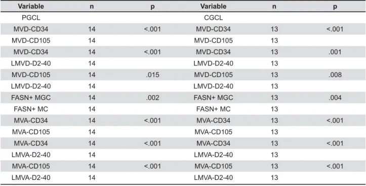
All vascular structures in all cases were highlighted by CD34. By comparing the MVD within < % + CD105-positive vessels than CD34-positive vessels (P<0.001) was observed (Table 2). In addition, a Table 2- Comparison between PGCL and CGCL in relation to (L)MVD, (L)MVA, and percentage of FASN-positive multinucleated giant cells and mononuclear cells
V?: n p V?: n p
PGCL CGCL
MVD-CD34 14 MVD-CD34 13
MVD-CD105 14 MVD-CD105 13
MVD-CD34 14 MVD-CD34 13 .001
LMVD-D2-40 14 LMVD-D2-40 13
MVD-CD105 14 .015 MVD-CD105 13 .008
LMVD-D2-40 14 LMVD-D2-40 13
FASN+ MGC 14 .002 FASN+ MGC 13 .004
FASN+ MC 14 FASN+ MC 13
MVA-CD34 14 MVA-CD34 13
MVA-CD105 14 MVA-CD105 13
MVA-CD34 14 MVA-CD34 13
LMVA-D2-40 14 LMVA-D2-40 13
MVA-CD105 14 MVA-CD105 13
LMVA-D2-40 14 LMVA-D2-40 13
% +!/#' than CD105-positive vessels in PGCL (P''&*Q
and CGCL (P'''Q$ @
percentage indicates that 31% and 13% of the vessels in PGCLs, and 33% and 8% of the vessels in CGCLs were positive for CD105 and D2-40, respectively.
The number of CD34-positive vessels was
%%<|P'''/Q
(Figures 1a, 1b; Figure 2a). However, no statistical difference regarding the number of CD105- and D2-40-positive vessels was found when comparing both lesions (Figures 1c, 1d; Figure 2a; Figures 3a, 3b).
% greater MVA for all vascular markers used in <$ |!"#'''& !&'* ''#& !/#' ''"Q |6% 2b). Moreover, when comparing the MVA within < % of CD105-positive vessels than CD34-positive vessels (P<0.001) was observed. In addition, it $ % + D2-40-positive vessels than CD105-positive vessels in both lesions (P<0.001) (Table 2).
Moreover, through consecutive section analysis, we found focal areas showing vessels stained with
P# Comparison between PGCL and CGCL in relation to MVD and LMVD (a), MVA and LMVA (b) and percentage of FASN-positive MGC and MC (c)
J Appl Oral Sci. CD105 and D2-40 in both lesions (Figures 3c, 3d), supporting the idea that some CD105-stained vessels represent newly formed lymphatic vessels.
All cases present FASN-positive multinucleated giant cells (PGCL, mean 39%; CGCL, mean 36%) and mononuclear cells (PGCL, mean 7%; CGCL, mean 10%) (Figure 2c; Figures 4a, 4b), without %++ $% both lesions (P'&*P'"/' Q We also observed focal areas showing weak FASN immunoexpression on endothelial cells in 46% and 21% of the CGCL and PGCL cases, respectively. % $ found between MVA-CD105 with FASN-positive |'*'''"Q
DISCUSSION
Both CGCL and PGCL of the jaws exhibit similar histopathological features, being characterized by numerous ovoid and spindle-shaped mononuclear cells, admixed with multinucleated giant cell, within a rich vascularized stroma11,16. Despite this, it is well known that these lesions may have different clinical behaviors23. In the current study, without % ++ $ <
all cases presented FASN-positive multinucleated giant cells and mononuclear cells. Nevertheless, we observed a higher number of multinucleated giant cells than mononuclear cells (ratio 4:1) presenting FASN immunoreactivity. It is probable that cell ++ may help to explain our results. Moreover, as a %J + %$+ multinucleated giant cells and mononuclear cells16 is most probably associated with angiogenesis in GCL, on the other hand the lack of correlation between FASN-positive multinucleated giant cells with vascular markers observed in the current study can suggest maintenance and/or remodeling role for FASN expression in multinucleated giant cells.
Angiogenesis, which is the formation of new vessels from pre-existing vessels, is thought to be of crucial importance to the growth and maintenance of proliferative tissues24. In the oral cavity, it has been shown that the number and size of blood vessels increase from normal oral epithelium through dysplastic epithelium to reach a maximum in invasive carcinoma1, while no significant difference seems to occur between normal mucosa, hyperplasia, and dysplasia for LMVD. Conversely, invasive carcinomas presented higher LMVD P# Immunoexpression of D2-40 in CGCL (a, 400x) and PGCL (b, 400x), notice that the lymphatic vessels are of larger diameter in PGCL than in CGCL. This case of CGCL on parallel consecutive sections presented vessels positive for both CD105 (c, 400x) and D2-40 (d, 400x), suggesting neolymphangiogenesis
P#D Immunoexpression of FASN in CGCL (a, 400x) and PGCL (b, 400x), note immunopositivity for FASN in the multinucleated giant cells and mononuclear cells in both lesions
than normal mucosa and precancerous lesions17. Regarding benign and reactive oral lesions, several studies have been performed to quantify MVD by using CD34 and CD1059,10,12,18,25-27, but none of them have assessed GCL considering both markers. In addition, although D2-40 expression has been demonstrated in normal odontogenic tissues as well as in cystic and tumor odontogenic lesions30, it is noteworthy that in GCL a stromal lymphatic vessel characterization has not been performed. Like CD105, D2-40 expression in GCL is unknown.
In the current study, we have observed uniform positivity for CD34 in all vascular structures in both lesions. Almost all our cases (93% and 92% of the PGCL and CGCL, respectively) exhibited CD105-positive vessels (about 32%, in relation to CD34-positive vessels), while only 78% and 69% of the PGCL and CGCL, respectively, showed D2-40-positive vessels (about 10%, in relation to CD34-positive vessels). Thus, the constitutive expression of CD105 in GCL supports angiogenic activity and tissue remodeling. This is further supported by FASN expression in endothelial cells, detected focally in about 46% and 21% of CGCL and PGCL cases, respectively. Moreover, the expression of endothelial markers in GCL is consistent with the presence of various angiogenic factors and matrix metalloproteinases (MMPs), such as tumor necrosis factor-alpha (TNF-α), transforming growth +|@6Q %$+ (b-FGF), vascular endothelial growth factor (VEGF), and MMP-915,16,28. Since it has been shown that aggressive rather than nonaggressive subtypes of CGCL present higher MVD-CD3410,18,25, further studies are needed in order to compare whether there are CD105 expression differences between aggressive and nonaggressive subtypes of CGCL. In relation to stromal lymphatic vessel characterization, it is interesting that lymphangiogenesis in reactive or benign oral lesions have been little studied. In the current study, we have shown that about 8% and 13% of the vessels in CGCL and PGCL group, respectively, were immunoreactive for !/#' 7% $ % ++$<|''/Q is suggested that the higher number of lymphatic vessels detected in PGCL (Figure 2) seems to be influenced by anatomical location, since a %+!/#' be observed at the periphery of the lesions in close association with the lamina propria. Furthermore, through consecutive section analysis, we found focal areas showing vessels stained with CD105 and D2-40, in both lesions. In order to validate % $ J + CD105 and D2-40 in oral GCL should be compared $ %% neolymphangiogenesis in these lesions.
< $ % greater MVA than CGCL for all vascular markers used. Similar results were shown27, which favored a reactive nature for PGCL. It is likely that, in addition to the anatomical location of these lesions, differences in the expression pattern of angiogenic growth factors1,16 may help to explain the differences found in our study. It has been shown that FASN inhibition by orlistat may reduce endothelial cell proliferation and angiogenesis4. As previously commented, to our knowledge, the FASN immunoexpression in giant cell lesions is unknown. However, in our study, different from FASN-positive multinucleated giant cells, it is % %+ between FASN-positive mononuclear cells and MVA-!&'* + % is suggested that FASN-positive mononuclear cells may contribute with new vessel formation. Future % determinate possible interactions between FASN expression and angiogenic proteins in GCL.
CONCLUSION
In summary, our results suggest that greater MVD-CD34, and greater MVA-CD34, CD105 and D2-40, in PGCL rather than in CGCL, might be associated with a reactive inflammatory process. Moreover, similar levels of FASN expression, neoangiogenesis (MVD-CD105), and lymphangiogenesis (LMVD-D2-40) between PGCL and CGCL indicate constitutive processes to regulate tissue maintenance and remodeling in both lesions.
ACKNOWLEDGMENTS
The authors would like to thank the University of Vales do Jequitinhonha e Mucuri for the master´s scholarship; Victor Toral Rizo for training in applied methodology; Patrícia Correia Faria for helping with graphics; Piracicaba Dental School, Oral Diagnosis Department, for performing the immunohistochemistry; São Paulo Research Foundation (FAPESP - 2009/53839-2).
CONFLICTS OF INTEREST
@ of interest in this manuscript.
REFERENCES
J Appl Oral Sci. 2- Alò PL, Visca P, Framarino ML, Botti C, Monaco S, Sebastiani V, et al. Immunohistochemical study of fatty acid synthase in ovarian neoplasms. Oncol Rep. 2000;7(6):1383-8.
3- Andrade BA, León JE, Carlos R, Delgado-Azañero W, Mosqueda-Taylor A, Graner E, et al. Expression of fatty acid synthase (FASN) in oral nevi and melanoma. Oral Dis. 2011;17(8):808-12. 4- Browne CD, Hindmarsh EJ, Smith JW. Inhibition of endothelial cell proliferation and angiogenesis by orlistat, a fatty acid synthase inhibitor. FASEB J. 2006;20(12):2027-35.
5- Cardoso SV, Souza KC, Faria PR, Eisenberg AL, Dias FL, Loyola AM. Assessment of angiogenesis by CD105 antigen in epithelial salivary gland neoplasms with diverse metastatic behavior. BMC Cancer. 2009;9:391.
6- Cheifetz S, Bellón T, Calés C, Vera S, Bernabeu C, Massagué J, et al. Endoglin is a component of the transforming growth factor-beta receptor system in human endothelial cells. J Biol Chem. 1992;267(27):19027-30.
7- Chirala SS, Wakil SJ. Structure and function of animal fatty acid synthase. Lipids. 2004;39(11):1045-53.
8- Chuong R, Kaban LB, Kozakewich H, Perez-Atayde A. Central giant cell lesions of the jaws: a clinicopathologic study. J Oral Maxillofac Surg. 1986;44(9):708–13.
9- Davey KJ, Perrier S, Ohe G, Gilbert AD, Bankfalvi A, Saunders WP, et al. Assessment of vascularity as an index of angiogenesis in periradicular granulomas: comparison with oral carcinomas and normal tissue counterparts. Int Endod J. 2008;41(11):987-96. 10- Dewsnup NC, Susarla SM, Abulikemu M, Faguin WC, Kaban LB, August M. Immunohistochemical evaluation of giant cell tumors of the jaws using CD34 density analysis. J Oral Maxillofac Surg. 2008;66(5):928-33.
11- Flórez-Moreno GA, Henao-Ruiz M, Santa-Sáenz DM, Castañeda-Peláez DA, Tobón-Arroyave SI. Cytomorphometric and immunohistochemical comparison between central and peripheral giant cell lesions of the jaws. Oral Surg Oral Med Oral Pathol Oral Radiol Endod. 2008;105(5):625-32.
12- Gadbail AR, Hande A, Chaudhary M, Nikam A, Gawande M, Patil S, et al. Tumor angiogenesis in keratocystic odontogenic tumor assessed by using CD-105 antigen. J Oral Pathol Med. 2011;40(3):263-9.
13- Kahn HJ, Marks A. A new monoclonal antibody, D2-40, for detection of lymphatic invasion in primary tumors. Lab Invest. 2002;82(9):1255-7.
14- Lima SC, Rizo VH, Silva-Sousa YT, Almeida LY, Almeida OP, León JE, et al. Immunohistochemical evaluation of angiogenesis Endod. 2011;37(12):1642-6.
15- Matos FR, Moraes M, Nonaka CF, Souza LB, Almeida Freitas R. Immunoexpression of TNF-α@6
giant cell lesions of the jaws. J Oral Pathol Med. 2012;41(2):194-9. 16- Matos FR, Nonaka CF, Miguel MC, Galvão HC, Souza LB, Freitas RA. Immunoexpression of MMP-9, VEGF, and vWF in central and peripheral giant cell lesions of the jaws. J Oral Pathol Med. 2011;40(4):338-44.
17- Palomba A, Gallo O, Brahimi A, Franchi A. Evaluation of lymphangiogenesis in premalignant conditions of the head and neck mucosa. Head Neck 2010;32(12):1681-5.
18- Peacock ZS, Jordan RC, Schimidt BL. Giant cell lesion of the jaws: does the level of vascularity and angiogenesis correlate with J+8%/'&/'|Q~&'
19- Pizer ES, Jackisch C, Wood FD, Pasternack GR, Davidson NE, Kuhajda FP. Inhibition of fatty acid synthesis induces programmed cell death in human breast cancer cells. Cancer Res. 1996;56(12):2745-7.
20- Rossi S, Graner E, Febbo P, Weinstein L, Bhattacharya N, Onody @6 J signatures in prostate cancer. Mol Cancer Res. 2003;1(1):707-15. 21- Seguin F, Carvalho MA, Bastos DC, Agostini M, Zecchin KG, Alvarez-Flores MP, et al. The fatty acid synthase inhibitor orlistat reduces experimental metastases and angiogenesis in B16-F10 melanomas. Br J Cancer. 2012;107(6):977-87.
22- Silva SD, Cunha IW, Nishimoto IN, Soares FA, Carraro DM, $ < % % + / |8</Q + |6789Q ErbB2 expression in oral squamous cell carcinomas. Oral Oncol. 2009;45(10):e134-9.
23- Souza PE, Mesquita RA, Gomez RS. Evaluation of p53, PCNA, Ki-67, MDM2 and AgNOR in oral peripheral and central giant cell lesions. Oral Dis. 2000;6(1):35-9.
24- Spiegelaere W, Casteleyn C, Van den Broeck W, Plendl J, Bahramsoltani M, Simoens P, et al. Intussusceptive angiogenesis: a biologically relevant form of angiogenesis. J Vasc Res. 2012;49(5):390-404.
25- Susarla SM, August M, Dewsnup N, Faquin WC, Kaban LB, Dodson TB. CD34 staining density predicts giant cell tumor clinical behavior. J Oral Maxillofac Surg. 2009;67(5):951-6.
26- Vasconcelos MG, Alves PM, Vasconcelos RG, Silveira EJ, Medeiros AM, Queiroz LM. Expression of CD34 and CD105 as markers for angiogenesis in oral vascular malformations and pyogenic granulomas. Eur Arch Otorhinolaryngol. 2011;268(8):1213-7. 27- Vassilopoulos SI, Tosios KI, Panis VG, Vrostsos JA. Endothelial cells of oral pyogenic granulomas express eNOS and CD105/ endoglin: an immunohistochemical study. J Oral Pathol Med. 2011;40(4):345-51.
28- Vered M, Buchner A, Dayan D. Giant cell granuloma of the $ ~+ $ %$+ growth factor. J Oral Pathol Med. 2006;35(10):613-9.
29- Weiss L, Hoffmann GE, Schreiber R, Andres H, Fuchs E, Körber E, et al. Fatty-acid biosynthesis in man, a pathway of < organ distribution of fatty-acid synthase. Biol Chem Hoppe Seyler. 1986;367(9):905-12.
30- Zustin J, Scheuer HA, Friedrich RE. Podoplanin expression in human tooth germ tissues and cystic odontogenic lesions: an immunohistochemical study. J Oral Pathol Med. 2010;39(1):115-20.