Correlation and comparison of immunohistochemistry
for HER2/neu, using the antibody
SP3
and
chromogenic
in situ
hybridization in breast
carcinomas samples
Correlação e comparação de imuno-histoquímica para HER2/neu, utilizando o anticorpo
SP3
com a hibridização
in situ
cromógena em amostras de carcinomas mamários
Franciele F. Wolf1; Giuliano S. Bublitz1; Henrique R. Frigeri2
1. Serviços Integrados de Patologia (SIP), Joinville (SC). 2. Pontifícia Universidade Católica do Paraná (PUCPR).
First submission on 06/07/15; last submission on 01/08/15; accepted for publication on 04/08/15; published on 20/12/15
ABSTRACT
Introduction: Advances in the ield of molecular biology have provided the differentiation of molecular subtypes of breast tumors,
providing better prognosis and important tools for the treatment of patients with breast cancer. Among these subtypes, the changes in the human epidermal growth factor receptor 2 gene (HER2/neu), increase its copy number and generating HER2 protein ampliication. Studies show that patients with breast cancer HER2/neu ampliied tend to relapse earlier and have shorter survival time, the monoclonal
antibody Trastuzumab is the therapy indicated. The eligibility of patients for therapy is initially made by the immunohistochemistry (IHC)
technique, which evaluates the expression level of the HER2 protein. After this evaluation, the cases with equivocal diagnosis (score 2+), are referred to a more accurate technique, the chromogenic in situ hybridization (CISH). Objective: To analyze the sensitivity and speciicity
of the antibody SP3, and determine their level of agreement with the CISH technique. Material and methods: Retrospective study in the database of the anatomy-pathology laboratory, in CISH tests reports for HER2/neu. Conclusion: The results revealed that clone SP3
showed 100% speciicity and 92% sensitivity. IHC reveals variability in its results; however, it is known that the technique is an important tool in the daily routine of laboratories, contributing to the initial screening of patients with breast cancer, which later showed satisfactory results when compared with the CISH technique.
Key words: breast cancer; in situ hybridization; immunohistochemistry.
INTRODUCTION
Breast cancer is among the most common neoplasm affecting women, both in developing and developed countries(1, 2). In Brazil,
according to the Instituto Nacional de Câncer (INCA), it were estimated for the years 2014 and 2015, about 57,000 new cases of female breast cancer each year, making it the second most frequent in the female population (non-melanoma skin cancer is the irst one). It ranks third among cancers that affect the population in general (irst non-melanoma skin cancer and second prostate cancer)(1).
Breast cancer is a heterogeneous disease, with a variety of morphological appearance and molecular characteristics. Studies in the field of molecular biology have provided the differentiation of five molecular subtypes of breast cancer: 1) luminal A; 2) luminal B-HER2 negative; 3) luminal B-HER2 positive; 4) basal-like or triple negative; and 5) HER2 overexpressing. They assist in understanding the mechanisms that regulate differentiation and cell proliferation, and thus allowing a better prognosis that provides important tools for the choice of therapy(2, 3).
LITERATURE REVIEW
Human epidermal growth factor receptor-2
genotype (
Her2/neu
)
The HER2/neu gene is variously known as neu, HER2
and c-erb-B2. A mutated version of the gene was irst observed in a rat neuro glioblastoma and, therefore, named neu(4). The
HER2/neu comprises breast carcinoma with estrogen receptor (ER) and progesterone receptor (PR) negative, but that present overexpression of the protein also called HER2(2, 5). In normal
cells and in the majority of breast cancers, only two copies of the gene expressing low levels of p185 protein (protein HER2) are usually transported(6). The gene ampliication or overexpressing is
evaluated on 25% to 35% of invasive breast carcinomas(7, 8), which,
in most cases (around 90%), are attributed to the ampliication of this protooncogene located on long arm of chromosome 17 (17q12)(5, 7, 9, 10). A minority of cases where it is observed HER2/neu
gene ampliication is characterized by chromosome 17 polysomy, which leads to increased gene copy number, resulting in high expression and thus providing equivocal diagnostic(9).
There are reports in the literature on patients with breast cancer with ampliied HER-2/neu, they tend to have early
recurrence, and reduced survival(11). The therapy with humanized
monoclonal antibody Trastuzumab combined with adjuvant chemotherapy was initially used to prolonging survival in patients with HER2/neu-positive metastatic breast cancer. Moreover, it was also indicated to reduce the chances of developing distant metastases in patients who do not already have them(7, 12). This
beneit occurs because the treatment is directed to the antigen HER2/neu protein, inhibiting the growth of tumor cells with
HER2/neu overexpressing(13). The correct detection of HER-2/neu
oncogene ampliication is essential to enable eligibility of patients with breast cancer and lead them to treatment with Trastuzumab.
This product is indicated because it is a speciic targeted therapy, which acts blocking the extra cellular domain of receptor(12, 14, 15).
Protein overexpressing and
immunohistochemistry (IHC)
IHC is a technique used to analyze the overexpression of p185 protein in tissue of mammary carcinomas, and was incorporated to surgical pathology as a complementary method. It is considered a technique of low cost, besides being the most used in the daily routine of pathology laboratories(14, 16). The development
of monoclonal antibodies provided great source of highly speciic reagents for the demonstration of different tissue and cell antigens;
along with the advent of antigen retrieval they were considered milestones in the evolution of IHC(17).
Several mono and polyclonal antibodies are commercially available for use in IHC, with differences in sensitivity and speciicity, and may result in variations in the inal quality of the reactions(18). Among these antibodies options commonly used in
the method, the mouse monoclonal antibody CB11 and rabbit
monoclonal antibody SP3 stand out.
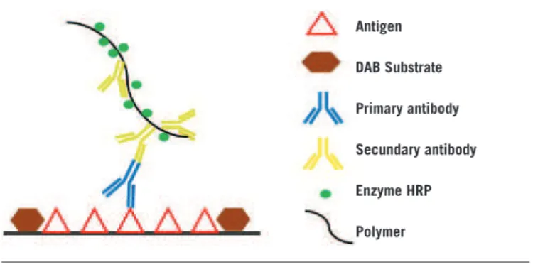
The principle of the IHC method lies in binding primary antibodies that recognize the speciic antigen. The limitations associated with the Avidin-Biotin complex system led to the development of systems detection with enhanced sensitivity and speciicity, using a polymer-based IHC method. This method uses a polymer structure with multiple secondary antibodies and enzyme molecules conjugated(19, 20) (Figure 1).
IHC provides the results of protein expression of the HER2 gene
using a scoring system ranging from zero to three(18). According
to the guidelines of the American Society of Clinical Oncology (ASCO) and the College of American Pathologists (CAP), the staining intensity of the cytoplasmic membrane must be observed, which has values ranging from negative (0), no immunoreactivity or immunoreactivity ≤ 10% of the tumor cells; negative (1+), weak or faint immunoreactivity in more than 10% of the tumor cells, but only part of the membrane is positive; equivocal (2+), weak to moderate complete membrane immunoreactivity in more than 10% of the tumor cells or intense circumferential membrane staining in ≤ 30% of the cells; and positive (3+), moderate to strong complete membrane immunoreactivity in more than 30% of the tumor cells(15, 21-23). The use of this scale has varying
interpretations that depend on the technique reaction quality, the type of antibody used, and the observer evaluation(18). These
discrepancies in interpretations of the results are, especially when the IHC revels score 2+, and this value was considered inconclusive
FIGURE 1 − Representation of IHC reaction
IHC: immunohistochemistry; DAB: diaminobenzidine; HRP: horseradish peroxidase. Antigen
DAB Substrate
Primary antibody
Secundary antibody
Enzyme HRP
for diagnosis. Based on this, the samples are sent to be tested in situ hybridization (ISH), since they are able to provide accurate
and reliable results(18, 24).
Overexpression of gene and ISH
The ISH techniques determine the number of copies of a gene. They use complementary deoxyribonucleic acid (DNA) probes, marked to the target genomic sequencing. Regarding the patients with breast tumors, the chromogenic in situ hybridization (CISH)
technique assesses the level of ampliication of the HER2/neu
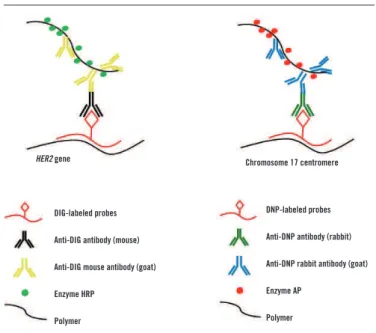
gene, measuring its copy number by combined ISH, as well as by a digoxigenin (DIG)-labeled probe, which targets the HER2 gene, and a dinitrophenol (DNP)-labeled centromeric probe, speciic for the centromeric alpha-satellite region of chromosome 17(25)
(Figure 2). The formation of double-stranded marked with probing may be visualized using primary antibodies (unlabelled), which are detected by secondary antibodies conjugated to polymerized enzyme. The enzymatic reactions of the substrates lead to the formation of strong red signals (chromosome 17)
and green (HER2 gene) permanent(25). This reaction creates
a chromogenic reaction similar to IHC staining, which enables viewing by light microscopy, as well as to avoid the need for luorescence (used in the ISH method by luorescence [FISH]), and provide a permanent staining record result. In addition, CISH has a lower cost compared with the luorescent method and is more easily interpreted by pathologists, since it can be correlated
DNP-labeled probes
Anti-DNP antibody (rabbit)
Anti-DNP rabbit antibody (goat)
Enzyme AP
Polymer
FIGURE 2 − Representation of CISH reaction
CISH: chromogenic in situ hybridization; DIG: digoxigenin; HRP: horseradish peroxidase; DNP: dinitrophenyl; AP: alkaline phosphatase.
HER2 gene
DIG-labeled probes
Anti-DIG antibody (mouse)
Anti-DIG mouse antibody (goat)
Enzyme HRP
Polymer
Chromosome 17 centromere
with tumor histology at the same time because they are more used to IHC labeling than the luorescent signal(6, 15, 16, 24).
CISH has emerged as a potentially promising alternative to FISH, showing high correlation between its results in the
literature(6, 15, 16, 26, 27). Both methods are approved by the Food and
Drug Administration (FDA) and considered the gold standard to assess the status (level) of gene ampliication when required for conirming the ambiguous results of IHC(16, 28). Furthermore,
CISH allows morphological tumor analyzing (interpreting the heterogeneity of it), the gene copies number of in different parts
of the tumor and the observation of invasive or in situ carcinoma ields, providing a special clinical meaning to HER2/neu(16, 26-28).
OBJECTIVE
This study aimed to analyze the SP3 antibody sensitivity and
speciicity and to determine their level of agreement with the CISH technique.
MATERIAL AND METHODS
This study was conducted through a survey of the Centro de Diagnóstico Anatomopatológico (CEDAP) database, located in the city of Joinville, Santa Catarina. This work is approved by the Research Ethics Committee of the Pontifícia Universidade Católica do Paraná (PUCPR) under CAAE record: 12717213.5.0000.0020. In the routine, the breast cancer samples are initially submitted
to HER-2 gene status search by IHC, using the CB11 clone (Cell Marque®). When the result of this reaction presents score 2+,
the assistant doctor responsible for the patient requests the CISH study (ZytoVision®) to deinition the case. For ratiication of the
results of the CISH, a new IHC is requested now using SP3 clone (Cell Marque®). In some special cases, the CISH research is still
required by the assistant doctor, even when satisfactory results by IHC technique are revealed (score 0, 1+ or 3+).
Thus, we selected all reports from January 2011 to March 2014 that showed research HER2/neu gene ampliication obtained by
CISH technique, accompanied by IHC tests with SP3 clone, totaling
98 cases.
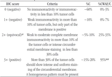
The parameters for interpretation of IHC score and evaluation of gene ampliication by CISH followed the recommendations of the ASCO/CAP, as shown in Tables 1 and 2, respectively(29). The
TABLE 1 − Report of the HER2 immunohistochemical results
IHC score Criteria %C %CWAF/C
0 (negative) No immunoreactivity or immunoreac-tivity in less than 10% tumor cells
~60% 0%-3%
1+ (negative) Weak immunoreactivity in more than 10% of tumor cells, but only part of the
membrane is positive
~10% 0%-7%
2+ (equivocal)# Weak to moderate complete membrane immunoreactivity in more than 10% of the tumor cells or intense circumfer-ential membrane staining in less than
30% cells
~5%-10% 25%-35%
3+ (positive) More than 30% of the tumor cells should show intense and uniform
stain-ing of the circumferential membrane. A homogeneous pattern must be present
~15%-20% 95%##
Adapted from: College of American Pathologists (CAP), 2012.
%C: percentage of cases; %CWAF/C: percentage of cases with amplification by FISH or CISH; #: The panel of expert recommended to test the doubtful cases to gene amplification by FISH; ##: The expert panel increased the fraction of cells with membrane staining from 10% to 30%, to increase the correlation with gene amplification by FISH. According to the panel, cancer with 3+ by IHC result should show gene amplification in at least 95% of cases.
IHC: immunohistochemistry; FISH: fluorescence in situ hybridization; CISH: chromogenic in situ hybridization.
The 98 cases were divided into two groups according to the expression level of the gene: a) positive CISH –
HER2/neu gene overexpression –; b) negative CISH – absence
of HER2/neu gene overexpression. After exposure of the data, it was performed the comparison and correlation of SP3 clone and CISH results.
IHC methodology
The methodology for samples preparation was performed according to the manufacturer’s instruction manual(19). After
preparing the slides with the selected sections, they were taken to the oven for 30 minutes at 70ºC. Antigen retrieval was performed on the PT link (Dako®) instrument for 20 minutes at 97ºC. The
slides were washed for 5 minutes in wash buffer (Wash Buffer 20× concentrate). Endogenous peroxidase was inactivated with Reagent Peroxidase Blocking (Dako®) for 5 minutes, adding
100 µl over the sections. Then the solution was withdrawn from the slides, proceeding again with wash buffer. Following, 100 µl of the antibody used (SP3 – dilution 1:500) was pipetted, maintained for 20 minutes at room temperature. The antibody was removed of the slides; and another washing was performed with wash buffer. Then the solution of horseradish peroxidase (HRP) was pipetted and left for 20 minutes at room temperature; then washed for 5 minutes in wash buffer. To the revelation, a
drop of Substrate Buffer + diaminobenzidine (DAB) Chromogen on the section was added for 5 minutes. Then it was washed with wash buffer. The counterstaining was performed with hematoxylin for 10 minutes and washed twice under running water and then kept in Scott solution for 1 more minute. Finally, a inal wash under running water was performed, followed by absolute ethanol for 30 seconds (twice) and xylene for also 30 seconds (twice).
CISH methodology
The methodology for samples preparation was performed according to the manufacturer’s instruction manual(25).
Pretreatment (first day)
The sections were made at 3 µm thick and laid to dry in an oven at 70ºC for 1 hour. Following they were incubated for 10 minutes in xylene at 10 minutes at 70ºC. Then slides were incubated for 5 minutes into xylene at room temperature (twice), followed by 3 minutes in 100% ethanol (three times) and for 5 minutes in 3% hydrogen peroxide solution. They were rinsed in distilled water for 1 minute (twice), and then incubated at 95ºC in ethylenediaminetetraacetic acid (EDTA) solution for 15 minutes. Subsequently they were transferred to distilled water for 2 minutes (twice). The pepsin solution was applied and incubated for 5 minutes in a moist chamber at room temperature. After this period of time, washing the slides in distilled water was conducted. Dehydration was performed with 70%, 90% and 100% ethanol for 1 minute each; the slides were air dried.
TABLE 2 −Report of the results of study with FISH and CISH for HER2
ISH result Critera Comments
Positive for ampliication
> 6 copies of the gene or > 2.2 ratio
> 6 copies of the gene may be present in a ratio < 2.2, if the polysomy is present. Besides the relation, the number
of genes and number of 17 chromosome must be provided when they are determined as part of the test
Equivocal for ampliication
4 to 6 gene or 1.8 to 2.2 ratio#
The guidelines suggest counting additional cells by FISH, retest,
or performing IHC Negative for
ampliication
< 4 gene or < 1.8 ratio
Adapted from: College of American Pathologists (CAP), 2012.
Denaturing and hybridization
The probe was homogenized by vortexing. Pipette 8 µl into the sections, which were covered with a cover slip. Preparations denaturation was performed in hybridizer for 5 minutes at 78ºC-80ºC. The slides were left overnight, hybridizing at 37ºC.
Detection (second day)
Cover slips were removed by submerging them in Wash Buffer SSC for 5 minutes at 25ºC, and then washed for 5 minutes in the same buffer at 75ºC-80ºC. Following the slides were washed in distilled water for 1 minute (twice) and again in buffer. Anti-DIG/DNP-Mix (room temperature) was applied over the sections and incubated for 15 minutes at 37ºC in a moist chamber. Washed for 1 minute in the buffer above mentioned and applied horseradish (HRP)/alkaline phosphatase (AP)-Polymer-Mix on cuts, and incubated for 15 min at 37ºC in a moist chamber. During incubation of the slides, a AP-Red Solution (adding
one drop of AP-Red Solution A in a graduated test tube, adding 1 ml of AP-Red Solution B) was prepared. It is noteworthy that the prepared solution is photosensitive. The slides were washed three times more for 1 minute in Wash Buffer
tris-buffered saline (TBS)(20× concentrate); AP-Red Solution was
applied incubating them for 10 minutes at room temperature, in the dark, between 7 and 15 minutes. During incubation a HRP-Green Solution (two drops of HRP-Green Solution A into a graduated tube, adding 1 ml of HRP-Green Solution B, mix thoroughly) was prepared. The slides were washed for 2 minutes in distilled water, counterstained for 2 minutes with Nuclear Blue Solution (room temperature), washed with running water and lowing three times of 30 seconds in 100% ethanol. Finally, they were lowed twice 30 seconds in xylene and mounted with
mounting medium.
RESULTS
From the 98 cases studied, 48 showed positive CISHand 50 were negative. Assessing the agreement of IHC with positive CISH group, 77% (37 cases) showed agreements using the SP3 clone (score 3+). In the same group, 6% (three cases) were negative (score 0; 1+) by IHC, and 16% (8 cases) showed inconclusive result (score 2+). Regarding the negative CISH group, 94% (47 cases) showed agreement with the gold standard assessment, while
6% (three cases) still remained uncertain in its results (Table 3).
In this same group, there were no cases presenting positive results by IHC.
The sensitivity and speciicity of clone SP3 clone were also evaluated, using CISH as standard test. For this analysis, we used only the cases that showed negative (0/1+) and positive (3+) results by IHC (Table 4). After performing the calculations, SP3
showed 92% sensitivity and 100% speciicity. The positive predictive value (PPV) was 100% (NPV) was 94%; 96% accuracy.
TABLE 4 − Viable results of IHC and CISH to calculate
the sensitivity and specificity
SP3 scores CISH positive CISH negative Total
SP3 3+ 37 0 37
SP3 0/1+ 3 47 50
Total 40 47 87
IHC: immunohistochemistry; CISH: chromogenic in situ hybridization.
TABLE 3 −Correlation of results between IHC and CISH
Score IHC SP3 CISH positive CISH negative
0/1+ 3 47
2+ 8 3
3+ 37 0
Total 48 50
IHC: immunohistochemistry; CISH: chromogenic in situ hybridization.
DISCUSSION
A few years ago, the rabbit monoclonal antibodies have been increasingly popular. This is due to the fact they have higher afinity for the desired epitope, revealing a more intense positive staining with lower background staining, and therefore it has higher afinity and speciicity than rat monoclonal
antibodies(22, 28). The rabbit monoclonal antibodies are directed to
the extracellular domain of the HER2 receptor, whereas the same therapy with Trastuzumab also targets the extramembranar
epitope of HER2; antibodies that detect this portion can produce results with greater clinical relevance regarding treatment response(14).
by Nunes et al.(23) showed that the rabbit mono or polyclonal
antibodies are more sensitive and ensure adequate treatment for patients with gene overexpression, including SP3. However,
in our study, using CISH as a reference test, the SP3 was 92% sensitivity and 100% speciicity. Other studies also show greater
SP3 speciicity in tendency, and less sensitivity, when compared
with other clones, they were evaluated by FISH or CISH standard
test(14, 22, 33). These results are less sensitive due to the number
of false negative cases (0/1+) by SP3, which were ampliied by CISH.
The discrepancy results in IHC is reported in the literature, although this technique widely used and well accepted in pathology laboratories. There are several possible explanations for these differences, such as the use of different antibodies available on the market, the different antibody staining techniques, including dilution and antigen retrieval, the experience of individuals involved in the technique, the lack of internal quality control, the time it takes for the sample to be submerged in formalin, the duration of ixing and also the use of inappropriate fasteners(14, 21, 22, 24, 31, 33, 34). The choice of an ideal
clone to detect the protein overexpression of HER2/neu gene
seems to have not yet been found and fully supported by a group of. According to Gown and Goldstein(31), the choice between
either clones may be the less important when comparing the procedures related to the IHC in the pre-analytical and analytical phase in which these, in turn, are even less important than the ability of the pathologist in evaluating the sample and itting it in the categories 0/1+, 2+ e 3+, knowing that interpreting variations between evaluators may occur(14, 22, 24, 31, 34).
CONCLUSION
The study found that the SP3 clone shows good results
compared to CISH technique/method. The sensitivity and speciicity of SP3 also reached satisfactory levels.
Realizing the possible differences between the results of IHC, even though this is an important and valuable tool in the daily routine of pathology laboratories, it is suggested a new analysis, with another clone, when the result of the first is not clearly obtained. Sending the inconclusive samples for genetic evaluation by ISH, whether chromogen or fluorescent, seems to be the most reliable way to ensure proper treatment of patients with breast cancer HER2/neu overexpressing.
CISH group showed the best correlation, in which 94% (47/50) of the cases in agreement between CISH and SP3. In the same group, 6% (3/50) of cases were equivocal by SP3 clone. There were no cases of de SP3 score 3+, which revealed negativity for
gene ampliication.
Studies show high concordance between the SP3 clone with FISH or CISH tests(30). When compared with the rabbit
polyclonal antibody A0485, some authors have noted a
signiicant reduction of 2+ with SP3(31). Manion et al.(22)
reported that using the rabbit monoclonal antibody anti-SP3
instead of A0485 reduced about 50% the number of doubtful cases sent to the FISH test.
But the positive CISH group had a lower percentage of agreement, showing 77% (37/48) of the cases correlating results between SP3 and CISH. We still must point out the cases
which had false negative result and/or equivocal (one case 0, two cases 1+ and eight cases 2+), since the possible exclusion of these patients for proper treatment with Trastuzumab, once they are truly positive for HER2/neu gene ampliication, further worse their life’s expectation, which is already limited.
Studies report the variability of scores among observers. The scores 0 and 3+ have high agreement, in contrast to the scores 1+ and 2+, which are often interpreted with variations(18, 26, 32).
Among these indicative, it is clear that despite IHC well-known advantages by pathology laboratory, this has the disadvantage of being a semiquantitative(18, 26, 28). This shows the need to
accurately verify the gene ampliication in samples that do not exhibit clear results.
The samples evaluated in this study derived from ambiguous results (score 2+) of CB11 clone, when tested in the initial
routine of the laboratory and then referred to the CISH technique for exact revealing the result through gene ampliication, along with the SP3 clone to corroborate this result. Unlike scores 0 and 3+ that have good deinition standard, the score 2+ is doubtful, and often score 1+ is also interpreted in the same way. The score 1+ overestimation may occur by the pathologist when it does not have a well-deined tumor histological section or a distinct staining of tissue structures, thus causing the referring for the gold standard technique – CISH –, which enables the patient has the opportunity for a more reliable diagnosis when only subjected to IHC.
REFERENCES
1. Ministério da Saúde. Instituto Nacional de Câncer José de Alencar Gomes da Silva. Coordenação de Prevenção e Vigilância. Estimativa 2014: incidência de câncer no Brasil. Rio de Janeiro: INCA; 2014. Available
at: http://www.inca.gov.br/estimativa/2014/estimativa-24042014.pdf.
[Acessed on: 23 jul 2014].
2. Cintra JRD, Teixeira MTB, Diniz RW, et al. Peril imuno-histoquímico e variáveis clinicopatológicas no câncer de mama. Rev Assoc Med Bras. 2012; 58(2): 178-87.
3. Rakha EA, Filho JSR, Baehner F, et al. Breast cancer prognostic classiication in the molecular era: the role of histological grade. Breast Cancer Res. 2010; 12(4): 207.
4. Gouvêa AP, Fernandes JRM, Olson SJ, Brandão EC, Leite MT, Gobbi H. Her-2/neu immunoreactivity in invasive mammary carcinomas: a comparative study using monoclonal and polyclonal antibodies including the HercepTestTM. J Bras Patol Med Lab. 2004; 40(1): 27-32.
5. Robbins SL, Cotran RS. Base patológica das doenças. 8th ed. Rio de Janeiro: Elsevier; 2010.
6. Gobbi H, Rocha RM, Nunes CB. Predictive factors of breast cancer evaluated by immunohistochemistry. J Bras Patol Med Lab. 2008; 44(2): 131-40.
7. Wang S, Saboorian MH, Frenkel E, Hynan L, Gokaslan ST, Ashfaq R. Laboratory assessment of the status of Her-2/neu protein and oncogene in breast cancer specimens: comparison of immunohistochemistry assay with luorescence in situ hybridisation assays. J Clin Pathol. 2000; 53: 374-81. 8. Vogel CL, Cobleigh MA, Tripathy D, et al. Efficacy and safety of Trastuzumab as a single agent in first-line treatment of HER-2
RESUMO
Introdução: Avanços no campo da biologia molecular têm proporcionado a diferenciação dos subtipos moleculares das
neoplasias mamárias, fornecendo melhor prognóstico e ferramentas importantes para a terapêutica de pacientes com câncer de mama. Entre esses subtipos, as alterações ocorridas no gene receptor tipo 2 do fator de crescimento epidérmico humano (HER2/
neu) amplificam o seu número de cópias e geram o aumento da proteína HER2. Estudos mostram que pacientes portadoras de câncer de mama HER2/neu amplificado tendem a ter recaída mais cedo e tempo de sobrevida menor, sendo o anticorpo monoclonal Trastuzumab a terapia indicada. A elegibilidade das pacientes para a terapia é feita inicialmente pela técnica de imuno-histoquímica (IHQ), que avalia o nível de expressão da proteína HER2. Após essa avaliação, os casos que apresentam diagnósticos equívocos (escore 2+) são encaminhados para uma técnica mais precisa, a hibridização cromógena in situ
(CISH). Objetivo: Analisar a sensibilidade e a especificidade do anticorpo SP3, além de determinar o seu nível de concordância com a técnica de CISH. Material e métodos: Estudo retrospectivo no banco de dados de um laboratório anatomopatológico, em laudos de exames de CISH para HER2/neu. Conclusão: Os resultados revelaram que o clone SP3 apresentou 100% de especificidade e 92% de sensibilidade. A IHQ revela variabilidade em seus resultados, porém é sabido que a técnica é uma importante ferramenta na rotina diária dos laboratórios, contribuindo na triagem inicial das pacientes portadoras de câncer de mama, que, posteriormente, mostram resultados satisfatórios quando comparados com a técnica de CISH.
Unitermos: neoplasias da mama; hibridização in situ; imuno-histoquímica.
overexpressing metastatic breast cancer. J Clin Oncol. 2002; 20(3): 719-26.
9. Bempt IV, Loo PV, Drijkoningen M, et al. Polysomy 17 in breast cancer: clinicopathologic signiicance and impact on HER-2 testing. J Clin Oncol. 2008; 26: 4869-74.
10. Rosenberg CL. Polysomy 17 and Her-2 ampliication: true, true, and unrelated. J Clin Oncol. 2008; 26(30): 4856-8.
11. Carr JA, Havstad S, Zarbo RJ, Divine G, Mackowiak P, Velanovich V. The association of HER-2/neu ampliication with breast cancer recurrence. Arch Surg. 2000; 135.
12. Dekker TJA, Borg ST, Hooijer GKJ, et al. Determining sensitivity and speciicity of HER2 testing in breast cancer using a tissue micro-array approach. Breast Cancer Res. 2012; 14: R93.
13. Rhodes A, Jasani B, Anderson E, Dodson AR, Balaton AJ. Evaluation of HER-2/neu immunohistochemical assay sensitivity and scoring on formalin-ixed and parafin-processed cell lines and breast tumors. Am J Clin Pathol. 2002; 118: 408-17.
14. Ricardo SAV, Milanezi F, Carvalho ST, Leitao DRA, Schmitt FCL. Her2 evaluation using the novel rabbit monoclonal antibody SP3 and CISH in tissue microarrays of invasive breast carcinomas. J Clin Pathol. 2007; 60: 1001-5.
18. Nunes CB, Rocha RM, Gouvêa AP, et al. Concordância interobservador na interpretação imuno-histoquímica da superexpressão do Her2 detectada por cinco diferentes anticorpos em array de carcinomas mamários. J Bras Patol Med Lab. 2007; 43(5): 373-9.
19. Dako Denmark A/S. EnVision FLEX, High pH, (Link). Denmark. 4th ed. 2011.
20. Dako Denmark A/S. Immunohistochemical Staining Methods. 6th ed. 21. Salles MA, Curcio VS, Perez AA, Gomes DS, Gobbi H. Contribuição da imuno-histoquímica na avaliação de fatores prognósticos e preditivos do câncer de mama e no diagnóstico de lesões mamárias. J Bras Patol Med Lab. 2009; 45(3): 213-22.
22. Manion E, Hornick JL, Lester SC, Brock JE. A comparison of equivocal immunohistochemical results with anti-HER2/neu antibodies A0485 and SP3 with corresponding FISH results in routine clinical practice. Am J Clin Pathol. 2011; 135: 845-51.
23. Nunes CB, Rocha RM, Reis-Filho JS, et al. Comparative analysis of six different antibodies against (SP3) and chromogenic in situ hybridisation in breast carcinomas. J Clin Pathol. 2008; 61: 934-8.
24. Chang E, Lee A, Lee E, et al. Her-2/neu oncogene ampliication by chromogenic in situ hybridization in 130 brest cancers using tissue microarray and clinical follow-up studies. J Korean Med Sci. 2004; 19: 390-6.
25. Zytovision. ZytoDot2C SPEC HER2/CEN 17 Probe Kit. Germany. February, 2011; (5.0).
26. Madrid MA, Lo RW. Chromogenic in situ hybridization (CISH): a novel alternative in screening archival breast cancer tissue samples for HER-2/ neu status. Breast Cancer Res. 2004; 6: R593-R600.
27. Vocaturo A, Novelli F, Benevolo M, et al. Chromogenic in situ hybridization to detect HER-2/neu gene ampliication in histological and thinprep®-processed breast cancer ine-needle aspirates: a sensitive
and practical method in the Trastuzumab era. Oncologist. 2006; 11: 878-86. 28. Vegt BVD, Bock GH, Bart J, Zwartjes NG, Wesseling J. Validation of the 4B5 rabbit monoclonal antibody in determining Her2/neu status in breast cancer. Mod Pathol. 2009; 22: 879-86.
29. College of American Pathologists. Protocol for the examination of specimens from patients with invasive carcinoma of the breast [Internet]. Invasive Breast. 2012. Available at: http://www.cap. org/apps/docs/committees/cancer/cancer_protocols/2012/ BreastInvasive_12protocol_3100.pdf. [Acessed on: 14 fev 2014]. 30. Rhodes A, Sarson J, Assam EE, Dean SJR, Cribb EC, Parker A. the reliability of rabbit monoclonal antibodies in the immunohistochemical assessment of estrogen receptors, progesterone receptors, and HER2 in human breast carcinomas. Am J Clin Pathol. 2010; 134: 621-32. 31. Gown AM, Goldstein LC. The knowns and the unknowns in HER2 testing in breast cancer. Am J Clin Pathol. 2011; 136: 5-6.
32. Thomson AT, Hayes MM, Spinelli JJ, et al. HER-2/neu in breast cancer: interobserver variability and performance of immunohistochemistry with 4 antibodies compared with luorescent in situ hybridization. Mod Pathol. 2001; 14(11): 1079-86.
33. Wludarski SCL, Bacchi CE. High Concordance of SP3 rabbit monoclonal antibody with FISH to evaluate HER2 in breast carcinoma. Appl Immunohistochem Mol Morphol. 2008; 16 (5): 466-70.
34. Bilous M, Dowsett M, Hanna W, et al. Current perspectives on HER2 testing: a review of National Testing Guidelines. Mod Pathol. 2003; 16(2): 173-82.
MAILING ADDRESS
Franciele Ferreira Wolf