UNIVERSIDADE FEDERAL DO CEARÁ
FACULDADE DE FARMÁCIA, ODONTOLOGIA E ENFERMAGEM PROGRAMA DE PÓS-GRADUAÇÃO EM ODONTOLOGIA
ROBERTO HANIERY PONTE ALVES
EFEITOS DO PLASMA DE BAIXA TEMPERATURA SOBRE OS SUBSTRATOS DE ESMALTE E DENTINA
ROBERTO HANIERY PONTE ALVES
EFEITOS DO PLASMA DE BAIXA TEMPERATURA SOBRE OS SUBSTRATOS DE ESMALTE E DENTINA
Dissertação apresentada ao Programa de Pós-Graduação em Odontologia da Universidade Federal do Ceará, como requisito do exame de defesa de Mestrado em Odontologia.
Área de concentração: Clínica Odontológica
Orientadora: Profª Drª. Iriana Carla Junqueira Zanin dos Santos
Coorientadora: Profª Drª. Lidiany Karla Azevedo Rodrigues
“Dedico esta dissertação a Deus e aos meus
Pais, Francisco Adauto e Antônia Maria,
por estarem sempre ao meu lado e terem me
apoiado, incondicionalmente, durante a
AGRADECIMENTOS
À Universidade Federal do Ceará, na pessoa do Magnífico Reitor Prof. Dr. Henry
de Holanda Campos.
À Faculdade de Farmácia, Odontologia e Enfermagem (FFOE), em nome da
Profª. Drª. Maria Goretti Rodrigues de Queiroz.
Ao Curso de Odontologia da Universidade Federal do Ceará, na pessoa do
Coordenador, Prof. Dr. Fabrício Bitu Sousa.
Ao Programa de Pós-Graduação em Odontologia da FFOE/UFC, em nome da
coordenadora, Profa Dra Lidiany Karla Azevedo Rodrigues.
À minha orientadora, Profa Dra Iriana Carla Junqueira Zanin dos Santos, que é um
exemplo de dedicação e competência. Deus foi muito generoso quando a colocou no meu
caminho, com toda sua experiência, paciência e generosidade. Muitos dos desafios que eu
tive, nos últimos dois anos, não teriam sido superados sem o incentivo dela.
Às funcionárias da coordenação do Programa de Pós-graduação, Janaine Marques
Leal e Lúcia Ribeiro Marques Lustosa, pela facilitação na resolução de questões burocráticas
referentes à pós-graduação.
Ao Prof. Alejandro Pedro Ayala, pela disponibilidade e pelo interesse em
contribuir cientificamente no desenvolvimento dos experimentos. Agradeço também por ele
ter facilitado o acesso ao Laboratório (LabRam) do Departamento de Física (UFC).
À técnica do Laboratório (LabRam) do Departamento de Física (UFC) Silmara
Alves, pelos ensinamentos durante os experimentos.
À Dra. Simone Duarte, pela disponibilidade e parceria no desenvolvimento da
pesquisa junto ao laboratório da Universidade de Nova York.
À Central Analítica do Departamento de Física (UFC), por disponibilizar o
AGRADECIMENTOS ESPECIAIS
A Deus, por iluminar a minha vida e estar sempre presente, transmitindo-me calma
nos momentos mais difíceis.
À minha família, por acreditar no meu sucesso desde criança e fazer parte da minha
trajetória em todos os momentos. Em especial, aos meus pais, Francisco Adauto Moreira
Alves e Antônia Maria Ponte, que não mediram esforços durante a minha formação. A
realização dos meus sonhos foi possível devido aos inúmeros incentivos que eles me
transmitiam diariamente. Às minhas irmãs, Roberta Michele Ponte Alves, Neyllysie Micaelly
Ponte Alves, Romênia Rafaela Ponte Alves e Maria Herbênya Naiara Ponte Alves, pelo
companheirismo.
Ao novo integrante da família, Jeronimo Alves Coelho, que veio em uma época muito
especial.
Às minhas tias, Cândida Maria e Ana Maria, por participarem da nossa criação e
sempre disponibilizarem do seu tempo a nos ajudar. Ao meu primo e irmão Miguel Franklin
da Ponte, que, apesar de sua seriedade, tem muita alegria no coração.
Ao meu amigo, Dr. Francisco César Barroso Barbosa, pelo companheirismo, apoio,
compreensão, paciência e presença, mesmo distante.
Ao Instituto Federal do Ceará Campus Juazeiro do Norte, na pessoa do Prof. Antônio
Adhemar Sousa, por conceder minha dedicação ao programa de mestrado e pela atenção
concedida em tempo integral.
À Drª. Sônia Luque Peralta, por ser tão prestativa, paciente e atenciosa. Agradeço
pelas contribuições no meu trabalho referentes às análises estatísticas e muitas dúvidas que
foram surgindo ao longo do trabalho. Além de uma ótima profissional, é uma grande amiga
que espero ter sempre por perto.
Às amigas do grupo de pesquisa, Ramille Araújo Lima e Karla Shangela, pela
seriedade e responsabilidade na execução dos trabalhos, mas sempre com simpatia e total
atenção. A união cultivada durante o mestrado foi essencial para o fortalecimento dos laços de
amizade.
A todos os colegas do Programa de Pós-Graduação em Odontologia, em especial,
Victor Pinheiro, Marcelo Sidou, Jacqueline Santiago Norjosa, Weslanny de Adrade Morais,
Ana Catarina Martins, Jamila Ricarte Alexandrino, Janaína Câncio, Bruna Albuquerque,
Rulioglésio Rocha, pela convivência diária nos laboratórios de pesquisa. A presença, a ajuda,
os ensinamentos e a dedicação foram fundamentais para os dias difíceis de experimentos e
aprendizado. Obrigado por serem pessoas positivas, incentivadoras e prestativas.
Aos amigos Pedro Henrique Acioly, Guedes Peixoto Viera, Renata Prado Vasconcelos
e Mirella Maria Soares Veras, que contribuíram significativamente.
Aos alunos de Iniciação Científica, Maria Tayara Marques de Freitas, Talyta Teixeira
Soares, Victor Aragão Abreu de Freitas e Bianca Marilena Teixeira da Costa, que foram
importantes na execução dos experimentos e contribuíram para o desenvolvimento das
atividades laboratorias.
Ao técnico do Laboratório de Pesquisa da Pós-Graduação, David Queiroz de Freitas,
pelas valiosas contribuições profissionais e pessoais. Obrigado por ser tão generoso, sempre
“Ninguém pode voltar atrás e fazer um novo
começo, mas qualquer um pode recomeçar e
Chico Xavier
RESUMO
O plasma de baixa temperatura (PBT) tem tido seu uso investigado na destruição dos biofilmes orais, no entanto,seu efeito sobre os substratos dentários ainda não está completamente esclarecido. O objetivo deste trabalho foi avaliar o efeito do PBT na dureza superficial, no ângulo de contato, na morfologia das superfícies e composição química do esmalte e dentina bovinos. Para tanto, foram analisados a dureza de superfície Knoop (DS), as alterações nos ângulos de contato (AC), a morfologia de superfície por Microscopia Eletrônica de Varredura (MEV) e sua composição química através da Espectofotometria Raman (ER). Inicialmente, foram confeccionados blocos de esmalte e dentina com dimensões de 4x4x2 mm preparados a partir de dentes bovinos. Todos os blocos foram polidos e dividos em três grupos (n=6): Plasma de Argônio, Gás de Argônio e Controle. A MS foi avaliada antes e depois de cada tratamento, realizando-se cinco penetrações em cada espécime (50g/5s para esmalte e 25g/5s para dentina). A determinação do AC foi realizada dispersando uma gota de água sobre a superfície tratada e observando o ângulo de contato obtido em 1 min e 1 hora, utilizando fotografias digitais de alta resolução e o programa Image J. As alterações físicas na morfologia do esmalte e dentina submetidos ao PBT foram observadas a partir da MEV. Adicionalmente, a proporção relativa do conteúdo orgânico e mineral do esmalte e dentina após os tratamentos foram analisados por ER. Os dados foram avaliados por análise de variância (ANOVA) – 1 via para MS e 2 vias (tratamento e tempo) para AC, após Tukey teste (α < 0,05). Os resultados da MS demostraram não haver diferença significativa no esmalte (p = 0,087) após os tratamentos. No entanto, a MS da dentina diminuiu após o tratamento de plasma de argônio (p = 0,005). Os resultados indicaram que os valores de ângulo de contato da água diminuíram após 1 minuto da aplicação de plasma de argônio no esmalte (p < 0,05) e após 1 minuto e 60 minutos da aplicação de plasma de argônio na dentina (p < 0,05). Os resultados da ER demonstraram que os tratamentos com o plasma de argônio e gás de argônio apresentaram menores intensidades nas bandas da matriz orgânica para o esmalte e para a dentina. Em adição, a MEV demonstrou que, além de remover a smear layer externa aos túbulos dentinários, o tratamento com PBT promoveu rachaduras na superfície do esmalte e da dentina. Em conclusão, o PBT diminuiu a dureza superficial da dentina e melhorou potencialmente o molhamento da superfície do esmalte e dentina. Alterações morfológicas superficiais e diminuição da matrix orgânica foram observadas para ambas as estruturas dentais, depois dos tratamentos com plasma de argônio e gás de argônio.
ABSTRACT
Low-temperature plasma (LTP) is a promising technology with the potential benefit of
destroying the matrix of the oral biofilm and also produce antimicrobial effect on the bacterial cell. However, the effect of LTP on dental structures is not clear. The objective of this study was to evaluate the effect of LTP on enamel and dentin substrates. Therefore, we analyzed
Knoop microhardness to calculate the percentage of surface hardness (SH), changes in contact
angles (CA), the surface topography by Scanning Electron Microscopy (SEM) and also the chemical composition by Raman spectrometry (RS). Initially, 4x4x2 mm enamel and dentin blocks were prepared from bovine incisors. All blocks were polished and divided into three groups (n = 6); argon plasma, argon flow rate and control. The SH was assessed before and after each treatment, performing five penetrations in each specimen (50g / 25g and 5s enamel / dentin 5s). Contact angles changes were performed by dispersing water drop on the treated surfaces and observing the contact angle obtained after 1 min and 1 hour. For that, high-resolution digital photos and the Image J program was used. Physical changes in the structure of enamel and dentin submitted to LTP were observed from the SEM. Additionally, the relative proportion of the organic and mineral content of the enamel and dentin after treatment were analyzed by RS. Data were evaluated by analysis of variance (ANOVA) - 2-way
(treatment and time) and Tukey test (α <0.05). For the SH the results showed no significant difference in the enamel (p=0.087), however, the SH of dentin decreased after treatment with argon plasma (p=0.005). The results indicated that water contact angle values in enamel decreased after 1min of the application of argon plasma (p<0.05). In the dentin the contact angle decreased after 1 and 60 min of the application of argon plasma (p<0.05). The RS results showed than argon plasma and argon gas presented lower organic matrix bands,
similar results were observated for both enamel and dentin. The SEM showed that in addition
to removing the external smear layer to the dentinal tubules, treatment with LTP promoted cracks in the enamel and the dentin surfaces. In conclusion, the use of argon plasma decreased the superficial hardness in dentin and, potentially increased the wetting in booth substract. Superficial morphological change and decreased of the organic matrix were observed in booth estructures after treatment with argon plasma and argon flow rate. !
SUMÁRIO
1 INTRODUÇÃO GERAL ... 12!
2.1 OBJETIVOS GERAIS ... 14!
3 CAPITULO ... 15!
3.1 INTRODUCTION ... 18!
3.2 MATERIALS AND METHODS ... 19!
3.3 RESULTS ... 22!
3.4 DISCUSSION ... 23!
3.5 CONCLUSION ... 25!
4. CONCLUSÃO GERAL ... 36!
1 INTRODUÇÃO GERAL
O plasma é considerado o quarto estado da matéria, apresentando uma grande
quantidade de espécies altamente reativas, como íons, elétrons, radicais livres e nêutrons
excitados eletronicamente. Existem plasmas que podem chegar a uma temperatura média de
10.000 0C (plasmas térmicos), enquanto os plasmas de baixa temperatura chegam a uma temperatura média de 40 0C (YASUDA et al., 2005; TENDERO et al., 2006). No caso dos equipamentos de baixa temperatura, os plasmas podem ser gerados a partir de diversos gases,
como o hélio (SHASHURIN et al., 2008; JIANG et al., 2009), o argônio (ZHANG et al.,
2012; ERMOLAEVA et al., 2012), o nitrogênio (ARDHAOUI et al., 2013), misturas
oxigenadas (HELLER et al., 2012) e ar ambiente (KOLB et al., 2008).
O plasma de baixa temperatura (PBT) é uma tecnologia inovadora que está sendo
extensivamente estudada devido à sua aplicabilidade clínica, biológica em biomaterias.
Alguns tratamentos com o PBT têm amplo sucesso nas engenharias de superfície no
processamento de materiais no estado sólido, especialmente na limpeza e condicionamento,
proporcionando melhorias no desenvolvimento de biomaterias (SHOHET, 1991; SHI et al.,
2002), na cicatrização de feridas (ERMOLAEVA et al., 2012), na dermatologia (HEINLIN et
al., 2011), na esterilização de materias cirúrgicos (SU-JIN et al., 2013; AHMAD NOUR et al., 2013) na descontaminação bacteriana (HELLER et al., 2012), na adesão de materias
adesivos e restauradores (CHEN et al., 2013; TEIXEIRA et al., 2015), apresentando bons resultados, mesmo com poucos tempo de aplicação.
A rede química resultante envolve mais de 200 reações, com produtos incluindo O3,
NO, NO2, etc. As principais moléculas reativas produzidas pelo PBT são geradas por
diversas reações transitórias, vias de colisões e dissociação de elétrons do plasma com o
oxigênio atmosférico. Supõe-se que os radicais de oxigênio e à base de nitrogênio são os
contribuintes mais significativos para os efeitos de esterilização do plasma. Essas espécies
reativas de oxigênio têm fortes efeitos sobre as estruturas externas das células, seja sobre o
revestimento de endosporos ou sobre a membrana celular, resultando na oxidação e
degradação das proteínas (LEVINE, 2011). A presença de O2 no plasmade ar conduz uma
geração de O3, que interfere com a respiração celular proporcionando forte efeito bactericida
(MORRIS, 2009).
O tratamento com o PBT, utilizando o gás argônio, pode ser utilizado na Odontologia
na destruição da matriz do biofilme, expondo as células bacterianas, em conjunto com o efeito
várias situações de cuidados orais, tanto na remoção mecânica do biofilme quanto na
prevenção da cárie dentária e em adição aos tratamentos dentários oferecidos aos pacientes na
prática odontológica geral (JIANG et al., 2009; PAN et al., 2010; SUNG et al., 2013).
Entre as vantagens do PBT, observa-se que este pode ser usado para um tratamento
localizado antibacteriano ou antisséptico em superfícies vivas e inanimadas (GURSOY et al.,
2013), fornecendo uma resposta bactericida instantânea, com segundos ou poucos minutos de
aplicação (JIANG et al., 2009), diminuindo a probabilidade do desenvolvimento de
resistência bacteriana e servindo como uma alternativa para a prevenção das infecções. Além
disso, a terapia PBT tem demonstrado efeitos colaterais mínimos compatíveis com células de
mamíferos e sem muita oscilação térmica, incentivando a sua utilização in vivo (FLUHR et al., 2012; PARTECKE et al., 2012; LAROSSI et al., 2015).
Além do efeito antimicrobiano, o PBT pode alterar as características das superfícies
poliméricas, fibras de colágeno e superfícies dentinárias, uma vez que aumenta a energia de
superfície do substrato, aumentando a hidrofilia das superfícies tratadas (D’AGOSTINO et al., 2005; CHEN et al., 2013; DONG et al., 2013; YASUDA, 2005; TEIXEIRA et al., 2015). O PBT parece alterar a estrutura química das fibras de colágeno aumentando a hidrofilicidade
da superfície da dentina, permitindo uma melhor penetração do adesivo e aumentando em
cerca de 60% a força de união na interface adesivo-dentina (RITTS et al., 2010). Assim, o Plasma de Baixa Temperatura (PTB) tem sido estudado com diversos propósitos na
Odontologia. No entanto, o efeito desse tratamento na estrutura do esmalte e dentina ainda
não está esclarecido. Dessa forma, o objetivo deste trabalho foi avaliar o efeito do PBT na
dureza superficial, no ângulo de contato, na morfologia das superfícies e composição química
do esmalte e dentina bovinos.
2.1 OBJETIVOS GERAIS
Avaliar as propriedades dos substratos de esmalte e dentina bovina após exposição ao
tratamento com plasma de argônio de baixa temperatura. Inicialmente, as alterações na
microdureza e na morfologia de superfície bem como na molhabilidade dos substratos serão
avaliadas. Adicionalmente a composição química do esmalte e da dentina tratada com plasma
3 CAPITULO
Esta Dissertação de Mestrado está baseada no artigo 46 do Regimento Interno do
Programa de Pós-graduação em Odontologia da Universidade Federal do Ceará, que
regulamenta o formato alternativo para Dissertações de Mestrado e Teses de Doutorado, e
permite a inserção de artigos científicos de autoria ou coautoria do candidato. Dessa forma,
esta tese é composta por um capítulo contendo artigo a ser submetido para publicação em
revista científica, conforme descrito a seguir:
Artigo
“Effecs of low temperature plasma on bovine enamel and dentin substrates”
Roberto Haniery Ponte Alves, Sônia Luque Peralta, Lidiany Karla Azevedo Rodrigues,
Simone Duarte, Iriana Carla Junqueira Zanin.
Este artigo será submetido ao periódico “Physics of plasma” disponível
Title: “Effecs of low temperature plasma on bovine enamel and dentin substrates”
Roberto Haniery Ponte Alves1, Sônia Luque Peralta1, Lidiany Karla Azevedo Rodrigues1, Simone Duarte2, Iriana Carla Junqueira Zanin3.
1
Department of Dental Clinics, Faculty of Pharmacy, Dentistry and Nursing, Federal
University of Ceara, Fortaleza, Ceara, Brazil.
2
Department of Basic Science and Craniofacial Biology, College of Dentistry, New York
University, New York, USA.
3
Department of Microbiology, College of Dentistry, Federal University of Ceara, Sobral, CE,
Brazil
# Corresponding Author:
Iriana Carla Junqueira Zanin, DDs, Ms, PhD
College of Dentistry, Federal University of Ceara, Sobral, CE, Brazil.
Coronel Estanislau Frota Street, s/n
Sobral – CE- Brazil
Zip code: 62010-560
Phone/fax: ++ 55 88 36132603
Mobile: ++ 55 88 97159496
ABSTRACT
The aim of this study was to evaluate the effect of low-temperature plasma (LTP) on the
dental enamel and dentine. Surface hardness Knoop (SH), contact angle (CA), Scannig
Electron Microscopy (SEM) and Raman spectrophotometry (RS) were used. Thus, enamel
and dentin slabs were prepared from bovine teeth divided in three groups (n=6); argon
plasma, flow control and negative control. The SH was assessed before and after in each
treatment. The wettability was evaluated by measuring the contact angle of a drop of water on
the substrates 1 and 60 minutes after treatment. The relative proportions of mineral and
organic content present in the enamel and dentin were analyzed by RS. Morphologic changes
were analyzed using SEM. Data were evaluated by ANOVA- one way for SH and two ways
(treatment and time) for CA followed by a Tukey test (p<0.05). Surface microhardness results
showed no significant difference in the enamel (p=0.087). However, SH of dentin decreased
after treatment with argon plasma (p=0.05). Contact angle values decreased in enamel and
dentin after 1min (p<0.05) and remain lower in dentin after 60 min of argon plasma treatment
(p<0.05). Raman spectrophotometry demonstrated that argon plasma and flow control
presented lower organic matrix bands both to enamel and dentin. Scanning electron
microscopy showed that LTP treatment only removes the external smear layer but also
produced cracks in the enamel and the dentin surfaces. In conclusion, the use of argon plasma
decreased the superficial hardness in dentin, and potentially increased the wetting in booth
substracts. Morphological changes and decreased in organic matrix were observed after
treatment with argon plasma and argon gas.
3.1 INTRODUCTION
Plasma, considered “the 4th state” of matter, is a partially ionized gas with ions,
electrons, and uncharged particles such as atoms, molecules, and radicals. There are two types
of plasma: thermal and non-thermal also called Low Temperature Plasma (LTP). Thermal
plasma has electrons and heavy particles (neutrals and ions) at higth temperature. LTP is a
non-thermal, since the gas phase may be controlled at an ambient temperature1.
Different studies showed that Low Temperature Plasma (LTP) represent a technology
that can improve interactions between materials and biological systems2,3. During the last decades, the interest in using alternative therapies for prevention and treatment of dental
caries has increased. Many treatments have been employed to alter the substrate to improve
the surfaces properties such as adhesive ability, wettability, surface cleaning and
permeability4,5,6,7.
In addition to the surface modification, several studies demonstrate other biological
effect of LTP. Data in the literature studied the effectiveness of LTP against oral bacteria,
including S. mutans 8. Many studied indicated that LTP could be a promising technique in various dental clinical applications such as bacterial disinfection and early caries
prevention9,10,11. Other studies in dentistry have investigated eradicate oral pathogens12 and disinfect root canals13. Previous studies indicate that LTPs were highly effective in oral bacterial disinfection14, interfacial bonding improvement of composite restorations14,3. Thus, LTPs are a promising therapy to prevent and control oral diseases. In addition, LTP can be
used for localized treatment providing an instantaneous bactericidal response12,15 which makes the likelihood of the development of bacterial resistance low and with minimal side
effects16. Moreover, the temperature of the LTPs is compatible with mammalian tissue, which
encourages their use in vivo17,18.
The effects of plasma treatment on enamel and dentin structure have not been
completely understood yet. Both substances have different complex and inhomogeneous
structures, which contain organic and inorganic components and need more investigations
with respect to their responses on plasma treatment. Enamel which is derived from ectoderm,
is a very brittle tissue composed of mostly hydroxyapatite mineral (∼96%), water (∼3%), and
trace of organic matrix (∼1%), the dentin is derived from the mesoderm, is a flexible,
mineralized tissue composed of 70% (weight%) inorganic material, 20% organic material,
within the dentin pose challenges for dental treatment. Therefore, the purpose of this study
was to evaluate the effect of low-temperature plasma (LTP) on the dental enamel and dentine
through the techniques of surface hardness (SH), contact angle (CA), Electron Microscopy
(SEM) and Raman spectrophotometry (RS).
3.2 MATERIALS AND METHODS
Experimental Design
For the in vitro experiment, enamel and dentin polished slabs were randon selected and
divided in three groups (n=6); argon plasma, flow control, and negative control (0.04 cm2). The SH was assessed before and after each treatment. The wettability was twice evaluated
during 1minute with a 60minute interval by measuring the contact angle of a water drop on
the substratum. In order to assess the chemical and physical changes promoted by the
treatments, the specimens of each group were analyzed by Raman spectroscopy followed by
Low Temperature Plasma (LTP): The LTP used in this study (KinPen 09, INP Greifswald, Germany) (Foest et al., 2005) consists of a hand-held unit (170 mm length, 20 mm diameter,
weighing 170 g) connected to a high-frequency power supply (frequency 1.1MHz, 2–6 kV
peak-to-peak, 10 W system power) for the generation of a plasma jet at atmospheric pressure.
The handheld unit has a pin-type electrode (1 mm diameter) surrounded by a 1.6 mm quartz
capillary. An operating gas consisting of Argon at a flow rate of 5 standard liters per minute
was used. The plasma plume emerging at the exit nozzle is about 1.5 mm in diameter and
extends into the surrounding air for a distance of up to 10 mm20. Time of argon plasma or flow control treatment was 1 minute.
Sample preparation: For the present study, 18 bovine incisors stored in a 0.01% (v/v) thymol solution at 4ºC for thirty days were used21,22. Slabs with 4x4x2 mm were cut from the center
portion of crowns using water-cooled diamond disks (Extec Corp., Enfield, CT, USA) and a
cutting machine (IsoMet Low Speed Saw, Buehler, Lake Bluff, IL, USA). The necessary adjustments in the slabs were perfomed using a low-speed water-cooled polishing machine
with 320 grit paper (Carbimet Paper Discs). Eighteen slabs of both enamel and dentin were
made. Afterwards, the specimens were polished using three different silicon carbide
waterproof papers (320, 600 and 1200 grit) as well as polishing cloths with 1 µm diamond paste (Buehler, Lake Bluff, IL, USA). The samples were cleaned in an ultrasonic bath for 15
min. In order to avoid dehydration, the specimens were stored in 100% humidity until being
used.
Microhardness evaluation: Microhardness measurements were performed in a hardness tester (Shimadzu HMV-2000 Shimadzu Corporation, Kyoto, Japan) using a Knoop diamond under a
50 g load for 5 s to select enamel slabs with similar hardness (314 to 387) and a Knoop
diamond under a 25 g load for 5 s to select dentin slabs with similar hardness (53 to 65). After
initial selection of slabs, another microhardness measurement was performed after argon
plasma or flow control treatments in the center area of the slab. For that, immediately after
treatments 5 penetrations (100 µm distant between them) were performed. The percentage of
surface hardness loss (%SHL) was obtained by subtraction of initial surface hardness (SHi)
Surface contact angle measurements: The wettability of the surfaces to water was determined with static contact angle measurements. The evaluation of the contact angle was performed
using 3 µl of distilled and deionized water droplet on each surface of enamel and dentin. For
all slabs, contact angles were measured before any treatment (t=0), 1 minute after (t=1 min)
and 60 minutes after (t=60 min) for negative control, flow control and argon plasma groups.
In each point, images were captured using a digital camera (Nikon D3000, Nikon Inc,
Chiyoda-ku, Tokyo, Japan) fixed with objective lens (Medical Nikkor 120mm). The contact
angle (θ) measurements were obtained using the computer software Image J 1.46r (National
Institutes of Health, Bethesda, MD, USA) and the statistical analysis was performed using the
average of angles obtained from the six pictures in eacth group24.
Raman Spectroscopy: Raman spectroscopy is a vibrational spectroscopy technique and qualitative analysis used to assess light scattered from biological molecules and ions, so that
the wavelength difference between scattered and incident light corresponds to molecular
vibration, leading to frequency shift bands (labeled in cm-1) in the Raman spectrum25. Raman spectroscopy has been widely used to investigate the molecular structure of biological tissues
and has many benefits: no special treatment is required, analysis can be performed at room temperature, the technique has good reproducibility, and the presence of a molecule to which
a spectrum is assigned can be appraised by calculating the spectrum peak26. The relative amounts of organic and mineral material in enamel and dentin were obtained for argon
plasma, flow control and negative control treatments. Spectra of the specimens were obtained
using an FT-Raman Spectrometer (RFS 100/S – Bruker Inc., Karlsruhe, Germany) withone
Ge diode detector cooled by liquid N2. In order to excite the spectra, the focused λ = 1064.1
nm line of an air cooled Nd:YAG laser source was used. The maximum laser power incidence
on the sample surface was about 500 mW and the spectrum resolution was 2 cm-1. The specimens were positioned in the sample-holder located in the sample compartment and the
IR354 lens collected radiation scattered at 180°. The FT-Raman spectra were obtained using
150 scans in three different points of the exposed slab area. The first point was located in the
central region of the dental slab, and the others were located to the right or to the left of this
first point. The distance between the points was 1 mm. The explored frequency ranged from
20 to 4000 cm-1 and allowed a characterization of both mineral content (hydroxyapatite) and
organic (essentially collagen) constituents. In order to normalize measurements and allow
curve for the “n” analyzed band27. The bands at 962 (v1), 1070 (v2) and 2943 (v3) cm-1 were analyzed, in the selected slabs, after treatments. These bands correspond to phosphate,
carbonate and organic matrix28, respectively. The curve was identified for each band using an
OriginPro 8.6 32 bit software system (Operating System:7, Copyright©2012 by OriginLab
Corporation, Northampton:MA. USA).
Scanning electron microscopy (SEM): The slabs were fixed on stubs with carbon tape or adhesive containing powdered graphite (Ceil, São Paulo, SP, Brazil) and sputter-coated with
gold in an ion coater (Denton Desk II, Denton Vacuum LLC, Moorestown, NJ, USA). Particle
size and morphology were examined under vacuum with a SEM (Inspect™ S50, Jeol, Tokyo,
Japan). The images were obtained using an specific software (EDS Software for SEM,Oxford
instruments) and 3 different magnifications were used (500X, 1000X and 5000X)29. The images were analysed visually.
Statistical analysis: Surface microhardness values (Knoop hardness number) and contact angle were evaluated for equal variances and normal distributions of erros by Shapiro-Wilk
test. Therefore, the data were analysed by a one-way ANOVA for microhardness and
two-way ANOVA test (time and treatment) for contact angle, followed by a Tukey test. The
significance level was set at 5%. The software SigmaStat for Windows, (version 3.5 2007,
Chicago, Illions, USA) was used.
3.3 RESULTS
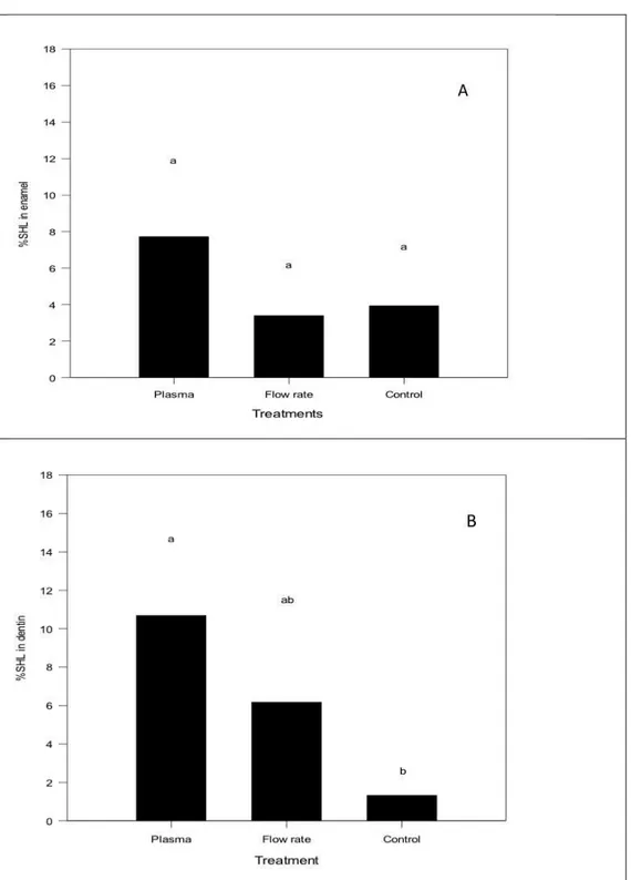
Figure 1 shows the difference between the initial and final microhardness in control,
argon flow rate and argon plasma group shows in percentage. The mean values of initial
surface (SH) and post surface (SH1) hardness were similar in enamel (p=0,087) (Fig 1A)
among the three groups. The microhardness measurements decreased in dentin compared
argon plasma and argon flow rate treatments demonstrated statistical significant differences
between argon plasma and control (p= 0.005) (Fig 1B).
The mean values and the standard deviations of enamel contact angle are expressed in
Figure 2A. Two-way analysis of variance (treatment type and time) showed a significant
effect for the factor time (p=0.025), no significant effect for the treatment type (p=0.092) and
different times tested (p>0.005). Argon plasma treatment showed statistically lower mean
than control after 1 min and 60 min (p<0.05). When the type of treatment was compared,
statistical differences were fond after 60 min (p<0.05).
The water contact angle results for dentin are shown in Figure 2B. Two-way analysis
of variance (treatment type and time) showed no statistical difference for the factor time
(p=0.087). Statistical significant difference was observed for the treatment type (p<0.001)
and for the interaction between them (p=0.042). Argon flow rate results demonstrated similar
contact angle at the different tested times. When the two treatments were compared at the
different times, statistically significant differences are found after 1 min and 60 min (p<0.05).
For the argon plasma treatment, the contact angle results for control group was statistically
higher than after plasma treatment 1min and 60 min (p<0.05).
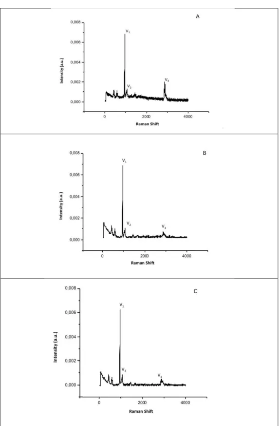
Selected Raman bands related to phosphate, carbonate and organic matrix and their
positions are shown in Figures 3-4. The bands observated are; at v1=962, v2=1070 and
v3=2943 cm-1. Both treatments with argon flow rate and argon plasma showed a decrease in
organic matrix bands for the enamel and dentin substrate.
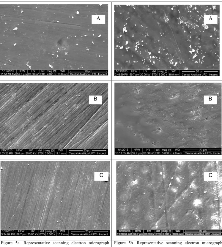
The surface morphology of enamel and dentin submitted to different treatments are
presented in Figure 5. Representative SEM images of control, argon flow rate and argon
plasma treatments visually demonstrate that both treatments remove smear layer and also
promote cracks on enamel and dentin surfaces, with more evident alterations in dentin
surfaces.
3.4 DISCUSSION
The gas utilized in this experiment was the argônio, a commonly used operating gas in
NTP (Norma Temperature and Pressure) technology because of its relatively low cost and
high sputtering yield30. The alterations in microhardness can be related to the loss or gain of minerals (demineralization and remineralization) of the dental structure. The results obtained
in the present study demonstrated that the enamel microhardness was not affected by argon
flow rate and argon plasma treatment. On the other hand, the results demonstrated that argon
plasma decrease significantilly the microhardness of dentin. These results might be an
indication, that LTP is able to etch organic substrate31 and to remove organic sheaths of the
surface, being a promosing technique in various dental clinical applications. Further studies
clinically relevante once recent researches demonstrated that the bleaching process, regardless
of whether or not it was light activated or different concentration, led to relevant changes in
the chemical composition of enamel and dentin in different concentration32,33.
Contact angle evaluation has been widely used to measure surface wettability of
different materials. Specifically, water contact angle is considered to be useful indication of
the surface tension and to reflect the wetting of the substrate by water4,34. The results indicated that contact angle values decreased considerably after 1min argon plasma treatment
for both enamel and dentin. For enamel, argon plasma treatment showed significant lower
contact angle after 1min. For dentin, this effect lasted for the 60 minutes of the experiment.
Sumarizing, the argon plasma treatment was able to significantty increase of surface wetting
with water which effect on dentin more pronounced than on enamel. The reduction of the
amount of carbon compounds on the surfaces which are transformed by chemical reactions is
assumed to be the main reason for better wetting. A strong fluid penetration into the surface
was also seen for dentin. These results are similar to those found in other studies on the same
substrate14,35,36 the X-ray Photoelectron Spectroscopy (XPS) analysis showed that after 30 seconds of plasma treatment, the atomic percent of carbon (C) decreased in the substrates,
suggesting that the C-containing materials were removed by the treatment14. It is also known
that the plasmas are able to etch the surface and remove organic matter via breaking of C-C
and C-H bonds. Polymeric matrix on the surface can be degraded into loosely-bonded
oligomers, low-molecular-weight oxidized material, or even volatile species by plasmas37.
Based on the concept that the intensity of the phosphate, carbonate and organic matrix
bands are related to the amount of each component in the analyzed substrate, it seems
reasonable to assume that the bands intensity reduction (organic matrix) could be that LTP is
able to etch the surface and remove organic matter via breaking of C-C and C-H bonds,
because this bonds are the most chemically energizable in the molecule38.
The SEM results of enamel indicated a decreased of the smear layer after application
of the argon plasma and argon gas, and morphological change type cracks were visible after
plasma application. These results are different to those found by34 using a different type of plasma. This could be explained because those authors used a helium gas flow of 3.525
standard liters per minute and a system power of 2W while in our study a argon plasma with a
higher flow rate (5 standard liters per minute) and power source (10W) was used. For the
dentin, similar SEM results were observed, with a decrease in smear layer and morphological
dentin. Dentinal tubules are filled with smear plugs and the intertubular areas appear slightly
roughened. No collagen fibers are visible on this surface after argon plasma treatment and the
orifices of dentine tubules appear irregularly, some of these results are similar to those
founded by34.
3.5 CONCLUSION
The use of argon plasma indicated decreased the superficial hardness of dentin and
increased the surface energy, wettability in both tested subtracts. It was demonstrated that
water contact angle values decreased after 1 min plasma treatment in dentin. Morphological
changes and decrease in the organic matrix were observed in enamel and dentin after argon
plasma and argon gas treatments. Summarising, the argon plasma is able to modify tooth
surfaces under tested conditions and showed who may offer possibility to integrate the
treatment as an additional step to optimize the adhesion of dental materials to enamel and
REFERENCES
1. Yasuda, H.,“Luminous chemical vapor deposition and interface engineering”,(2005).
2. D’Agotino, R., Favia, P., Oehr, C., and Wertheimer, M. R., “Low-temperature plasma
processinh of materials: Past, present, and future.” Plasma Process Polym.; 2 :7-15
(2005).
3. Penkov, O. V., Khadem, M., Lim, W., and Kim, D., “A review of recent applications
of atmosphericpressure plasma jets for materials processing. J. Coat. Technol. Res., 12
(2): 225–235 (2015). DOI 10.1007/s11998-014-9638-z .
4. Dowling, D. P., O’Neill, F. T., Langlais, S. J., and Law, V. J., “Influence of DC
Pulsed Atmospheric Pressure Plasma Jet Processing Conditions on Polymer
Activation”, Plasma Process. Polym., 8 (8) 718–727 (2011).
5. Ionita, E., Ionita, M., Stancu, E., Teodorescu, M., and Dinescu, G., “Small Size
Plasma Tools for Material Processing at Atmospheric Pressure”, Appl. Surf. Sci.;
255(10): 5448–5452, (2009).
6. Donegan, M., Milosavljevic, V., Dowling, D. P, “Activation of PET Using an RF
Atmospheric Plasma System”. Plasma Chem. Plasma Process. 33(5):941–957, 2013.
7. Ying, J., Chunsheng, R., Liang, Y., Jialiang, Z., and Dezhen, W., “Atmospheric
Pressure Plasma Jet in Ar and O2/Ar Mixtures: Properties and High Performance for
Surface Cleaning. Plasma Sci. Technol. 15 (12) 1203–1208 (2013).
8. Yang, B., Chen, J., Yu, Q., Li, H., Lin, M., and Mustapha, A., et al. Oral bacterial
deactivation using a low-temperature atmospheric argon plasma brush. J Dent. 39 (1):
48-56 (2011).
9. Shen, J., Cheng, C., Fang, S., Xie, H., Lan, Y., Ni, G., Meng, Y., Luo, J., Xiangke.
“Sterilization of Bacillus subtilis Spores Using an Atmospheric Plasma Jet with Argon
and Oxygen Mixture Gas”, Appl. Phys. Express. 5 :(3) 036201–036203 (2012).
10.Kang, S. K., Choi, M. Y., Koo, I. G., Kim, P. Y., Kim, Y., Kim, G.J., Mohamed, A.
A., Collins, G. J., and Lee, J.K., “Reactive Hydroxyl Radical-Driven Oral Bacterial
Inactivation by Radio Frequency Atmospheric Plasma”, Appl. Phys. Lett.2011, 98(14)
143702–143703 (2011).
11.Molnar, I., Papp, J., Alpar Simon, A., and Dan Anghel S., Deactivation of
No.6.(2013)
12.Duarte, S., Kuo, S. P., Murata, R. M., Chen, C. Y., Saxena, D., Huang, K. J., and
Popovic, S. Air plasma effect on dental disinfection. Physics of Plasmas, (2011).
13.Lu, X., Cao, Y., Yang, P., Xiong, Q., Xiong, Z., Xian, Y., and Pan, Y., “A Plasma
Device for Sterilization of Root Canal of Teeth”, IEEE T. Plasma Sci.;37 (5):668–673
(2009).
14.Chen, M., Zhang, Y., Driver, M. S., Caruso, A.N., Yu, Q., and Wang, Y., “Surface
modification of several dental substrates by non-thermal, atmospheric plasma brush”
Dent Mater. 29(8): 871-80 (2013).
15.Xiaohu, L., Feng, H., Ying, G., Jing, Z., and Jianjun, S., “Sterilization of
Staphylococcus Aureus by an Atmospheric NonThermal Plasma Jet”, Plasma Sci.
Technol., 15(5): 439–442 (2013).
16.Lademann, J., Richter, H., Alborova, A., Humme, D., Patzelt, A., and Kramer, A., et
al. “Risk assessment of the application of a plasma jet in dermatology,” J Biomed Opt.
14(5):054025, (2009).
17.Fluhr, J. W., Sassning, S., Lademann, O., Darvin, M. E., Schanzer, S., and Kramer, et
al., “In vivo skin treatment with tissue-tolerable plasma influences skin physiology
and antioxidant profile in human stratum corneum”, Exp Dermatol., 21(2):130-4
(2012).
18.Partecke, L. I., Evert, K., Haugk, J., Doering, F., Normann, L., and Diedrich, S., et al.
“Tissue tolerable plasma (TTP) induces apoptosis in pancreatic cancer cells in vitro
and in vivo”, BMC Cancer. 12:473 (2012).
19.Xu, C., Yao, X., Walker, M. P., and Wang, Y., “Chemical/molecular structure of the
dentin–enamel junction is dependent on the intra tooth location”, Calcified Tissue
Internat. 84(3): 221–8 (2009).
20.Teixeira, H. S., Coelho, P. G., Duarte, S., Janal, M. N., Silva, N., and Thompson, V.P.,
“Influence of atmospheric pressure plasma treatment on mechanical proprieties of
enamel and sealant bond strength”, J Biomed Mater Res B Appl Biomater.
103(5):1082-91 (2015).
21.White, D. J., and Featherstone, J. D., “A longitudinal microhardness analysis of
fluoride dentifrice effects on lesion progression in vitro”, Caries Res. 21(6): 502-12
(1987).
P. Biochemical composition and cariogenicity of dental plaque formed in the presence
of sucrose or glucose and fructose,” Caries Res., 34: 491-7 (2000).
23.Meredith, N., Sherriff, M., Setchell, D. J., and Swanson, S. A., “Measurement of the
microhardness and Young's modulus of human enamel and dentine using an
indentation technique”, Arch Oral Biol., 41(6): 539-45(1996).
24.Benetti, P., Della, Bona, A., and Kelly, J.R., “Evaluation of thermal compatibility
between core and veneer dental ceramics using shear bond strength test and contact
angle measurement,” Dent Mater. 26(8): 743-50 (2010).
25.Kinoshita, H., Miyoshi, N., Fukunaga, Y., Ogawa, T., Ogasawara, T. and Sano, K.J.,
Functional mapping of carious enamel in human Raman spectroscopy. Raman
Spectrosc, (2008).
26.Levallois, B et al. Molecular structural analysis of carious lesions using micro-Raman
spectroscopy. European Journal of Oral Sciences (0909-8836)2012 vol:120 iss:5
pg:444 -51, (2012).
27.Tramini, P., Plissier, B., Valcarcel, J., Bonnet, B., and Maury, L., “A Raman
spectroscopic investigation of dentin and enamel structures modified by lactic acid”,
Caries Res. 34 (3): 233-40 (2000).
28.Tsuda, H., Ruben, J., and Arends J., “Raman spectra of human dentin mineral”. Eur J
Oral Sci. 104:123-131, (1996).
29.Cruz, G. A., Toledo, S., Sallum, E. A., and Lima, A. F., “Morphological and chemical
analysis of bone substitutes by scanning electron microscopy and microanalysis by
spectroscopy of dispersion energy,” Braz Dent J., 18 (2): 129-33 (2007).
30.Chu, P.K., Chen, J.Y., Wang, L.P., and Huang, N., “Plasma-surface modification of
biomaterials”, Mater Sci Eng R-Report 36: 143–206 (2002).
31.Fricke, K., Steffen, H., Von Woedtke, T., Schröder, K., Weltmann, K. D., “High rate
etching of polymers by means of an atmospheric pressure plasma jet.” Plasma
Processes and Polymers. 8(1): 51–8 (2012).
32.Klaric, E., Rakic, M., Sever. I., Milat, O., Par, M., and Tarle, Z., “Enamel and Dentin
Microhardness and Chemical Composition After Experimental Light-activated
Bleaching” Operative Dentistry, 40-3, (2015).
33.Mondelli, L. R. F., Gabriel, G. T. R., Rizzante, P. F. A., Magalhães, A. C.,
Bombonatti, S. J. F., and Ishikiriama, S.K., “Do different bleaching protocols affect
34.Moritzer, E., Budde, C., and Leister, C., “Effect of Atmospheric Pressure Plasma
Pre-treatment and Aging Conditions on the Surface of Thermoplastics”, Weld World., 1–
10 (2014).
35.Lehmann, A., Rueppell, A., Axel Schindler, A., Isabella-Maria Zylla, I. M., Seifert, H.
J., Nothdurft, F., Hannig, M., Rupf, S., “Modification of Enamel and Dentin Surfaces
by Non-Thermal Atmospheric Plasma” Plasma Processes and Polymeres, 2013, 10,
262–270 (2013).
36.Silva, N. R., Coelho, P. G., Valverde, G. B., Becker, K., Ihrke, R., and Quade, A., et
al. Surface characterization of Ti and Y-TZP following non-thermal plasma exposure.
J Biomed Mater Res B Appl Biomater. 99(1): 199-206, (2011).
37.Gilliam, M., and Yu, Q., “Surface modification of a group of polymers using a low
temperature cascade arc torch”, J Appl Polym Sci., 105: 360–372 (2007).
38.Dong, X., Ritts, A. C., Staller, C., Yu, Q., Chen, M., and Wang, Y., “ Evaluation of
plasma treatment effects on improving adhesive-dentin bonding by using the same
tooth controls and varying cross-sectional surface areas. Eur J Oral Sci. 121(4):
Figure 1. Microhardness (A) Enamel polished and (B) Dentin polished
submitted to different treatments. Data represent mean values (n = 6) of the
percentage of enamel surface hardness loss (%SHL) according to the
treatments and the bars show standard deviations. Data followed by
different letters differ statistically (p < 0.05). Low case letters show the
Figure 2. Contact angle (A) Enamel polished and (B) Dentin polished submitted to different
treatments during 1min and 60min. Data represent mean values (n = 6) and standard
deviations. Data followed by different letters differ statistically (p < 0.05). Capital letters
compare treatments; Low case letters compare different times. Groups tested Plasma Argon En a m e l c o n ta c t a n g le 0 20 40 60 80 100 Control T=1mn T=60mn Aa Ab
Ba Aa Aa
Figure 5a. Representative scanning electron micrograph images of the surfasse morfology after treatments (5000X). Enamel polished (A) Control; (B) Argon flow rate; (C) Argon plasma.
Figure 5b. Representative scanning electron micrograph images of the surfasse morfology after treatments (5000X). Dentin polished (A) Control; (B) Argon flow rate; (C) Argon plasma.
A A
B B
4. CONCLUSÃO GERAL
A utilização de plasma de argônio diminuiu a dureza superficial de dentina e
aumentou a molhabilidade da superfície do esmalte e dentina. Mudanças na morfologia das
superfícies tratadas e diminuição na matriz orgânica foram observadas no esmalte e dentina
depois dos tratamentos com plasma de argônio e gás de argônio. Resumindo, o plasma de
argônio foi capaz de modificar as superfícies dos dentes nas condições testadas
REFERÊNCIAS GERAIS
AHMAD-NOUR, I.; FUAD, A.; MATTHIAS, H. Destruction of oral biofilms formed in situ on machined titanium (Ti) surfaces by cold atmospheric plasma. Biofouling, v.29, n.4, p.369-79, 2013.
ARDHAOUI, M.; ZHENG, M.; PULPYTEL, J.; DOWLING, D.; JOLIVALT, C.; KHONSARI, F.A. Plasma functionalized carbon electrode for laccase-catalyzed oxygen reduction by direct electron transfer. Bioelectrochemistry, n.91, p.52–61, 2013.
CHEN, M.; ZHANG, Y.M.; DRIVER, S. Surface modification of several dental substrates by non-thermal, atmospheric plasma brush. Dental Materials. v.29, p.871-80, 2013.
D’AGOTINO, R.; FAVIA, P.; OEHR, C.; WERTHEIMER, M.R. Low-temperature
plasma processinh of materials: Past, present, and future. Plasma Process Polym. v.2, p.7-15, 2005.
DONG, X.; RITTS, A.C.; YU, Q.; CHEN, M.; WANG, Y. Evaluation of plasma treatment effects on improving adhesive-dentin bonding by using the same tooth controls and varying cross-sectional surface areas. European Journal Oral Science. v.121, p.355-62, 2013.
DUARTE, S.; KUO, S.P.; MURATA, R.M.; CHEN, C.Y.; SAXENA, D.; HUANG, K.J.; POPOVIC, S.Air plasma effect on dental disinfection. Physics of Plasmas 18, 073503, 2011.
ERMOLAEVA, S.A.; SYSOLYATINA, E.V.; KOLKOVA, NATALIA, K. I. Non-thermal argon plasma is bactericidal for the intracellular bacterial pathogen Chlamydia trachomatis. Journal Medical Microbiology. V.61, p.793–99, 2012.
FLUHR, J.W.; SASSNING, S.; LADEMANN, O.; DARVIN, M.E.; SCHANZER, S.; KRAMER, S.; RICHTER, H.; STERRY, W.; LADEMANN, J. In vivo skin treatment with tissue-tolerable plasma influences skin physiology and antioxidant profile in human
stratum corneum. Experimental Dermatology. v.21, p.130-34, 2012.
GURSOY, H.; OZCAKIR-TOMRUK, C.; TANALP, J.; YILMAZ, S. Photodynamic therapy in dentistry: a literature review. Clinical Oral Investigations. v. 17, p.1113-1125, 2013.
HEINLIN, J.; ISBARY , G.; STOLZ, W.; MORFILL, G.; LANDTHALER, M.;
SHIMIZU, T.; STEFFES, B.; NOSENKO, T.; ZIMMERMANN, J.L.; KARRER, S.
Plasma applications in medicine with a special focus on dermatology. Journal European
Acadademy Dermatology Venereolgy. v.5, p.1–11, 2011.
HELLER, L.C.; EDELBLUTE, C.M.; MATTSON, A.M.; HAO, X.; KOLB, J.F.
Inactivation of bacterial opportunistic skin pathogens by nonthermal DC‐operated
JIANG, C.; CHEN, M.; GORUR, A.; SCHAUDINN, C.; JARAMILLO, D.E.; COSTERNON, J.W.; SEDGHIZADEH, P.P.; VERNIER, P.T.; GUNDERSEN, M.A.
Nanosecond Pulsed Plasma Dental Probe. Plasma Processes and Polymers. v.6, p.479–
83, 2009.
LAROUSSI, M. Low-Temperature Plasma Jet for Biomedical Applications: A Review.
IEEE Transactions On Plasma Science. v.43, n.3, p. 703, March 2015.
LEVINE, R. and EART R. Stadtman. Oxidative modification of proteins during aging E. Experimental Gerontology. v.36, n.9, p 1492-1502, September, 2001.
RITTS, A.C.; LI, H.; YU, Q.; XU, C.; YAO, X.; HONG, L.; WANG, Y. Dentin surface treatment using a non-thermal argon plasma brush for interfacial bonding improvement in
composite restoration. European Journal Oral Sciences. v. 118, p.510-516, 2010.
PAN, J, PENG SUN, P.; TIAN, Y.; ZHOU, H.; WU, H.; BAI, N.; LIU, F.; ZHU, W.; ZHANG, J.; KURT H. BECKER, K.H.; FANG, J. A novel method of tooth whitening using cold plasma microjet driven by direct current in atmospheric-pressure air. Plasma Sciences. IEEE Trans. v.38, p. 3143–3151, 2010.
PARTECKE, L.I; EVERT, K.; HAUGK, J.; DOERING, F.; NORMANN, L.; DIEDRICH, S.; WEISS, F.; EVERT, M.; HUEBNER, N.O.; GUENTHER, C.; HEIDECKE, C.D.; KRAMER, A.; BUSSIAHN, R.; WELTMANN, K.; PATI, O.; BENDER, C.; BERNSTORFF, W. Tissue Tolerable Plasma (TTP) induces apoptosis in
pancreatic cancer cells in vitro and in vivo. BioMedCentral Cancer. 12, 2012.
SHI, D.; HE, P.; LIAN, J.; WANG, L.; VANOOIJ, W.J. Plasma deposition and characterization of acrylic acid thin film on ZnO Nanoparticles. Journal of Materials Research. v.17, p.2555–2560, 2002.
SHOHET, J. L. Plasma aided manufacturing. IEEE Trans Plasma Sciences. v.19, p.725–
734, 1991.
SUHG, S.; HUH, J.; YUN, M.; CHANG, B.M.W.C.; J, C.; JEON, Y. Sterilization effect
of atmospheric pressure non-thermal air plasma on dental instruments. Journal
Advanced Prosthodontics. v.5, p. 2–8, 2013.
TEIXEIRA, H.S.; COELHO, P.G.; DUARTE, S.; JANAL, M.N.; SILVA, N. Influence of atmospheric pressure plasma treatment on mechanical proprieties of enamel and sealant
bond strength. Journal Biomedical Materials Research Part B-Applied Biomaterials.
v.103, n.5, p.1082-91, 2015.
TENDERO, C., TIXIER, C.; TRISTANT, P.; DESMAISON, J.; LEPRINCE, P.;
Atmospheric pressure plasmas: a review. Spectrochimica Act Part B. v.61, n.1, p.2-30,
2006.
YASUDA, H. Luminous Chemical Vapor Deposition and Interface Engineering. Santa Barbara, California: Marcel Dekker, 2005.
ZHANG, Q.; SUN, P.; ,FENG, H.; WANG, R.; LIANG, Y.; ZHU, W.; BECKER, K. H.
Anexo
Physics of Plasmas (PoP)
Manuscript Length for PoP Letters and Brief Communications
Manuscripts for Letters in Physics of Plasmas, as well as for Brief Communications, should
not exceed 3500 words (approximately four printed journal pages). Abstract, title, author list,
references and acknowledgments are all excluded from the 3500-word limit. Figures, tables,
and equations, however, are included and must be accounted for by calculating a word count
equivalent to the space they occupy. Circumvention of the length limitation by dividing a long
article into smaller parts is contrary to the purpose of this journal.
Please use these guidelines for estimating length of PoP Letters and Brief Communications
TeX users
Authors are advised to use the REVTeX 4.1 PoP style file. If the double-column version of
the manuscript obtained using the “reprint” option fits on approximately four pages
(excluding abstract, title, author list, references and acknowledgements), the length is
acceptable.
Word users
Highlight the manuscript text, excluding abstract, author list, acknowledgements and
references, and note the word count at the bottom of the screen. Add to that the
word-count-equivalents for figures, tables and equations as follows:
Figures: An average single-column figure will displace 220 words. For a more accurate estimation, use the following: 150/aspect ratio + 20 words for single-column figures and
300/0.5 X aspect ratio + 40 words for double-column figures. Aspect ratio = width/height.
Tables: 6.5 words per line, plus 13 words for single-column tables. 13 words per line, plus 26 words for double-column tables.
Equations: 16 words per row for single-column equations. 32 words per row for double-column equations.
If the total number of words (text + figures + tables + equations) is 3500 or less, the length is
acceptable.
Acceptable file formats
Please use Microsoft Word® or REVTeX 4.1.
Meticulous attention to the following brief guidelines will help the author and ensure prompt
error-free publications that precisely reflect the author’s intent.
Equations need to be editable so we recommend that you create them with the built-in Microsoft® Equation Editor included with your version of Word. If you wish to use
Mathtype, check for compatibility at http://tinyurl.com/lzny753.
Users of the Windows version of Word: Please embed all fonts.
Users of Macintosh Word: Please save all files in DOCX format, as the use of DOC is not
supported. Additionally, because font embedding is not possible, Mac Word users should
limit their font selection to those available from the basic installation.
Tables should be created with Word’s Insert Table function. If the table has already been made, please be sure it has been made with Word’s Table features. Tables created with spaces
or tabs will create problems and may be improperly typeset. To assure your table is published
as you wish, you must use Word’s Table function.
Footnotes should be inserted using Word’s Insert Footnote command. REVTeX
The REVTeX 4.1 package has been updated with changes for AIP Publishing journals and is
available for download.
The AIP Publishing Style will only work with REVTeX 4.1 and the latest version of natbib,
natbib8.3.
We are now accepting BibTex files. For more information please click here.
English-language editing
AIP Publishing recommends that authors contact Edanz, a global editing service with offices
in Japan and China, to receive a quotation of price and time. Note that this is not a
requirement or a guarantee of acceptance for publication.
Manuscript guidelines
These are the general guidelines for the AIP Publishing journals. However, in addition to
these guidelines if you are preparing a manuscript for APL, APM, CHA, JCP, JPCRD, PHF,
RSI, or SDY there are specific requirements which only apply to each particular journal.
Therefore, it is important to also read the section, specific journal guidelines after reading
these general guidelines.
The manuscript, including the abstract, references, and captions, should be set up on a 21.6 x 28 cm (8-1/2 x 11 in. or A4) grid with ample margins. It is essential that the motivation forthe
intelligible to a broad audience. The manuscript must be in good scientific AmericanEnglish.
All pages need to be numbered consecutively.
The manuscript should be arranged in the following order: title, author(s), affiliation(s), abstract, text, acknowledgments, appendixes, and references. Figures, with figure captions,
may be embedded within the manuscript to assist the reviewers. In addition, please submit
separate figure source files.
Series publications should be submitted in sequential order (Part I or I, Part II or II, etc.) and properly identified in the references. For series publication of closely related papers, the
descriptor “Part I,” or simply “I,” will not be included in the title of an article unless Part II
has been submitted for publication in the journal.
The abstract should serve as an index (including all subjects, major and minor, about which new information is given) and as a summary (presenting the conclusions and all results of
general interest in the article). It should be one paragraph with approximately 250 words. The
abstract should not contain displayed mathematical equations, footnotes, references, graphics,
or tabular material.
Authors’ names should be presented consistently across all publications to facilitate indexing and avoid ambiguity.
You may choose to have your Chinese, Japanese, or Korean names published in your own
language alongside the English versions in the author list. For Chinese, you may use either
Simplified or Traditional characters. Chinese, Japanese, or Korean characters must appear
within the author list of the manuscript when you are submitting and resubmitting the article.
For further information, please read Guidelines for Chinese, Japanese, or Korean names.
Equations should be punctuated and aligned to bring out their structure and numbered on the right. Mathematical operation signs indicating continuity of the expression should be placed
at the left of the second and succeeding lines. Use × rather than a centered dot, except for scalar products of vectors. The solidus (/) should be used instead of built-up fractions in
running text, and in display wherever clarity would not be jeopardized. Use “exp” for
complicated exponents.
Notation must be legible, clear, compact, and consistent with standard usage.
Footnote to the title should be set as a “Note” above the byline footnotes. For footnotes to the bylines, use a), b), c), etc. Avoid textual footnotes by inserting the information in the text.
Footnotes within tables should be designated by lowercase roman letter superscripts and given
References should be set as a double-spaced list at the end of the text. The names, including initials, of all authors in each reference should be given (in the text, the use of “et al.” is
permissible). For footnotes to title and bylines, use a), b), c), etc. Avoid lengthy textual
footnotes by inserting the information in the text. Footnotes within tables should be
designated by lowercase roman letter superscripts and given at the end of the table. All
references to books and journal articles, listed at the end of the paper, are to appear in only
one of these three formats:
By number, in order of first appearance, presenting the names of the authors, the journal name, volume, first page number only, and year, as in:19L. M. Pecora, T. L. Carroll, G. A.
Johnson, D.J. Mar, and J. F. Heagy, Chaos 7, 520 (1997).
This paper will be listed as the 19th in the list of references and cited as 19 or Ref. 19.
In alphabetical order according to the first author’s last name, including, in addition to the name, the title of the paper cited, journal name, volume, first and last page, and year, as in:
Pecora, L. M., Carroll, T. L., Johnson, G. A., Mar, D. J., and Heagy, J. F., “Fundamentals of
synchronization in chaotic systems, concepts, and applications,” Chaos 7, 520–543 (1997). This paper will be cited as “Pecora et al. (1997).” If there are several papers by the same
author(s) and the same year, they should be distinguished by letters, as in (1997a).
Alphabetically listed references (with full titles and page ranges) may be numbered according
to their alphabetical order and cited by their number.
Separate tables (numbered with roman numerals in the order of appearance in the text) should
be used for all but the simplest tabular material; they should have captions that make the
tables intelligible without reference to the text. The structure should be clear, with simple
column headings denoting all units. Unaltered computer output and notation are generally