Eicosapentaenoic acid and docosahexaenoic acid exert
anti-inflammatory and antinociceptive effects in rodents
at low doses
Maria Elizabeth Pereira Nobre
a,b, Alyne Oliveira Correia
a, Marília de Brito Borges
a,
Thayga Maria Araújo Sampaio
a, Solon Arcoverde Chakraborty
a,
Danilo de Oliveira Gonçalves
c, Gerly Anne de Castro Brito
c,
Luzia Kalyne Almeida Moreira Leal
c, Cícero Francisco Bezerra Felipe
a, Daniel Luna Lucetti
a,
Ricardo Mário Arida
b, Glauce Socorro de Barros Viana
a,c,⁎
aFaculty of Medicine Estácio of Juazeiro do Norte
–FMJ, Juazeiro do Norte, Brazil bFederal University of São Paulo
–UNIFESP, São Paulo, Brazil cFederal University of Ceará
–UFC, Fortaleza, Brazil
A R T I C L E I N F O A B S T R A C T
Article history:
Received 21 April 2012 Revised 17 February 2013 Accepted 28 February 2013
In the present study, we evaluated omega-3 polyunsaturated fatty acid (PUFA) (consisting of 20:5n-3 and 22:6n-3) properties on inflammation and nociception. Among the in vivo tests, writhing, formalin, and hot plate tests were conducted in mice, and carrageenan-induced paw edema, peritonitis, and Hargreaves tests were performed in rats. Following the carrageenan-induced edema, immunohistochemistry for tumor necrosis factor-α(TNF-α) was also carried out. We found that omega-3 PUFA treatment significantly decreased acetic acid–induced abdominal contortions as well as the first and second phases of the formalin test, which were reversed by naloxone. The carrageenan-induced rat paw edema was significantly reduced, along with neutrophil migration to the peritoneal cavity in the omega-3 PUFA treatment. In addition, there was a decrease in TNF-αimmunostained cells in the inflamed paw with the omega-3 treatment compared with no omega-3. Withdrawal threshold in response to the thermal stimulation was significantly increased by the omega-3 treatment in the Hargreaves and hot plate tests. The in vitro studies (myeloperoxidase, lactate dehydrogenase, MTT cell viability and lipid peroxidation assays) were performed in human neutrophils. These studies showed that omega-3 treatment significantly decreased myeloperoxidase release, presented no cytotoxicity, and did not alter lipid peroxidation. Our study suggests that omega-3 PUFA anti-inflammatory and antinociceptive actions may involve inhibition of cyclooxygenases and microglial activation, leading to a reduced release of proinflammatory cytokines such as TNF-α, among other factors. The omega-3 PUFAs are potential candidates used alone or in combination with conventional nonsteroidal anti-inflammatory drugs, for the treatment of diseases where inflammation plays an important role. © 2013 Elsevier Inc. All rights reserved.
Keywords:
Omega-3 polyunsaturated fatty acids
Anti-inflammatory and anti-nociceptive actions Cytotoxicity
Lipid peroxidation Antioxidant effects Neutrophils
Abbreviations:ANOVA, analysis of variance; BSA, bovine serum albumin; COX, cyclooxygenase; DHA, docosahexaenoic acid; EPA, eicosapentaenoic acid; MPO, myeloperoxidase; MTT, 3-(4 5-dimethylthiazol-2-yl)-2, 5-diphenyltetrazolium bromide; NSAID, nonsteroidal anti-inflammatory drug; PG, prostaglandin; PMA, phorbol myristate acetate; PUFA, polyunsaturated fatty acid; TBARS, thiobarbituric acid reactive substances; TNF-α, tumor necrosis factor-α.
⁎ Corresponding author.
E-mail address:[email protected](G.S.B. Viana).
0271-5317/$–see front matter © 2013 Elsevier Inc. All rights reserved.
http://dx.doi.org/10.1016/j.nutres.2013.02.011
A v a i l a b l e o n l i n e a tw w w . s c i e n c e d i r e c t . c o m
1.
Introduction
Omega-3 refers to a family of polyunsaturated fatty acids (PUFAs) among which the most nutritionally notable ones are α-linolenic acid (18:3n-3), eicosapentaenoic acid (EPA) (20:5n-3), and docosahexaenoic acid (DHA) (22:6n-3). The body converts α-linolenic acid to EPA and then to DHA. The formulation used in the present work is a combination of EPA to DHA, that is, a direct and efficient way to increase long-chain omega-3 PUFA body levels (eg, by consuming EPA- and DHA-rich foods or supplements)[1]. Eicosapentaenoic acid is converted to pros-taglandins (PGs) in the body to regulate cell activity and maintain healthy cardiovascular functions. Docosahexaenoic acid is one of the products of omega-3 PUFA metabolism and the main n-3 PUFA in the brain and nervous tissues[2].
The anti-inflammatory properties of EPA as well as DHA have been supported by numerous clinical studies, such as those focusing on maintaining normal blood cholesterol levels, controlling depression, and preventing stroke and cancer[3]. Docosahexaenoic acid is another long-chain omega-3 PUFA, found in abundance in fish and some algae. It is the predominant omega-3 PUFA, not only in the brain, but also in the retina, and an adequate supply of DHA is essential for proper brain, eye, and nerve functions. Low levels of DHA have been associated with Alzheimer disease and dementia[4].
Anti-inflammatory drugs consist of nonsteroidal anti-inflammatory drugs (NSAIDs) and glucocorticoids. Nonsteroi-dal anti-inflammatory drugs include nonselective cyclooxy-genase inhibitors (COX-1 and COX-2 inhibitors) and COX-2 selective inhibitors, also referred to as coxibs. Although COX-2 inhibitors present less upper gastrointestinal toxicity than traditional NSAIDs, both selective and nonselective COX inhibitors (excluding aspirin) can exert deleterious effects on the cardiovascular system[5,6]. Continued studies on inflam-matory diseases are needed because the side effects of available anti-inflammatory drugs pose a major problem for clinical use. The development of newer and more powerful anti-inflammatory drugs with fewer side effects is greatly needed.
Most of the literature on omega-3 PUFA deals with their anti-inflammatory effects and actions on cardiovascular diseases and diabetes[7-9]. Some literature states their effects on inflammatory bowel diseases and rheumatoid arthritis [10-13]. Recent studies also report omega-3 PUFA actions on inflammation in rodents[14-19], but few deal with nocicep-tion[20,21]. Furthermore, with few exceptions that adminis-tered omega-3 PUFA intravenously[17,19], most were carried out by adding omega-3 to the diet[15,16,18,21,22].
Based on these observations, we hypothesized that long-chain omega-3 PUFA exerts anti-inflammatory properties in the rat. To test this hypothesis, EPA and DHA were adminis-tered orally to rodents at low doses to determine the actions on nociception. In addition, our specific research objectives included conducting immunohistochemistry assays for TNF-α in rat paws with carrageenan-induced edema as well as myeloperoxidase (MPO), lactate dehydrogenase (LDH), 3-(4, 5-dimethylthiazol-2-yl)-2, 5-diphenyltetrazolium bromide (MTT), and thiobarbituric acid reactive substances (TBARS) assays in human neutrophils in vitro, to examine the extent of omega-3 PUFA anti-inflammatory and antioxidant actions.
2.
Methods and materials
2.1. Drugs
The omega-3 supplement used for this study was obtained from Proepa, Aché Laboratórios Farmacêuticos, São Paulo, Brazil, and each 1000-mg capsule contained 180 mg EPA, 120 mg DHA, and 2 mg tocopherol. Carrageenan (λ type IV), naloxone, and indomethacin were purchased from Sigma Chemical (St Louis, MO). Dexamethasone was obtained from Aché Laboratories; heparin, from Wyeth (São Paulo, Brazil); and morphine, from Cristália (São Paulo, Brazil). The omega-3 supplement (administered orally) was dissolved in an aque-ous suspension prepared with 0.4% Tween 80 (as vehicle and always used as control) immediately before use. All other reagents were of analytical grade.
2.2. Animals
Male Swiss mice (25-30 g) or male Wistar rats (180-200 g) were provided by the Animal House of the Federal University of Ceará, Brazil. The animals were housed in plastic cages with sawdust as bedding and kept in a room with controlled temperature (25°C ± 2°C) under a 12 hours/12 hours light/dark cycle. Food and water were provided ad libitum. The experiments were carried out according to the Guide for the Care and Use of Laboratory Animals of the US Department of Health and Human Services (NIH publication no. 85-23, revised 1985). The project was approved by the Animal Ethics Committee of the Faculty of Medicine of the Federal University of Ceará.
2.3. Pharmacological testing in vivo
2.3.1. Acetic acid–induced abdominal contortions
(writhing test) in mice
This method is used to evaluate peripheral analgesic actions. Acetic acid causes analgesia by liberating endogenous sub-stances, including serotonin, histamine, PGs, bradykinin, and substance P that stimulate pain in nerve endings. Local peritoneal receptors (acetylcholine and histamine receptors) and their mediators are postulated to be partly involved in the abdominal constriction (writhing) response[23]. The method has been associated with increased levels of PGE2and PGF2αin peritoneal fluids as well as lipoxygenase products. Male Swiss mice (8-12 animals per group) were used in this experiment. The animals were treated with the omega-3 supplement (1, 2.5, and 5 mg/kg, orally) 60 minutes before receiving a 0.6% acetic acid injection (10 mL/kg, IP), and the number of contractions was recorded for 20 minutes after a 10-minute interval.
2.3.2. Formalin test in mice
was recorded from 0 to 5 minutes (phase 1, neurogenic) and from 20 to 25 minutes (phase 2, inflammatory) after the formalin injection. The animals (8-15 per group) were treated with saline (0.1 mL/10 g, IP), morphine (4 mg/kg, IP), omega-3 supplement (0.5, 1, and 5 mg/kg, orally), morphine + nalox-one (4 and 1 mg/kg, IP, respectively), or naloxnalox-one + omega-3 (5 mg/kg, orally, respectively), 30 minutes before the formalin injection. Naloxone was injected 15 minutes before morphine or the omega-3 administrations. Morphine was used as the reference drug, whereas naloxone, an opioid antagonist, was used to assess a possible participation of the opioid system in the omega-3 PUFA effect.
2.3.3. Carrageenan-induced paw edema in rats
Paw swelling or footpad edema is a convenient method for assessing inflammatory responses to irritants such as carra-geenan. Typically, the drugs are assessed for acute anti-inflammatory activity by examining their ability to reduce or prevent the development of carrageenan-induced paw swell-ing. This model has long been used to evaluate the anti-inflammatory properties of agents such as NSAIDs that inhibit PG production[25]. The animals were randomly selected and divided into groups of 5 to 17 animals. The omega-3 supplement was dissolved in 1% Tween 80 and orally administered at the doses of 1, 2.5, and 5 mg/kg. The other groups were injected with either the reference drug (indo-methacin, 20 mg/kg, orally) or vehicle (Tween 80). Thirty minutes later, edema was induced by the injection of 0.1 mL of a 1% carrageenan solution into the animal's right hind paw. Measurements of the paw volume were done by means of a plethysmometer (Ugo Basile, Comerio, VA, Italy), immediately before the carrageenan injection and 1, 2, 3, 4, and 24 hours after. The paw edema volume was determined by the difference between the final and initial volumes.
2.3.4. Immunohistochemistry analyses for tumor necrosis
factor-α
Tumor necrosis factor-α (TNF-α) is a central regulator of inflammation, and its antagonists may be effective in treating inflammatory disorders in which TNF-αplays an important pathogenic role[26]. For immunohistochemistry assays of the TNF-α, the streptavidin-biotin-peroxidase method was used. Three groups of rats were treated with either the vehicle (0.4% Tween 80 in distilled water as normal controls) or omega-3 (2.5 or 5 mg/kg, orally). After 30 minutes, an intraplantar injection of 1% carrageenan was administered to the animals, and 3 hours later, all animals were euthanized, and 5-mm plantar region sections were immersed in a buffered formalin solution for 24 hours. The sections were then deparaffinized, dehy-drated in xylol and ethanol, and immersed in 0.1 M citrate buffer (pH 6) under microwave heating for 18 minutes to allow for antigen recovery. After cooling at room temperature for 20 minutes, the sections were washed with a phosphate-buffered saline (PBS) and followed by a 15-minute blockade of endogenous peroxidase with a 3% H2O2solution. The sections were incubated overnight (4°C) with rabbit primary antibodies (anti–TNF-α) as 1:200 in PBS–bovine serum albumin (BSA). The next day, the sections were washed in PBS and incubated for 30 minutes with the secondary biotinylated rabbit antibody (anti-IgG), 1:200 dilution in PBS-BSA. After washing in PBS, the
sections were incubated for 30 minutes, with the conjugated streptavidin peroxidase complex (ABC Vectastain complex; Vector Laboratories, Burlingame, CA). After another washing with PBS, the sections were stained with 3,3′ -diaminobenzi-dine-peroxide (DAB) cromophore, counterstained with Mayer hematoxylin, dehydrated, and mounted in microscope slides for analyses.
2.3.5. Carrageenan-induced peritonitis in rats
The carrageenan-induced peritonitis is a well-characterized experimental model of acute inflammation, largely used to test new anti-inflammatory drugs that permit the quantifica-tion of the leukocytes migraquantifica-tion into the peritoneal cavity[27]. Groups of 5 animals were treated with either the omega-3 supplement (2.5, 5, and 10 mg/kg, orally), dexamethasone (5 mg/kg, IP), or vehicle, 30 minutes before the induction of inflammation by means of a 2% carrageenan (500 μg/mL) intraperitoneal injection. All drugs were administered at a 10 mL/kg volume, and then the animals were returned to their cages with free access to water. After 5 hours, the peritoneal fluid was collected by abdominal laparoscopy. For this, all animals were pretreated with a heparinized saline (5 IU/mL, IP). A sample of the peritoneal fluid was diluted (1:10) in Turk liquid for quantification of cell numbers, using a Neubauer chamber. For differential counting of leukocytes, the exudate was centrifuged at 1000 rpm for 5 minutes, and 3% BSA (200μL) was added to the pellet for the preparation of slides. The cells were stained by a conventional fast pigment, and the results were expressed by the number of cells/mm3 (total and differential leukocyte counts in the wash fluid).
2.3.6. Hargreaves test in rats
The plantar test (Hargreaves' method) enables the researcher to discern the drug effect on a peripherally mediated response to thermal stimulation in the unrestrained rat. It measures the response to an infrared heat stimulus by focusing the infrared source below the rat plantar surface[28]. By pressing a button, the latency to paw withdrawal and infrared intensity are recorded automatically. The rats (180-200 g) were distributed into the following groups (5-6 animals per group): normal controls (animals received only distilled water, orally), carra-geenan group (animals received water, orally, and subse-quently an intraplantar injection of 100μL 1% carrageenan into the right hind paw), Indo20 (animals received 20 mg/kg indomethacin, orally, before the injection of carrageenan), and 3 groups were treated with the omega-3 supplement (1, 5, and 10 mg/kg, orally). With the exception of normal controls, the treatments of all other groups were immediately followed by the administration of carrageenan. One hour after treat-ments, the animals were submitted to a focused thermal stimulation for provoking peripheral hyperalgesia. The with-drawal threshold (time in seconds to move the injected paw) was measured. The data were analyzed by analysis of variance (ANOVA) and the Student-Newman-Keuls test, with
P< .05 considered significant.
2.3.7. Hot plate test in rats
the nociceptive responses of mice when placed on a warmed metal plate, at a standard constant temperature. The latency to a nociceptive response was recorded at the baseline and 30, 60, 90, and 120 minutes after administration of the tested drugs. It was defined as the time elapsed until the subject licked or flicked its hind paw. The omega-3 supplement was orally administered at doses of 2.5 and 5.0 mg/kg, and morphine (4 mg/kg, IP) was used as reference.
2.4. In vitro studies
2.4.1. Myeloperoxidase release from human neutrophils
Myeloperoxidase is a hemeprotein abundantly expressed in polymorphonuclear leukocytes (neutrophils) and secreted during their activation [30]. The MPO release test from human neutrophils was performed according to a previously described method[31]. Myeloperoxidase is widely used as a biomarker for inflammation. In the present work, human leukocytes (2.5 × 10⁶ cells/mL) were suspended in buffered Hank's balanced solution with calcium and magnesium. The preparations predominately contained neutrophils (85.0% ± 2.8%), and the cell viability, as determined by the trypan blue test, was 97.7% ± 0.94%. The cells were preincubated with the omega-3 supplement (1, 10, 50, and 100μg/mL) for 15 minutes at 37°C. Indomethacin (Indo, 35.7 μg/mL) was used as the positive control and reference drug. Human neutrophils were stimulated by the addition of phorbol myristate acetate (PMA) (0.1 μg/mL) for 15 minutes at 37°C. The suspension was centrifuged for 10 minutes at 2000g, 4°C. Aliquots (50μL) of the supernatants were added to PBS (100μL), phosphate buffer (50μL, pH 7.0), and H2O2 (0.012%). After 5 minutes at 37°C, 3,3′,5, 5′-tetramethylbenzidine (1.5 mM, 20μL) was added as substrate, and the reaction was stopped by 30-μL sodium acetate (1.5 M, pH 3.0). The absorbance was determined spectrophotometrically at 620 nm.
2.4.2. Lactate dehydrogenase assay in human neutrophils
The assay was performed according to the manufacturer instructions (Labtest, Minas Gerais, Brazil). The LDH is a simple and accurate colorimetric assay for dead and plasma membrane damaged cells. Lactate dehydrogenase present in human neutrophils (due to plasma membrane damage) participates in a coupled reaction, which converts a yellow tetrazolium salt into a red formazan dye measured at 492 nm. The concentrations of the omega-3 supplement used were 1, 10, 50, and 100μg/mL. The amount of formazan formed in the reaction is directly proportional to the amount of LDH present in the samples, which, in turn, is directly proportional to the number of dead or damaged cells[32].
2.4.3. MTT assay for cell viability measurements in
human neutrophils
Another parameter used for cytotoxicity is the metabolic activity of viable cells. Tetrazolium salts are reduced only by metabolically active cells. Thus, the yellow salt MTT is reduced by the mitochondrial enzymatic system succinate-tetrazole reductase to a blue-purple–colored formazan salt. The absence of MTT reduction is an indication of cytotoxicity
[33]. Human neutrophils (2.5 × 10⁶cells/mL) were incubated in plaques of 96 wells for 30 minutes at 37°C under a 5% CO2
atmosphere, in the presence of omega-3 (1-100μg/mL), Hanks (untreated cells), or Triton X-100 (0.2% as reference). The plaques were then centrifuged at 2000 rpm for 15 minutes at 25°C, and the supernatants discarded. A 10% MTT solution (200μL, 10 mg/mL) was added to each well. After 3 hours, the cells were centrifuged again under the same conditions, and the supernatants discarded. Then, dimethyl sulfoxide (150μL) was added, the plaques were shaken for 15 minutes, and the absorbance was measured in the plaque reader at 540 nm. The experiments were carried out fivefold and repeated on 3 different days.
2.4.4. Determination of TBARS levels in human neutrophils
Oxidative stress in the cellular environment results in the formation of highly reactive and unstable peroxides derived from PUFAs. Their decomposition results in the formation of malondialdehyde, which can be colorimetrically quantified, following its reaction with thiobarbituric acid. The measure-ment of TBARS is a well-established method for screening and monitoring lipid peroxidation. This method was previ-ously described[34]. Briefly, a suspension of neutrophils (2.5 × 10⁶ cells/mL) was incubated with omega-3 (1, 10, 50, and 100μg/mol), vitamin E (50μg/mL), distilled water (vehicle), or Hanks' solution [Hanks’ balanced salt solution (HBSS) untreated cells]. Next, 10-μM citochalasin B was added, followed by 100-nM N-formyl-methionyl-leucyl-phenyl-ala-nine. After 20 minutes, the mixture was then centrifuged, and trichloroacetic (20% in HBSS) and tiobarbituric (1.2%) acids were added to the supernatant (500μL). The reaction medium was heated for 30 minutes at 100°C, and the absorbance determined at 560 nm.
2.5. Statistical analyses
All results are presented as means ± SEM. A One-way ANOVA followed by the Student-Newman-Keuls test were used for
Control
Omega3 1 Om
ega 3 2.
5
Omeg a3 5
0 10 20 30
***
***
***
N
o
. abd
om
inal con
to
rt
io
n
s
/20
m
in
comparing the results among treatments. However, in the case of tests evaluating effects at different time points (carrageenan-induced paw edema and hot plate tests), the Two-way ANOVA andP<0.05.
3.
Results
3.1. Pharmacological testing in vivo
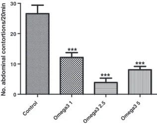
3.1.1. Writhing test
The acetic acid–induced writhing test in mice is used for detecting both central and peripheral analgesia. The intraper-itoneal administration of acetic acid provokes a stereotype behavior characterized by abdominal contractions, among other effects. In the writhing test, the oral administration of the omega-3 supplement decreased the acetic acid-induced
abdominal contortions by 48%, 85%, and 71% at doses as low as 1, 2.5, and 5 mg/kg, respectively (Fig. 1).
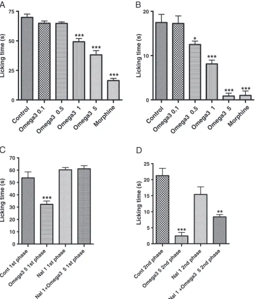
3.1.2. Formalin test
The omega-3 supplement doses at 1 and 5 mg/kg decreased the response of the first phase of the formalin test by 30% and 46%, respectively, whereas no effect was seen with the 0.5 mg/ kg dose. However, inhibitions of the order of 29%, 54%, and 95% were observed with the doses of 0.5, 1, and 5 mg/kg in the second phase of the test. Morphine, as expected, significantly decreased both phases of the formalin test (77% and 94% for the first and second phases, respectively) (Fig. 2A). On the other hand, the naloxone (1 mg/kg, IP, 15 minutes before omega-3 administration) pretreatment significantly reversed the effect of the omega-3 supplement at both phases of the test, suggesting the involvement of the opioid system in the antinociceptive effect of the fatty acids (Fig. 2B).
Contr ol
Omega3 0.1Omega3 0.5Omega3 1Omega3 5 Morphine
Contr ol
Omega3 0.1Omega3 0.5Omega3 1Omega3 5 Morphine 0
25 50 75
***
***
***
Lic
king time (s)
A
0 10 20
*
***
*** ***
Lic
king time (s)
B
Cont 1st phase
Omega3 5 1st phase Nal 1 1st phase
Nal 1+Omega3 5 1st phase
0 10 20 30 40 50 60 70
***
Lic
king time (s)
Lic
king time (s)
Cont 2nd phase
Omega3 5 2nd phase Nal 1 2nd phase
Nal 1 +Omega3 5 2nd phase
0 5 10 15 20 25
***
**
C
D
3.1.3. Carrageenan-induced paw edema in rats
The omega-3 supplement at doses of 1, 2.5, and 5 mg/kg, orally, significantly decreased the edema volume at all periods of observation, with percentage inhibitions of 42, 46, and 63 (third hour) and 65, 48, and 40 (fourth hour), at the 3 doses studied, respectively. Indomethacin, which was used as the reference drug, decreased the edema volume by 75% and 63%, at the third and fourth hours, respectively (Fig. 3).
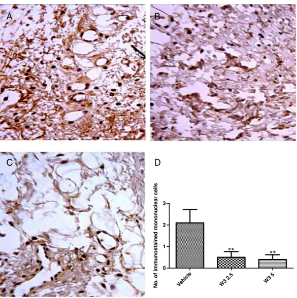
3.1.4. Immunohistochemistry for TNF-αin the
carrageenan-induced rat paw edema
InFig. 4A, an intense immunostaining was observed, indicat-ing a massive expression of TNF-α–positive cells in rat paws injected with 1% carrageenan (inflamed paw from the untreated group). It may also be inferred that TNF-αplays an important role in this inflammation model. It is possible to observe a strong brown staining in the cytoplasm of neutro-phils, eosinoneutro-phils, and macrophages because TNF-αis a well-known proinflammatory cytokine involved in carrageenan mechanisms. In the groups treated with omega-3 (2.5 and 5 mg/kg, orally), the immunostaining for TNF-αwas markedly reduced (Fig. 4B and C). The leukocytes infiltration was also reduced, and the small quantity of neutrophils present did not show any immunostaining. Therefore, it could be assumed that, at the doses tested, the omega-3 supplement reduces the expression of TNF-αin the model of paw edema induced by carrageenan.Fig. 4D shows the reduction in immunostained mononuclear cells after omega-3 treatment, as compared with the carrageenan-treated group (vehicle).
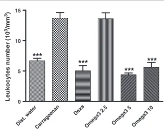
3.1.5. Carrageenan-induced peritonitis in rats
Although carrageenan significantly increased the neutrophil migration to the rat peritoneal cavity by 82%, as compared with negative controls, this increase was only 51% with omega-3 at the dose of 2.5 mg/kg, orally. The values returned to normal at omega-3 supplement doses of 5 and 10 mg/kg, and a similar effect was observed with dexamethasone (10 mg/kg, orally), used as the reference drug (Fig. 5).
3.1.6. Hargreaves test in rats
The omega-3 supplement at doses of 1, 5, and 10 mg/kg, orally, increased more than 2-fold the latency to withdrawal from the thermal stimulus (eg, withdrawal threshold), as compared with controls injected with carrageenan only. These effects were very similar to those observed after indomethacin administration, which was used as reference drug (2.4 times increase) (Fig. 6).
3.1.7. Hot plate test in mice
The omega-3 supplement significantly increased the with-drawal threshold in a dose-dependent manner compared with controls. Thus, 30 minutes after omega-3 administration (2.5 and 5.0 mg/kg, orally), the latency time increased 1.1 and 1.9 times compared with controls at the same period of observa-tion. After 90 minutes, the increases were 1.2 and 2 times for the 2 doses. Morphine (4 mg/kg, IP), used as the reference, increased around 3 times and near to the cutoff time (45 seconds) of the animal's withdrawal threshold to thermal stimuli (Fig. 7).
3.2. In vitro testing
3.2.1. PMA-stimulated MPO release from human neutrophils
The cells incubated in the presence of positive controls (0.4% Tween 80) significantly had increased MPO release, a bio-marker for inflammation in human neutrophils, by more than 2 times when compared with cells exposed to Hanks' solution (negative controls). The cells exposed to the omega-3 PUFA, at concentrations of 1, 10, 50, and 100 μg/mL, brought MPO release values close to or even lower (with the higher concentration) than those observed with the negative control. Similar results were observed for indomethacin that was used as a reference drug (Fig. 8).
3.2.2. Lactate dehydrogenase assay in human neutrophils
Although 0.2% Triton used as a cytotoxic and positive control increased the LDH release 6-fold, indicating a cell membrane damage, values observed in the presence of omega-3 PUFA at concentrations ranging from 1 to 100μg/mL were close to and not significantly different from those of the negative controls (HBSS solution) (Fig. 9).
3.2.3. MTT assay in human neutrophils
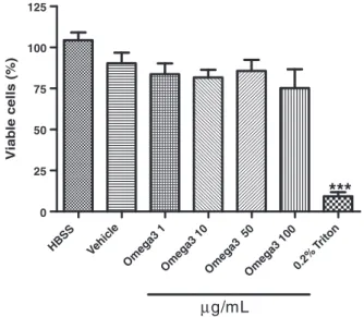
The omega-3 PUFA did not alter cell viability as evaluated by the MTT assay in human neutrophils in vitro. The observed values were close to those observed with the negative control (HBSS solution). The percentage of viable cells in the presence of 0.2% Triton (positive control) was very low (Fig. 10).
3.2.4. Determination of TBARS levels in human neutrophils
(lipid peroxidation assay)
The results presented inFig. 11show that the omega-3 PUFA at a low concentration range (1-50μg/mL) did not affect lipid peroxidation levels. However, at higher concentrations (100μg/mL), it increased TBARS levels, which is indicative of lipid peroxidation as compared with the negative control (HBSS solution). Use of 0.2% Triton significantly increased lipid peroxidation levels 9-fold. Vitamin E (50 μg/mL) pre-sented values close to those of negative controls.
1 h 2 h 3 h 4 h
0.0 0.5 1.0 1.5 2.0 2.5
Control Omega-3 1 Omega-3 2.5
Omega-3 5 Indo 20
***
***
***
***
***
***
***
Edema v
olume (mL)
4.
Discussion
Dietary omega-3 PUFA are known to present anti-inflamma-tory and immunomodulating effects that may be of relevance to several diseases (such as atherosclerosis and stroke) as well as chronic diseases involving the inflammatory processes
[12,13,35,36]. Previously, omega-3 PUFA anti-inflammatory actions were attributed mainly to their suppressive effect on the formation of arachidonic acid–derived PGs and leukotri-enes. However, more recent studies demonstrated that those effects are due to omega-3–derived lipid mediators, resolvins and protectins, which present anti-inflammatory and inflam-mation-resolving properties[37,38].
Other findings have demonstrated that some resolvins, such as those belonging to the D and E series, can dampen pain of inflammatory and postoperative origin [39]. Chemi-cally, these compounds are hydroxylated derivatives of EPA (for E-resolvins) and of DHA (for D-resolvins and protectin D1)
[40]. A recent review[41]discussed the mechanisms by which resolvins act as antinociceptives on their receptors in immune and neuronal cells by regulating inflammatory mediators, transient receptor potential ion channels, and spinal cord synaptic transmission.
Nonsteroidal anti-inflammatory drugs are the most widely prescribed drugs for the treatment of pain and inflammation. Conventional NSAIDs inhibit COX-1 and COX-2 isoforms of the COX enzyme. The inhibition of COX-1 up-regulates COX-2
D
B
A
C
**
**
Vehic le
W3 2.5 W3 5
0 1 2 3
No.
of imm
unostained monon
uc
lear cells
Fig. 4–Omega-3 PUFA decreased TNF-αimmunostainings in the rat inflamed paw, as evaluated by the carrageenan-induced edema model. The animals were treated with distilled water or the omega-3 supplement (2.5 and 5 mg/kg, PO) and 60 minutes later injected with carrageenan at the right hind paw. The animals were euthanized 3 hours later, and their paws processed for immunohistochemistry studies. A, Inflammation positive control (TNF-α). B, Omega-3 (2.5 mg/kg). C, Omega-3 (5 mg/kg). The
brown staining indicates interaction of primary and secondary antibodies and, as a consequence, the presence of TNF-α. The
expression in association with gastric hypermotility, and PGs produced by COX-2 counteract the deleterious effect of COX-1 inhibition[42]. In addition, the suppression of COX-2–derived prostacyclin (PGI2) is sufficient to explain most adverse cardiovascular effects of NSAIDs, which are likely to be augmented by secondary mechanisms such as suppression of nitric oxide production [43]. Newer NSAIDs have been
introduced in recent years, and although they present better safety, efficacy, and tolerability, the full spectrum of adverse reactions of these drugs is yet to be known[44]. Furthermore, there is a need for safer and more tolerable drugs. Natural products, alone or in combination with NSAIDs, would amplify the potency of anti-inflammatory drugs and reduce their side effects as already suggested[45].
Although the literature presents several studies on the inflammatory effects of omega-3 PUFA in humans [46-53], there are only a few based on their antinociceptive effects[20]
and almost none in rodents. Such findings induced us to conduct this work, relating the anti-inflammatory activity of omega-3 PUFA to their antinociceptive effects on experimental models of inflammation and nociception, at low doses. Although we did not perform pharmacokinetic experiments, there are several earlier and more recent works[54-57]on the
Dist. water Carrageenan
Dexa
Omeg a3 2
.5
Omega3 5 Omeg a3 10
0 5 10 15
***
***
***
***
L e uk oc y te s nu m b e r ( 1 0 3/m m 3)Fig. 5–Omega-3 PUFA (2.5, 5, and 10 mg/kg, orally) reduced leukocytes migration into the peritoneal cavity, as evaluated by the carrageenan-induced peritonitis model in rats. Dexamethasone (Dexa, 10 mg/kg, orally) was used as reference. Values are expressed as means ± SEM from 3 to 5 animals per group. Cell counting was performed 5 hours after the intraperitoneal injection of carrageenan. ***P< 0.001 vs carrageenan only (One-way ANOVA followed by Student-Newman-Keuls as the post hoc test).
Contr ol
Indo
Omega3 1 Om ega3
5
Om ega
3 10
0 10 20
**
***
***
**
R e ac ti o n t im e ( s )Fig. 6–Omega-3 PUFA (1, 5, and 10 mg/kg, orally) increased the latency to withdrawal (seconds) from the thermal stimuli, in the Hargreaves method in rats. Indomethacin (Indo 20 mg/ kg, orally) was used as reference. Measurements were performed 1 hour after administration of carrageenan at the right hind paw. Values are expressed as means ± SEM from 5 to 6 animals per group. **P< 0.01 and ***P< 0.001 vs control (One-way ANOVA followed by Student-Newman-Keuls as the post hoc test).
0 30 min 60 min 90 min 120 min
0 10 20 30 40 50 Control 2.5 mg/kg 5 mg/kg Morphine ** ** ** *** *** *** ** R e a c ti o n t im e (s )
Fig. 7–Omega-3 PUFA (2.5 and 5 mg/kg, orally) increased the reaction time (seconds) only at the higher dose, as evaluated by the hot plate test in mice. Morphine (4 mg/kg, IP) was used as reference. Measurements were performed at indicated times and the cutoff time was 45 seconds. Values are expressed as means ± SEM from 7 to 8 animals per group. *P< 0.05 and **P< 0.01 vs control, at the same period (Two-way ANOVA followed by the Bonferroni test).
HB SS
0.2% T riton
Om ega3 1
Omega3 10Omega3 50 Omega3 100 Indo 37.5 0 250 500 750
µ
g/mL
***
***
***
***
***
***
M P O r el eas e ( IU /m L )Fig. 8 – Omega-3 PUFA (1–100 μg/mL) inhibited
PMA-stimulated MPO (a biomarker for inflammation) release from human neutrophils in vitro. Indomethacin (Indo 37.5
μg/mL) was used as reference. Triton 0.2% was the positive
bioavailability of omega-3 PUFA in humans, and some in rodents as well[58,59]. We observed, even at very low doses, that omega-3 PUFAs were tolerated and absorbed in rats, but we cannot assume that the effects are entirely due to EPA and DHA or from their lipid metabolites. However, the evidence for the safety and efficacy of omega-3 PUFA are compelling[60]
and appear to be beneficial in the current study.
In the writhing test, we found that omega-3 PUFA decreased mice writhes values significantly and dose depen-dently. Different nociceptive mechanisms are known to be
involved in this rather nonselective model, such as the release of biogenic amines (as histamine and serotonin) and in-hibitions of COXs and their metabolites (as PGE2and PGF2α) as well as of the opioid system[61,62]. It is also established that the nociceptive response caused by acetic acid is dependent upon the release of cytokines, such as TNF-α, interleukin-1β, and interleukin-8, via modulation of macrophages and mast cells in the peritoneal cavity[63].
Although omega-3 PUFA significantly inhibited both phases of the formalin test, its effect occurred predominantly at the second phase of the test. The first phase of the formalin test corresponded to acute neurogenic pain, whereas the second phase corresponded to inflammatory pain. They are believed to reflect the excitation of peripheral afferent nociceptors and central sensitization [64,65]. Substance P and bradykinin participated in the first phase, whereas serotonin, histamine, nitric oxide, and PGs were involved in the second phase [66]. The first and second phases were attenuated by opioids, whereas COX inhibitors were known to attenuate only the second phase. Interestingly, we found that the naloxone pretreatment partially reversed the omega-3 PUFA effects in both phases of the formalin test, suggesting that the opioid system participated in that action. A recent work [20]also showed that antinociceptive effects of DHA were abolished after the naloxone pretreatment, which confirms our results. Thus, we could assume that omega-3 PUFA acts by both peripheral and central mechanisms because the formalin test encompasses inflammatory, neu-rogenic, and central mechanisms of nociception[24].
Carrageenan-induced inflammation is a classical model of edema formation and hyperalgesia, extensively used in studies of NSAIDs. It is known that peripheral inflammation involves an increase in COX-2–mediated PG synthesis in the central nervous system, contributing to allodynia and
HB SS
Omega3 1 Omega 3 10
Om ega3
50
Omega 3 10
0
0.2% Triton
0 50 100 150 200 250
µ
g/mL
***
***
***
***
***
LDH activity (U/L)
Fig. 9–Omega-3 PUFA (1-100μg/mL) effects evaluated by the LDH assay in human neutrophils in vitro. HBSS and 0.2% Triton are negative and positive controls, respectively. Values are expressed as means ± SEM. ***P< 0.001 vs 0.2% Triton (One-way ANOVA followed by the Student-Newman-Keuls as the post hoc test).
HBSS Vehic le
Omega3 1Omega 3 10
Ome ga3 5
0
Ome ga3 1
00
0.2% T riton
0 25 50 75 100 125
µ
g/mL
***
Viab
le cells (%)
Fig. 10–Omega-3 PUFA (1-100μg/mL) effects evaluated by
the MTT assay in human neutrophils in vitro. HBSS and 0.2% Triton were used as negative and positive controls, respectively. Values are expressed as means ± SEM. ***P< .001 vs all other samples (1-way ANOVA followed by the Student-Newman-Keuls as the post hoc test).
HBSS
0.2% T riton
Omega3 1 µg/
mL
Omega3 10 µg/mL
Omega3 50µ
g/mL
Ome ga3 100
µg/ mL
VitE 50 µg/
mL
0.00 0.25 0.50 0.75 1.00
a,b
a,b
a,b
b
b
Absorbance at 560 nm
Fig. 11–Omega-3 PUFA (1-100μg/mL) effects assessed by the
TBARS assay in human neutrophils in vitro. HBSS and 0.2% Triton are the negative and positive controls, respectively. Vitamin E (Vit E, 50μg/mL) was used as reference. Values are
hyperalgesia[25]. We observed that omega-3 PUFA reduced the carrageenan-induced edema that leads us to assume that these PUFA inhibit COX-2, thus decreasing PG concentrations. We also found that the omega-3 PUFA decreased MPO release to the peritoneal cavity, as evaluated by the carrageenan-induced peritonitis. In addition, the number of neutrophils was significantly reduced. This is a model of acute inflammation
[28] characterized by a rapid influx of polymorphonuclear neutrophils, followed by mononuclear cell infiltration. It is often used to assess the anti-inflammatory effects of drugs and as an in vivo model to study inflammatory mediators[67,68].
The Hargreaves method used mild radiant heat to measure thermal nociception in cutaneous hyperalgesia, and the time for foot withdrawal characterized the pain response [28]. It can be used as an in vivo model where inflammation is produced by a chemical agent as carrageen-an. This test enabled one to discern the drug effect on a peripherally mediated thermal stimulation, in the unre-strained rat, and is sensitive to COX inhibitors. We demon-strated that the omega-3 PUFA significantly increased the withdrawal threshold, not only in the plantar test, but also in the hot plate test. This last test is considered to be suitable for measuring the effects of opioid analgesics and is not sensitive to analgesic effects of NSAIDs [69]. Like the Hargreaves method, the hot plate is a common test that measures the response to thermal nociception of drugs acting by a central mechanism. Thus, as far as the antinociceptive effects are concerned, our data from both the Hargreaves method and the hot plate test confirm that these omega-3 PUFAs act by both peripheral and central mechanisms.
It is assumed that omega-3 PUFA may have antinociceptive properties, in part by inhibiting microglial release of matrix metalloproteinases[70]. Microglial cells appear to play a vital role in the initiation of processes promoting persistent pain states [71]. Glial activation can be induced by C-fiber nociceptive input from the sciatic nerve, and thus, the nociceptive-induced glial activation appears to be crucial in contributing to acute and inflammatory pain in rodent models
[72]. The pretreatment of RAW 264.7 cells with omega-3 was shown to significantly attenuate TNF-αproduction in lipo-polysaccharide-stimulated macrophages[73].
Clinical trials show that omega-3 PUFA may be of benefit in the management of patients with neuropathic pain[74]. A significant factor in neuropathic pain is the activation of spinal cord glial cells[75]. Activated glial cells are character-ized by the proliferation, hypertrophy, and increased produc-tion of inflammatory cytokines, such as interleukin-1β, interleukin-6, and TNF-α. Eicosapentaenoic acid and DHA could possibly reduce the production of these cytokines. In the present work, we demonstrated that the omega-3 treatment reduced immunostaining for TNF-αin the inflamed rat paw, as evaluated by the carrageenan-induced edema.
Our in vitro studies showed that the omega-3 supplement significantly decreased MPO release from PMA-stimulated human neutrophils, confirming the anti-inflammatory action of EPA and DHA. In addition, it did not present any cytotoxic effect on those cells at the concentration range used as assessed by the LDH and MTT assays. However, EPA and DHA increased lipid peroxidation at higher concentrations, as demonstrated by the TBARS assay.
Although we did not explore some other molecular/ cellular targets for EPA and DHA actions, these may involve inhibition of COXs and microglial activation, leading to a reduced release of proinflammatory cytokines, such as TNF-α. The EPA and DHA could be combined to reduce the amount of NSAIDs for the management of inflammatory diseases and pain. However, some additional translational studies are needed to confirm the beneficial effects of EPA and DHA and/or their active metabolites, as previously reported [76], alone or in conjunction with NSAIDs, for their potential in the treatment of inflammation and pain-related processes.
Acknowledgment
The authors are grateful to the Brazilian National Research Council (CNPq) and to the Foundation for Research and Development of the State of Ceará (FUNCAP) for financial support. They also thank M.O.L. Viana for the orthographic correction of the manuscript and Ms. Xenia M. de Sousa Serra for technical assistance.
R E F E R E N C E S
[1] Le HD, Meisel JA, Meijer VE, Gura KM, Puder M. The essentiality of arachidonic acid and docosahexaenoic acid. Prostaglandins Leukot Essent Fatty Acids 2009;81:165–70. [2] Pick M, Chen P, Perez M, Michaud M, Véricel E, Guichardant M,
et al. DHA metabolism: targeting the brain and lipoxygena-tion. Mol Neurobiol 2010;42:48–51.
[3] Kris-Etherton PM, Harris WS, Appel LJ. Fish consumption, fish oil, omega-3 fatty acids, and cardiovascular disease. Arterioscler Thromb Vasc Biol 2003;23:20–30.
[4] Calon F. Omega-3 polyunsaturated fatty acids in Alzheimer's disease: key questions and partial answers. Curr Alzheimer Res 2011;8:470–8.
[5] Roubille C, Martel-Pelletier J, Davy JM, Haraoui B, Pelletier JP. Cardiovascular adverse effects of anti-inflammatory drugs. Antiinflamm Antiallergy Agents Med Chem 2012 (Epub ahead of print).
[6] Macdonald TM, Mackenzie IS, Wei L, Hawkey CJ, Ford I. Methodology of a large prospective, randomized, open, blinded end-point streamlined safety study of celecoxib versus traditional non-steroidal anti-inflammatory drugs in patients with osteoarthritis or rheumatoid arthritis: protocol of the standard care versus celecoxib outcome trial (SCOT). BMJ Open 2013,http://dx.doi.org/10.1136/bmj open-2012-002295.
[7] Russo GL. Dietary n-6 and n-3 polyunsaturated fatty acids: from biochemistry to clinical implications in cardiovascular prevention. Biochem Pharmacol 2008;77:937–46.
[8] Skulas-Ray AC, Kris-Etherton PM, Harris WS, Vanden Heuvel JP, Wagner PR, West SG. Dose-response effects of omega-3 fatty acids on triglycerides, inflammation, and endothelial function in health persons with moderate hypertriglyceridemia. Am J Clin Nutr 2011;93:243–52.
[9] Bellenger J, Bellenger S, Bataille A, Massey KA, Nicolaou A, Rialland M, et al. High pancreatic n-3 fatty acids prevent STZ-induced diabetes in fat-1′mice: inflammatory pathway inhibition. Diabetes 2011;60:1090–9.
and immune-mediated diseases: inflammatory bowel disease and rheumatoid arthritis. Curr Pharm Des 2009;15: 4135–48.
[11] Wann AK, Mistry J, Blain EJ, Michael-Titus AT, Knight MM. Eicosapentaenoic acid and docosahexaenoic acid reduce interleukin-1β–mediated cartilage degradation. Arthritis Res Ther 2010;12:R207.
[12] Mori TA, Beilin LJ. Omega-3 fatty acids and inflammation. Curr Atheroscler Rep 2004;6:461–7.
[13] Wall R, Ross RP, Fitzgerald GF, Stanton C. Fatty acids from fish: the anti-inflammatory potential of long-chain omega-3 fatty acids. Nutr Rev 2010;68:280–9.
[14] Hagi A, Nakayama M, Shinzaki W, Haji S, Ohyanagi H. Effects of the omega-6: omega-3 fatty acid ratio of fat emulsions on the fatty acid composition in cell membranes and the anti-inflammatory action. J Parent Enteral Nutr 2010;34:263–70.
[15] McNamara RK, Jandaccek R, Rider T, Tso P, Cole-Strauss A, Lipton JW. Omega-3 fatty acid deficiency increases constitutive pro-inflammatory cytokine production in rats: relationship with central serotonin turnover. Prostaglandins Leukot Essent Fatty Acids 2010;83:185–91.
[16] Rossmeisl M, Jilkova ZM, Kuda O, Jelenik T, Medrikova D, Stankova B, et al. Metabolic effects of n-3 PUFA as phospho-lipids are superior to triglycerides in mice fed a high-fat diet: possible role of endocannabinoids. PLoS One 2012;7:e38834,
http://dx.doi.org/10.1371/journal.pone.0038834.
[17] Qui YD, Wang S, Yang Y, Yan XP. Omega-3 polyunsaturated fatty acids promote liver regeneration after 90% hepatectomy in rats. World J Gastroenterol 2012;18:3288–95.
[18] Labrousse VF, Nadjar A, Joffre C, Costes L, Aubert A, Grégoire S, et al, Layé S. Short-term long chain omega-3 diet protects from neuroinflammatory processes and memory impairment in aged mice. PLoS One 2012;7:e36861,http:
//dx.doi.org/10.1371/journal.pone.0036861.
[19] Hall JC, Priestley JV, Perry VH, Michael-Titus AT. Docosahexaenoic acid, but not eicosapentaenoic acid, reduces the early inflammatory response following compression spinal cord injury in the rat. J Neurochem 2012;121:738–50.
[20] Nakamoto K, Nishinaka T, Mankura M, Fujita-Hamabe W, Tokuyama S. Antinociceptive effects of docosahexaenoic acid against various pain stimuli in mice. Biol Pharm Biol 2010;33: 1070–2.
[21] Veigas JM, Williams PJ, Halade G, Rahman MM, Yoneda T, Fernandes G. Fish oil concentrate delays sensitivity to thermal nociception in mice. Pharmacol Res 2011;63: 377–82.
[22] Robbez Masson V, Lucas A, Gueugneau AM, Macaire JP, Paul JL, Grynberg A, et al. Long-chain (n-3) polyunsaturated fatty acids prevent metabolic and vascular disorders in fructose-fed rats. J Nutr 2008;138:1915–22.
[23] Calixto JB, Beirith A, Ferreira J, Santos AR, Cechinel-Filho V, Yunes RA. Naturally occurring antinociceptive substances from plants. Phytother Res 2000;14:401–18.
[24] Tjolsen A, Hole K. Animal models of analgesia. In: Dickenson A, Besson J, editors. The Pharmacology of Pain. Berlin, Germany: Spring Verlag; 1997. p. 1–20.
[25] Guay J, Bateman K, Gordon R, Mancini J, Riendeau D. Carrageenan-induced paw edema in rat elicits a predominant prostaglandin E2 (PGE2) response in the central nervous system associated with the induction of microsomal PGE2 synthase-1. J Biological Chemistry 2004;279: 24866–72.
[26] Esposito E, Cuzzocrea S. TNF-alpha as a therapeutic target in inflammatory diseases, ischemia-reperfusion injury and trauma. Curr Med Chem 2009;16:3152–67.
[27] Murai N, Nagai K, Fujisawa H, Hatanaka K, Kawamura M, Harada Y. Concurrent evolution and resolution in an acute
inflammatory model of rat carrageenan-induced pleurisy. J Leukoc Biol 2003;73:456–63.
[28] Hargreaves K, Dubner R, Brown F, Flores C, Joris J. A new and sensitive method for measuring thermal nociception in cutaneous algesia. Pain 1988;32:77–88.
[29] Sabina EP, Chandal S, Rasool MK. Inhibition of monosodium urate crystal-induced inflammation by withaferin A. J Pharm Pharm Sci 2008;11:46–55.
[30] Klebanoff SJ. Myeloperoxidase: friend and foe. J Leukocyte Biol 2005;77:598–625.
[31] Lucisano YM, Mantovani B. Lysosomal enzyme release from polymorphonuclear leukocytes induced by
immune complexes of IgM and of IgG. J Immunol 1984;132: 2015–20.
[32] Haslam G, Wyatt D, Kitos PA. Estimating the number of viable animal cells in multi-well culture based on their lactate dehydrogenase activities. J Biol Chem 2000;32: 63–75.
[33] Mosmann TJ. Rapid colorimetric assay for cellular growth and survival: application to proliferation and cytotoxicity assays. Immunol Methods 1983;65:55–63.
[34] Chiang K, Parthasarathy S, Santanam N. Estrogen, neutrophils and oxidation. Life Sci 2004;75:2425–38. [35] Kremer JM. N-3 Fatty acid supplements in rheumatoid
arthritis. Am J Clin Nutr 2000;71:349S–51S.
[36] Simopoulos AP. Omega-3 fatty acids in inflammation and autoimmune diseases. J Am Coll Nutr 2002;21:495–505. [37] Xu ZZ, Ji RR. Resolvins are potent analgesics for arthritic pain.
Br J Pharmacol 2011;164:274–7.
[38] Lee HN, Surth YJ. Therapeutic potential of resolvins in the prevention and treatment of inflammatory disorders. Biochem Pharmacol 2012;84:1340–50.
[39] Sommer C, Birklein F. Resolvins and inflammatory pain. F1000 Med Rep 2011;3:19.
[40] Weylandt KH, Chiu CY, Gomolka B, Waechter SF, Wiedenmann B. Omega-3 fatty acids and their lipid mediators: towards an understanding of resolving and protectin formation. Prostaglandins Other Lipid Mediat 2012;97:73–82.
[41] Ji RR, Xu ZZ, Strichartz G, Serhan CN. Emerging roles of resolvins in the resolution of inflammation and pain. Trends Neurosci 2011;34:599–609.
[42] Takeuchi K. Pathogenesis of NSAID-induced gastric damage: importance of cyclooxygenase inhibition and gastric hypermotility. World J Gastroenterol 2012;18: 2147–60.
[43] Yu Y, Riccioti E, Scalia R, Tang SY, Grant G, Yu Z, et al. Vascular COX-2 modulates blood pressure and thrombosis in mice. Sci Transl Med 2012;4:132–54.
[44] Ong CKS, Lirk P, Tan CH, Seymour RA. An evidence-based update on nonsteroidal anti-inflammatorty drugs. Clin Med Res 2007;5:19–34.
[45] Whithouse MW, Butters DE. Combination anti-inflammatory therapy: synergism in rats of NSAIDs/corticosteroids with some herbal/animal products. Inflammopharmacol 2003;11: 453–64.
[46] Komatsu W, Ishihara K, Murata M, Saito H, Shinohara K. Docosahexaenoic acid suppresses nitric oxide production and inducible nitric oxide synthase expression in interferon-gamma plus lipopolysaccharide-stimulated murine macrophages by inhibiting the oxidative stress. Free Radic Biol Med 2003;15:1006–16.
[47] Rasic-Milutinovic Z, Perunicic G, Pljesa S, Gluvic Z, Sobajic S, Djuric I, et al. Effects of N-3 PUFAs supplementation on insulin resistance and inflammatory biomarkers in hemodi-alysis patients. Ren Fail 2007;29:321–9.
[49] Tull SP, Yates CM, Maskrey BH, ODonnell VB, Madden J, Grimble RF, et al. Omega-3 fatty acids and inflammation: novel interactions review a new step in neutrophil recruit-ment. PLoS Biol 2009;7:e1000177,http:
//dx.doi.org/10.1371/journal.pbio1000177.
[50] Hao W, Wong OY, Liu X, Lee P, Chen Y, Wong KK.ω-3 fatty
acids suppress inflammatory cytokine production by macrophages and hepatocytes. J Pediatr Surg 2010;45: 2412–8.
[51] van Bussel BC, Henry RM, Schalkwijk CG, Ferreira I, Feskens EJ, Streppel MT, et al. Fish consumption in healthy adults is associated with decreased circulating biomarkers of endo-thelial dysfunction and inflammation during a 6-year follow-up. J Nutr 2011;41:1719–25.
[52] Calder PC. Fatty acids and inflammation: the cutting edge between food and pharma. Eur J Pharmacol 2011;668:S50–58. [53] Araki Y, Matsumya M, Matsuura T, Oishi M, Kaibori M,
Okumura T, et al. Peroxidation of n-3 polyunsaturated fatty acids inhibits the induction of iNOS gene expression in proinflammatory cytokine-stimulated hepatocytes. J Nutr Metab 2011,http://dx.doi.org/10.1155/2011/374542Article ID 374542.
[54] Marsen TA, Pollok M, Oette K, Baldamus CA.
Pharmacokinetics of omega-3-fatty acids during ingestion of fish oil preparations. Prostaglandins Leukot Essent Fatty Acids 1992;46:191–6.
[55] Bryhn M, Hansteen H, Schanche T, Aakre SE. The
bioavailability and pharmacodynamics of different concen-trations of omega-3 acid ethyl esters. Prostaglandins Leukot Essent Fatty Acids 2006;75:19–24.
[56] Dyerberg J, Madsen P, Moller JM, Aadestrup I, Schmidt EB. Bioavailability of marine n-3 fatty acid formulations. Prostaglandins Leukot Essent Fatty Acids 2010;83:137–41. [57] Davidson MH, Johnson J, Rooney MW, Kyle ML, King DF. A
novel omega-3 free fatty acid formulation has dramatically improved bioavailability during a low-fat diet compared with omega-3-acid ethyl esters: The ECLIPSE (Epanova® compared to Lovaza® in pharmacokinetic single-dose evaluation) study. J Clin Lipidology 2012;6:573–84.
[58] Tang X, Li ZJ, Xu J, Li JZ, Wang JF, Yanagita T, et al. Short term effects of different omega-3 fatty acids on lipid metabolism in mice fed high or low fat diet. Lipids Health Dis 2012;11:20. [59] Stavrovskaya IG, Bird SS, Marur VR, Baranov SV, Greenberg
HK, Porter CL, et al. Dietary omega-3 fatty acids do not change resistance of rat brain or liver mitochondria to Ca+2and/or pro-oxidants. J Lipids 2012;http://dx.doi.org/10.1155/2012/ 797105.
[60] Howland RH. Dietary supplement drug therapies for depression. J Psychosoc Nurs Ment Health Serv 2012;50:13–6. [61] Duarte IDG, Nakamura M, Ferreira SH. Participation of the
sympathetic system in acetic acid-induced writhing in mice. Braz J Med Biol Res 1988;21:341–3.
[62] Collier HOJ, Dinneen JC, Johnson CA, Schneider C. The abdominal constriction response and its suppression by analgesic drugs in the mouse. Br J Pharmacol Chemother 1968;32:295–310. [63] Ribeiro RA, Vale ML, Thomazzi SM, Paschoalato AB, Poole S,
Ferreira SH, et al. Involvement of resident macrophages and mast cells in the writhing nociceptive response induced by zymosan and acetic acid in mice. Eur J Pharmacol 2000;387: 111–8.
[64] Dickenson AH, Sullivan AF. Peripheral origins and central modulation of subcutaneous formalin-induced activity of rat dorsal horn neurons. Neurosci Lett 1987;83:207–11.
[65] Yaksh TL, Ozaki G, McCumber D, Rathbun M, Svensson C, Malkmus S, et al. An automated flinch detecting system for use in the formalin nociceptive bioassay. J Appl Physiol 2001;90:2386–402.
[66] García MD, Fernandez MA, Alvarez A, Saenz MT. Anti-nociceptive and anti-inflammatory effect of the aqueous extract from leaves ofPimenta racemosavar. ozua (Mirtaceae). J Ethnopharmacol 2004;91:69–73.
[67] Cuzzocrea S, McDonald MC, Filipe HM, Costantino G, Mazzon E, Santagati S, et al. Effects of tempol, a membrane-permeable radical scavenger, in a rodent model of carrageenan-induced pleurisy. Eur J Pharmacol 2000;390:209–22.
[68] Frode-Saleh TS, Calixto JB. Synergistic anti-inflammatory effect of kappaB inhibitors and steroidal or non-steroidal anti-inflammatory drugs in the pleural inflammation induced by carrageenan in mice. Inflam Res 2000;49:330–7. [69] Le Bars D, Gozariu M, Cadden SW. Animal models of
nociception. Pharmacol Rev 2001;53:597–652.
[70] Liuzzi GM, Latronico T, Rossano R, Viggiani S, Fasano A, Riccio P. Inhibitory effect of polyunsaturated fatty acids on MMP-9 release from microglial cells- implications for
complementary multiple sclerosis treatment. Neurochem Res 2007;32:2184–93.
[71] Smith HS. Activated microglia in nociception. Pain Physician 2010;13:295–304.
[72] Fu KY, Light AR, Maixner W. Relationship between nociceptors activity, peripheral edema, spinal microglial activation in cutaneous hyperalgesia. Pain 2000;101:1127–35. [73] Babcock TA, Helton WS, Hong D, Espat NJ. Omega-3 fatty acid
lipid emulsion reduces LPS-stimulated macrophage TNF-alpha production. Surg Infect (Larchmt) 2002;3:145–9. [74] Ko GD, Nowacki NB, Arseneau L, Eitel M, Hum A. Omega-3
fatty acids for neuropathic pain: case series. Clin J Pain 2010;26:168–72.
[75] White FA, Jung H, Miller RJ. Chemokines and the
pathophysiology of neuropathic pain. Proc Natl Acad Sci USA 2007;104:20151–8.